
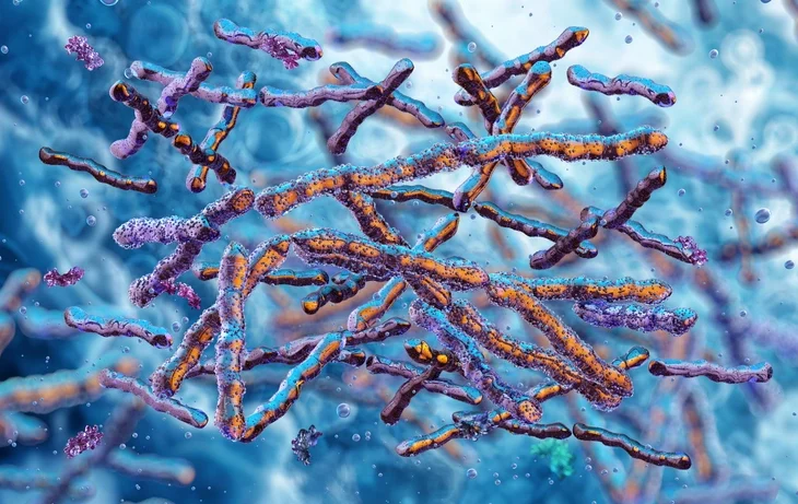
The oral cavity represents one of the most complex microbial ecosystems in the human body. It harbors a rich and dynamic microbial community composed of bacteria, fungi, viruses, protozoa, and archaea, existing in a delicate balance between health and disease. The field of oral microbiology explores these microorganisms, their ecological interactions, and their roles in oral and systemic pathology.
Understanding this balance is fundamental in periodontology, restorative dentistry, and preventive care. The microorganisms inhabiting the mouth are not merely passive colonizers; they are active participants in maintaining oral homeostasis, contributing to health, or promoting disease when equilibrium is disturbed.
Table of Contents
ToggleDevelopment of the Oral Microbiome
Initial Colonization
Colonization of the oral cavity begins within hours of birth. The initial microbial acquisition occurs through maternal and environmental exposure, primarily involving aerobic and facultative anaerobic bacteria such as Streptococcus salivarius, Streptococcus mitis, and Streptococcus oralis. These early colonizers establish themselves on the oral mucosa, tongue, and gingiva, creating a foundation for later microbial succession.
Ecological Succession
As teeth erupt, new non-shedding surfaces provide a stable substrate for biofilm formation, leading to the development of dental plaque, a structured biofilm that supports a more diverse microbial community. This process allows colonization by anaerobes and Gram-negative species, particularly in subgingival niches where oxygen tension is low.
By adulthood, over 700 bacterial species may colonize the oral cavity, with about 400 species commonly present in periodontal pockets. These organisms exist in a dynamic equilibrium, influenced by host factors (saliva, immune response, diet) and environmental conditions (oxygen levels, pH, temperature).
The Oral Biofilm: Structure and Function
Definition and Composition
A biofilm is a structured community of microorganisms enclosed within a self-produced extracellular polymeric matrix adherent to a surface. In the oral cavity, these surfaces include teeth, mucosa, and prosthetic materials.
Biofilm formation occurs through a sequence of events:
- Pellicle formation – salivary glycoproteins coat the tooth surface.
- Initial adhesion – pioneer bacteria such as Streptococcus sanguinis attach to the pellicle.
- Co-aggregation – secondary colonizers bind to primary ones through receptor–ligand interactions.
- Maturation – the biofilm develops a complex architecture with nutrient channels and communication networks.
- Detachment – cells or clusters disperse to colonize new sites.
Microbial Interactions and Communication
Biofilms exhibit quorum sensing, a process in which bacteria communicate using signaling molecules (autoinducers) to regulate gene expression collectively. This allows them to coordinate activities such as virulence factor production, biofilm maturation, and resistance to antimicrobials.
Fusobacterium nucleatum plays a pivotal role as a “bridge organism,” facilitating the integration of early colonizers (Streptococci) with late colonizers (anaerobes like Porphyromonas gingivalis and Treponema denticola).
Normal Oral Microflora and Their Beneficial Roles
Protective Functions
Commensal oral microorganisms provide numerous benefits:
- Colonization resistance – They prevent the establishment of exogenous pathogens by competing for nutrients and adhesion sites.
- Immune modulation – Normal flora stimulates the mucosal immune system, promoting IgA production and immune tolerance.
- Metabolic benefits – Certain bacteria produce beneficial metabolites, such as short-chain fatty acids and vitamins.
- Systemic interactions – Some bacterial products contribute to cardiovascular and gastrointestinal health through metabolic signaling.
Commensal–Pathogen Balance
In health, the oral microbiota remains in symbiosis, characterized by a balanced microbial community compatible with host tissues. When disturbed by factors such as poor hygiene, dietary changes, xerostomia, or immune suppression, a shift towards dysbiosis occurs, predisposing the host to diseases like dental caries, gingivitis, and periodontitis.
Pathogenic Biofilms and Disease Development
Caries-associated Biofilms
Caries is primarily driven by acidogenic and aciduric microorganisms capable of fermenting dietary carbohydrates into lactic acid, leading to enamel demineralization.
The major culprits include:
- Streptococcus mutans
- Streptococcus sobrinus
- Lactobacillus spp.
These organisms thrive in low pH environments and produce extracellular polysaccharides (dextran) that enhance biofilm cohesion and retention.
Periodontal Biofilms
Periodontal disease results from a host-mediated inflammatory response to a dysbiotic biofilm dominated by anaerobic, proteolytic Gram-negative species.
Key periodontal pathogens include:
- Porphyromonas gingivalis
- Tannerella forsythia
- Treponema denticola
- Aggregatibacter actinomycetemcomitans
These organisms produce virulence factors such as proteases, endotoxins (LPS), and enzymes that degrade host tissues, disrupt epithelial barriers, and modulate immune responses.
Major Microorganisms of Oral Importance
1. Streptococci
Streptococci dominate the oral flora, particularly the mitis, mutans, and salivarius groups.
Streptococcus mutans group
Includes S. mutans and S. sobrinus — key etiological agents in dental caries.
- Metabolism: Facultative anaerobes; ferment carbohydrates producing lactic acid.
- Virulence factors: Glucosyltransferases synthesize sticky dextrans aiding plaque adherence; acid tolerance mechanisms allow survival in low pH.
- Clinical relevance: Major contributors to enamel and dentin caries; high counts in plaque correlate with caries activity.
Streptococcus sanguinis, S. mitis, S. oralis
Members of the S. oralis group.
- Act as pioneer species in plaque formation.
- Associated with infective endocarditis when entering the bloodstream.
- Produce hydrogen peroxide, inhibiting pathogenic anaerobes.
Streptococcus salivarius
- Dominant in saliva and dorsal tongue.
- Produces bacteriocins that suppress pathogens like S. pyogenes.
- Generally non-pathogenic but important in maintaining oral balance.
Streptococcus anginosus, S. constellatus, S. intermedius
Formerly the S. milleri group.
- Microaerophilic; found in oral abscesses and purulent infections.
- Believed to contribute to periodontal disease progression.
2. Lactobacillus spp.
- Facultative anaerobic rods, secondary colonizers in carious lesions.
- Highly acidogenic and aciduric, promoting lesion progression rather than initiation.
- Commonly found in dentin caries, indicating advanced disease.
- Used as a diagnostic indicator of caries activity.
3. Actinomyces spp.
- Filamentous, branching bacteria resembling fungi.
- Actinomyces israelii causes actinomycosis, a chronic granulomatous infection producing sulfur granules.
- A. naeslundii and A. viscosus are associated with root surface caries and early plaque development.
- Thrive in low oxygen, high carbohydrate environments along the gingival margin.
4. Porphyromonas gingivalis
Gram-negative, obligate anaerobic rod.
A key pathogen in chronic and aggressive periodontitis.
Possesses numerous virulence factors:
Gingipains (proteases) that degrade host proteins and immune mediators.
Capsule and fimbriae aiding adhesion and immune evasion.
Endotoxins that trigger inflammation and bone resorption.
Often found in association with Tannerella forsythia and Treponema denticola forming the “red complex”, highly pathogenic in periodontal disease.
5. Aggregatibacter actinomycetemcomitans
- Formerly Actinobacillus actinomycetemcomitans.
- Capnophilic, microaerophilic Gram-negative coccobacillus.
- Major pathogen in aggressive periodontitis, particularly localized aggressive periodontitis (LAP) affecting adolescents.
- Produces leukotoxin targeting neutrophils and macrophages, impairing host defense.
- Invades epithelial cells, persisting in tissues and resisting antibiotics.
6. Tannerella forsythia
- Gram-negative, obligate anaerobe.
- Member of the red complex.
- Produces surface layer (S-layer) proteins aiding immune evasion.
- Frequently isolated from deep periodontal pockets.
- Correlates strongly with bleeding on probing and attachment loss.
7. Prevotella intermedia and Prevotella nigrescens
- Gram-negative anaerobes involved in necrotizing periodontal diseases and acute gingival infections.
- P. intermedia is often found in sites with severe gingival inflammation, especially in pregnancy gingivitis due to hormonal influence.
- P. nigrescens may be a more virulent variant, though its role is less defined.
8. Fusobacterium nucleatum
- Gram-negative, spindle-shaped anaerobe.
- Acts as a bridging organism, linking early and late colonizers within biofilms.
- Produces butyric acid and induces apoptosis in host cells.
- Involved in necrotizing ulcerative gingivitis (NUG) and systemic infections.
- Emerging evidence links it to colorectal cancer via hematogenous spread.
9. Spirochetes
- Motile, spiral-shaped organisms such as Treponema denticola and Borrelia vincentii.
- Highly invasive, capable of penetrating epithelial and connective tissues.
- Play critical roles in necrotizing periodontal diseases.
- Treponema denticola expresses proteases and adhesins enhancing virulence and tissue invasion.
- Borrelia vincentii is often considered a commensal but can participate in polymicrobial infections.
10. Candida albicans
- Opportunistic yeast-like fungus, commonly part of the normal flora.
- Becomes pathogenic in immunocompromised states, under antibiotic therapy, or with prosthetic appliances.
- Causes oral candidiasis, angular cheilitis, and denture stomatitis.
- Exhibits dimorphism (yeast and hyphal forms) aiding tissue invasion.
- Capable of co-aggregating with bacteria like Streptococcus mutans, enhancing pathogenicity.
Host–Microbe Interactions in Oral Health and Disease
Innate Defense Mechanisms
The oral cavity employs multiple innate defense systems:
- Saliva: Contains lysozyme, lactoferrin, histatins, and peroxidases with antimicrobial properties.
- Epithelial barriers: Provide physical protection and secrete cytokines and defensins.
- Crevicular fluid: Contains neutrophils and complement components that control subgingival microbes.
Adaptive Immunity
- Secretory IgA in saliva prevents bacterial adherence.
- IgG and IgM in gingival crevicular fluid neutralize bacterial antigens.
- Dysregulation of immune responses can lead to chronic inflammation and tissue destruction.
Symbiosis, Dysbiosis, and Periodontal Disease
The symbiosis–dysbiosis model describes the shift from a balanced commensal state to a pathogenic microbial community.
- In symbiosis, commensal bacteria promote tissue homeostasis and immune tolerance.
- In dysbiosis, ecological changes (e.g., inflammation, nutrient availability) favor pathogenic species such as P. gingivalis, which further exacerbate inflammation — a vicious cycle leading to periodontitis.
P. gingivalis acts as a keystone pathogen, capable of altering the microbial community structure and host response even at low abundance.
Clinical Implications and Management
Diagnosis
Understanding the microbial basis of disease guides:
- Microbiological testing (e.g., PCR, DNA–DNA hybridization).
- Plaque sampling to identify specific pathogens.
- Recognition of disease-specific microbial profiles.
Therapeutic Strategies
- Mechanical debridement (scaling and root planing) remains the cornerstone of therapy, disrupting biofilms.
- Antimicrobial agents (e.g., chlorhexidine, metronidazole, amoxicillin) target key pathogens.
- Probiotics and prebiotics aim to restore microbial balance.
- Host modulation therapies (e.g., subantimicrobial-dose doxycycline) reduce destructive inflammatory responses.
Preventive Measures
- Effective oral hygiene practices minimize biofilm accumulation.
- Dietary control of fermentable carbohydrates reduces caries risk.
- Fluoride use enhances remineralization and inhibits bacterial metabolism.
- Regular professional maintenance sustains microbial balance and periodontal health.
Conclusion
The oral microbiome represents a highly evolved, intricate ecosystem whose stability is essential for maintaining oral health. Disruption of this balance — through microbial shifts, host factors, or environmental influences — leads to pathological biofilms associated with caries, gingivitis, and periodontitis.
For the dental professional, an in-depth understanding of oral microbiology is indispensable. It forms the biological foundation for effective preventive, diagnostic, and therapeutic strategies in managing oral diseases. Future research focusing on microbiome modulation, precision diagnostics, and host–microbe interactions holds great promise for advancing periodontal and restorative care.
References
- Marsh, P. D., & Martin, M. V. (2009). Oral Microbiology (5th ed.). Elsevier Health Sciences.
- Newman, M. G., Takei, H., Klokkevold, P. R., & Carranza, F. A. (2019). Carranza’s Clinical Periodontology (13th ed.). Elsevier.
- Samaranayake, L. (2018). Essential Microbiology for Dentistry (5th ed.). Elsevier.
- Slots, J., & Slots, H. (2019). Periodontitis: Facts, fallacies and the future. Periodontology 2000, 79(1), 7–23.
- Dewhirst, F. E., Chen, T., Izard, J., Paster, B. J., Tanner, A. C. R., Yu, W.-H., Lakshmanan, A., & Wade, W. G. (2010). The human oral microbiome. Journal of Bacteriology, 192(19), 5002–5017.
- Hajishengallis, G., & Lamont, R. J. (2021). Polymicrobial communities in periodontal disease: Their quasi-organismal nature and dialogue with the host. Periodontology 2000, 86(1), 210–230.
- Kolenbrander, P. E., Palmer, R. J., Periasamy, S., & Jakubovics, N. S. (2010). Oral multispecies biofilm development and the key role of cell–cell distance. Nature Reviews Microbiology, 8(7), 471–480.
- Paster, B. J., & Dewhirst, F. E. (2006). The phylogenetic diversity of the oral microbiota. Periodontology 2000, 42, 44–58.
- Hajishengallis, G. (2014). The inflammophilic character of the periodontitis-associated microbiota. Molecular Oral Microbiology, 29(6), 248–257.
- Socransky, S. S., & Haffajee, A. D. (2005). Periodontal microbial ecology. Periodontology 2000, 38(1), 135–187.
- Lamont, R. J., Koo, H., & Hajishengallis, G. (2018). The oral microbiota: Dynamic communities and host interactions. Nature Reviews Microbiology, 16(12), 745–759.
- Takahashi, N., & Nyvad, B. (2011). The role of bacteria in the caries process: Ecological perspectives. Journal of Dental Research, 90(3), 294–303.
- Jenkinson, H. F., & Lamont, R. J. (2005). Oral microbial communities in sickness and in health. Trends in Microbiology, 13(12), 589–595.
- Darveau, R. P. (2010). Periodontitis: A polymicrobial disruption of host homeostasis. Nature Reviews Microbiology, 8(7), 481–490.
- Holmstrup, P., Klinge, B., & Matsson, L. (2017). Clinical Periodontology and Implant Dentistry (6th ed.). Wiley-Blackwell.