
Oral medicine frequently overlaps with dermatology and rheumatology because many systemic and skin disorders present with oral signs and symptoms. The oral cavity often reflects underlying systemic pathology, and in some cases, oral lesions may precede cutaneous or systemic manifestations. Recognition of these features is essential for early diagnosis, appropriate referral, and long-term monitoring.
Table of Contents
ToggleLichen Planus
Lichen planus is a chronic inflammatory mucocutaneous disorder that primarily affects adults. It involves both the skin and mucous membranes and affects approximately 1–2% of the general population. Oral lichen planus (OLP) is common: around 50% of patients with cutaneous lesions have oral involvement, and about 25% may present with oral lesions alone.
Etiopathogenesis
Lichen planus is considered an autoimmune condition. It is mediated by a T-lymphocyte–driven immune response directed against basal keratinocytes of stratified squamous epithelium. Cytotoxic CD8+ T cells trigger apoptosis of epithelial cells, leading to characteristic histopathologic changes. The condition may be associated with:
- Genetic predisposition
- Psychological stress
- Systemic diseases
- Hepatitis C infection (in some populations)
- Drug-induced lichenoid reactions (e.g., antihypertensives, NSAIDs, antimalarials)
- Contact hypersensitivity reactions (e.g., to amalgam restorations)
Clinical Features
Oral lichen planus is more common in females, with a female-to-male ratio of approximately 3:2. It is typically chronic and may persist for months or years.
The lesions are usually bilateral and symmetrical, most commonly affecting:
- Posterior buccal mucosa
- Tongue
- Gingiva
- Lips
- Floor of the mouth
It rarely involves the palate.
The most common presentation is the reticular form, characterized by lacy white lines known as Wickham striae. These represent areas of hyperkeratosis.
Other clinical patterns include:
- Papular
- Plaque-like
- Erosive (ulcerative)
- Atrophic
- Bullous
The erosive and atrophic forms are often symptomatic and may cause burning, soreness, or sensitivity to spicy foods.
A common gingival manifestation is desquamative gingivitis, where erythematous, friable gingivae may peel or ulcerate.
Cutaneous lesions classically appear on flexor surfaces of wrists, arms, and legs as purple, polygonal, pruritic papules with fine white lines.
Histopathology
Microscopically, the condition demonstrates:
- Hyperkeratosis
- Saw-tooth–shaped elongated rete ridges
- Dense band-like lymphocytic infiltrate beneath the epithelium
- Degeneration of basal keratinocytes
Malignant Potential
Although lichen planus is essentially benign, there is ongoing debate about malignant transformation, particularly in the erosive and atrophic types. Reported transformation rates range from 0.5% to 2%. Regular monitoring is therefore recommended.
Management
Treatment depends on symptom severity.
- Asymptomatic reticular lesions: reassurance and periodic review
- Symptomatic lesions: topical corticosteroids such as betamethasone, dexamethasone, or prednisolone mouthwash
- Severe or refractory cases: systemic corticosteroids, azathioprine, tacrolimus, or retinoids
- Biopsy: indicated to confirm diagnosis or exclude dysplasia
Erosive forms require follow-up every 6 months. Clinical photography and repeat biopsy may be indicated if suspicious changes occur.
Dyskeratosis Congenita
Dyskeratosis congenita is a rare inherited disorder, usually autosomal dominant, characterized by defective telomere maintenance.
Clinical Triad
- Oral leukoplakia
- Nail dystrophy
- Skin hyperpigmentation
Oral leukoplakia in this condition carries a high risk of malignant transformation. Patients are also at risk for bone marrow failure, leading to anemia, leukopenia, and thrombocytopenia.
Oral Manifestations
- Persistent white patches (leukoplakia)
- Increased risk of squamous cell carcinoma
- Periodontal disease
Management
Requires multidisciplinary care, including hematologic monitoring and regular oral cancer screening.
Vesiculo-Bullous Lesions
Vesiculo-bullous disorders are characterized by blister formation and may involve the oral cavity either primarily or secondarily. They are broadly classified into:
- Intraepithelial blistering disorders (e.g., pemphigus vulgaris)
- Subepithelial blistering disorders (e.g., mucous membrane pemphigoid)
Oral lesions may precede skin involvement, especially in pemphigus vulgaris. These conditions often present with fragile bullae that rupture to form painful erosions.
Management typically involves systemic corticosteroids and immunosuppressive therapy.
Ehlers–Danlos Syndrome
Ehlers–Danlos syndrome (EDS) is a heterogeneous group of inherited connective tissue disorders caused by mutations affecting collagen synthesis.
Clinical Features
- Hyperextensible skin
- Hypermobile joints
- Fragile blood vessels
- Easy bruising and bleeding
Oral Manifestations
- Severe early-onset periodontal disease
- Small teeth with short roots
- Pulp stones
- Temporomandibular joint hypermobility
- Delayed wound healing
- Excessive bleeding during dental procedures
Some types are associated with mitral valve prolapse and increased susceptibility to infective endocarditis. Others carry risk of vascular rupture and cerebrovascular accident.
Dental Considerations
- Gentle surgical technique
- Careful suturing
- Hemostasis monitoring
- Caution during root canal treatment
Management is largely supportive.
Rheumatoid Arthritis
Rheumatoid arthritis (RA) is a chronic autoimmune inflammatory disease primarily affecting synovial joints.
Associations
- Sjögren syndrome (~15%)
- TMJ involvement (~10%)
TMJ arthritis may cause:
- Pain
- Swelling
- Limited mouth opening
- Joint stiffness
Rarely, pannus formation can occur, leading to joint destruction.
Oral Considerations
- Xerostomia (if associated with Sjögren syndrome)
- Increased caries risk
- Difficulty maintaining oral hygiene
Management includes systemic immunosuppressive therapy such as methotrexate or TNF-α inhibitors.
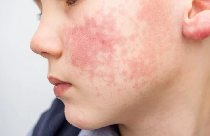
Systemic Lupus Erythematosus (SLE)
Systemic lupus erythematosus is a multisystem autoimmune disease of unclear etiology. It predominantly affects females.
Clinical Features
- Malar “butterfly” rash
- Photosensitivity
- Arthritis
- Anemia
- Renal involvement
Oral Manifestations
Present in about 30% of patients:
- Ulcers
- Erythematous patches
- Purpura
Antinuclear antibodies (ANA) are present. Diagnosis may involve histologic and immunofluorescent confirmation.
Management includes systemic corticosteroids and immunosuppressive agents.
Discoid Lupus Erythematosus (DLE)
DLE is a chronic cutaneous form of lupus limited to skin and mucosa.
Oral Features
- Disc-like white plaques
- Central atrophy
- Peripheral keratotic striae
- Lip lesions (potentially premalignant in women)
Approximately 30% of patients have oral lesions. DLE can occasionally progress to SLE but often remains localized.
Diagnosis
Distinguished from SLE by specific antibody profiles (e.g., double-stranded DNA antibodies).
Management
- Topical corticosteroids
- Dapsone or thalidomide in refractory cases
- Monitoring for malignant transformation
Systemic Sclerosis (Scleroderma)
Systemic sclerosis is characterized by progressive fibrosis of connective tissues.
Epidemiology
- More common in females
- Onset between 30–50 years
Clinical Features
- Raynaud’s phenomenon
- Mask-like facies (“Mona Lisa face”)
- Skin tightening
- Dysphagia due to esophageal involvement
Oral Manifestations
- Microstomia (reduced mouth opening)
- Xerostomia
- Difficulty wearing dentures
- Increased periodontal disease risk
Autoantibodies are present. Disease monitoring may involve biomarkers such as E-selectin.
Management
- Immunosuppressive therapy
- Cyclophosphamide and steroids in early disease
- Symptomatic care
Five-year survival is approximately 70%.
Polyarteritis Nodosa
Polyarteritis nodosa is a necrotizing vasculitis affecting small and medium-sized arteries.
Clinical Features
- Systemic inflammation
- Organ ischemia
- High mortality (up to 60% in first year untreated)
Oral Manifestations
Ulcerations due to vascular necrosis
Treatment with systemic steroids improves 5-year survival to approximately 40%.
Dermatomyositis
Dermatomyositis is an inflammatory disease affecting skin and skeletal muscles.
Clinical Features
- Proximal muscle weakness
- Heliotrope rash
- Gottron’s papules
Approximately 15% of cases are associated with internal malignancy.
Oral Manifestations
- Ulcers
- Soreness of tongue
- Palatal discomfort
- Gingival inflammation
Management includes corticosteroids and immunosuppressive therapy, along with cancer screening.
Reiter Syndrome (Reactive Arthritis)
Reactive arthritis is characterized by the triad:
- Arthritis
- Urethritis
- Conjunctivitis
Oral manifestations may include:
- Mucosal ulcerations
- Geographic tongue–like lesions
- Keratoderma blennorrhagicum (cutaneous lesions)
Management includes NSAIDs and immunomodulatory therapy.