
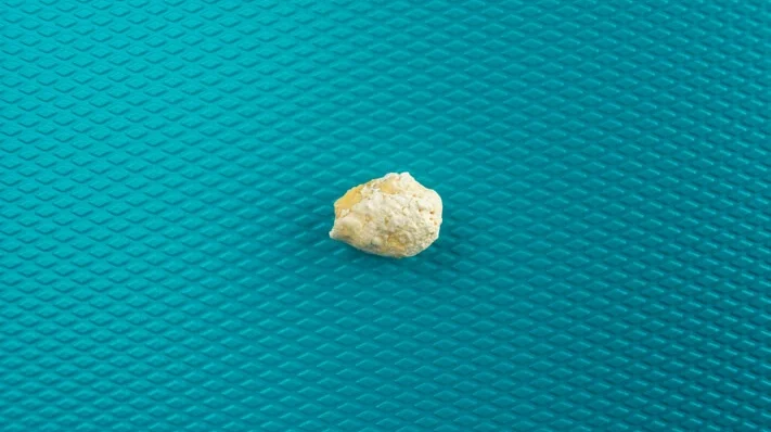
Sialolithiasis, commonly referred to as salivary gland stones, is a condition characterized by the formation of calcified masses or stones within the salivary glands. These stones primarily occur in the submandibular gland, although they can also be found in the parotid and sublingual glands. Sialolithiasis is one of the most common diseases affecting the salivary glands, accounting for about 50% of all major salivary gland pathologies. This article provides a comprehensive overview of sialolithiasis, including its causes, clinical presentation, diagnostic methods, and treatment options.
Table of Contents
ToggleAnatomy and Physiology of Salivary Glands
To understand sialolithiasis, it is crucial to first comprehend the anatomy and physiology of the salivary glands. There are three pairs of major salivary glands in the human body: the parotid, submandibular, and sublingual glands. Additionally, there are numerous minor salivary glands scattered throughout the oral cavity.
Parotid Glands
Located in front of and slightly below each ear, the parotid glands are the largest of the salivary glands. They secrete a serous, watery fluid rich in enzymes, particularly amylase, which aids in the digestion of starch.
Submandibular Glands
Situated beneath the lower jaw, the submandibular glands produce both serous and mucous secretions. These glands contribute significantly to the volume of saliva in the mouth, especially during meals.
Sublingual Glands
These are the smallest of the major salivary glands, located beneath the tongue. They predominantly secrete mucous, which acts as a lubricant for the oral cavity.
Saliva plays a critical role in maintaining oral health, aiding in digestion, and protecting teeth from decay. The glands produce saliva, which is then secreted into the mouth through ducts. The submandibular gland drains saliva through Wharton’s duct, while the parotid gland drains through Stensen’s duct. The sublingual glands drain through a series of smaller ducts called the ducts of Rivinus.
Pathophysiology of Sialolithiasis
Sialolithiasis occurs when minerals such as calcium phosphate and carbonate precipitate and form calculi (stones) within the salivary ducts or glands. The exact mechanism of stone formation is not entirely understood, but it is believed to be multifactorial. Several factors contribute to the development of sialolithiasis:
- Stasis of Saliva
- Altered Salivary Composition
- Ductal Abnormalities
- Infection
- Dehydration
- Medications
Stasis of Saliva
Reduced salivary flow, or stasis, is a significant factor in stone formation. When saliva flow decreases, minerals in the saliva can precipitate out and form stones.
Altered Salivary Composition
Changes in the chemical composition of saliva, such as increased calcium or phosphate levels, can promote stone formation.
Ductal Abnormalities
Anatomical abnormalities in the salivary ducts, such as strictures or kinks, can obstruct saliva flow, leading to stasis and stone formation.
Infection
Chronic infections of the salivary glands can lead to inflammation, which may contribute to stone formation. Infected or inflamed ducts may produce thicker, more viscous saliva, which is more likely to precipitate minerals.
Dehydration
Dehydration reduces saliva production, leading to a higher concentration of minerals in the saliva and increasing the likelihood of stone formation.
Medications
Certain medications, such as antihistamines, diuretics, and anticholinergics, can decrease saliva production, predisposing individuals to sialolithiasis.
Epidemiology
Sialolithiasis predominantly affects adults between the ages of 30 and 60 years, with a slight male predominance. The submandibular gland is the most commonly affected, accounting for approximately 80-90% of cases. This is likely due to the nature of the saliva produced by the submandibular glands, which is more mucous and contains higher levels of calcium and phosphate, as well as the longer and more tortuous course of Wharton’s duct. The parotid gland is affected in about 5-20% of cases, while the sublingual gland is rarely involved.
Clinical Presentation of Sialolithiasis
The clinical presentation of sialolithiasis can vary depending on the size and location of the stone. Common symptoms include:
- Pain and Swelling
- Recurrent Infections
- Dry Mouth (Xerostomia)
- Visible or Palpable Stone
- Difficulty Swallowing or Speaking
Pain and Swelling
The most frequent symptom is pain and swelling of the affected gland, particularly during meals when saliva production is stimulated. This is known as meal-time syndrome and occurs because the stone obstructs the flow of saliva, leading to increased pressure within the gland.
Recurrent Infections
Chronic obstruction of the salivary ducts can lead to recurrent episodes of sialadenitis (infection of the salivary glands), characterized by painful swelling, redness, and sometimes pus discharge from the duct orifice.
Dry Mouth (Xerostomia)
In some cases, patients may experience dry mouth due to decreased saliva flow from the affected gland.
Visible or Palpable Stone
In certain cases, the stone may be visible or palpable in the floor of the mouth, particularly if it is located near the duct orifice.
Difficulty Swallowing or Speaking
Large stones or significant gland swelling can cause difficulty swallowing (dysphagia) or speaking.
Diagnosis of Sialolithiasis
Diagnosing sialolithiasis involves a combination of clinical evaluation and imaging studies. The diagnostic process typically includes the following steps:
- Clinical Examination
- Imaging Studies
- Sialendoscopy
- Laboratory Tests
Clinical Examination
A thorough clinical examination is the first step in diagnosing sialolithiasis. The healthcare provider will palpate the affected gland and duct to detect any stones or tenderness. They may also examine the duct orifice for signs of pus or saliva flow obstruction.
Imaging Studies
Imaging is crucial for confirming the presence of a stone, determining its size, and identifying its location. Common imaging modalities used include:
- Plain Radiographs (X-rays): Traditional X-rays, particularly occlusal and panoramic views, can detect radiopaque stones. However, not all stones are radiopaque, making X-rays less sensitive for detecting non-calcified stones.
- Ultrasound: Ultrasound is a non-invasive and highly sensitive imaging technique for detecting sialoliths. It can identify stones that are not visible on X-rays and assess the size and location of the stone, as well as any associated glandular swelling.
- Sialography: Sialography involves injecting a contrast medium into the salivary duct, followed by X-ray imaging. It is particularly useful for visualizing the ductal system and identifying stones or strictures. However, it is contraindicated in acute infections.
- CT Scan (Computed Tomography): CT scans provide detailed cross-sectional images of the salivary glands and ducts, making them useful for identifying small or deeply embedded stones. CT is especially valuable in complex cases or when surgical intervention is being considered.
- MRI (Magnetic Resonance Imaging): MRI can be used to assess soft tissue structures and is particularly helpful in differentiating between stones and soft tissue masses. However, it is less commonly used for routine diagnosis of sialolithiasis.
Sialendoscopy
Sialendoscopy is a minimally invasive procedure that allows direct visualization of the salivary ducts using a small endoscope. It can be both diagnostic and therapeutic, as it allows for the removal of stones and treatment of ductal strictures.
Laboratory Tests
In cases of suspected infection, laboratory tests such as complete blood count (CBC) and C-reactive protein (CRP) levels may be ordered to assess the presence and severity of inflammation.
Differential Diagnosis
Several conditions can mimic the symptoms of sialolithiasis, and it is important to differentiate between them to ensure appropriate treatment. Differential diagnoses include:
- Sialadenitis
- Mucoceles and Ranulas
- Neoplasms
- Obstructive Sialadenopathy
- Lymphadenopathy
Sialadenitis
Inflammation of the salivary glands, often due to infection, can cause pain and swelling similar to sialolithiasis. However, sialadenitis may occur without the presence of stones and can be associated with systemic conditions such as Sjögren’s syndrome.
Mucoceles and Ranulas
Mucoceles are mucus-filled cysts that can develop in the minor salivary glands, while ranulas are similar cysts that occur in the sublingual gland. Both can cause swelling in the floor of the mouth but do not involve calcified stones.
Neoplasms
Salivary gland tumors, both benign and malignant, can present with gland swelling and pain. Imaging and biopsy may be required to distinguish tumors from sialolithiasis.
Obstructive Sialadenopathy
This refers to ductal obstruction without stone formation, which can be caused by strictures, scar tissue, or external compression.
Lymphadenopathy
Enlarged lymph nodes, often due to infection or malignancy, can mimic the swelling of the salivary glands.
Treatment of Sialolithiasis
The treatment of sialolithiasis depends on the size, location, and symptoms of the stone, as well as the presence of any complications such as infection. Treatment options range from conservative management to surgical intervention.
Conservative Management
- Hydration and Saliva Stimulation
- Massage and Warm Compresses
- Medications
- Monitoring
Hydration and Saliva Stimulation
Increasing fluid intake and using sialogogues (substances that stimulate saliva production, such as sour candies or lemon slices) can help promote saliva flow and potentially expel small stones.
Massage and Warm Compresses
Gentle massage of the affected gland and applying warm compresses can help relieve discomfort and promote the passage of smaller stones through the ducts. These measures are often effective in early or mild cases of sialolithiasis.
Medications
Pain relievers such as nonsteroidal anti-inflammatory drugs (NSAIDs) can be used to manage pain and inflammation associated with sialolithiasis. If an infection is present, antibiotics may be prescribed to treat or prevent sialadenitis.
Monitoring
In some cases, particularly when the stone is small and not causing significant symptoms, a watch-and-wait approach may be adopted. Regular follow-up visits are necessary to monitor the condition and ensure that the stone does not cause complications.
Minimally Invasive Procedures
- Sialendoscopy
- Extracorporeal Shock Wave Lithotripsy (ESWL)
- Ductal Dilation
Sialendoscopy
Sialendoscopy is a key minimally invasive technique used both for diagnosing and treating sialolithiasis. During this procedure, a tiny endoscope is inserted into the salivary duct, allowing the physician to visualize the stone. Small instruments can then be used to extract the stone, or it can be fragmented using laser lithotripsy, which breaks the stone into smaller pieces that can pass through the duct naturally. Sialendoscopy has a high success rate, particularly for stones located in the distal duct (near the duct opening).
Extracorporeal Shock Wave Lithotripsy (ESWL)
ESWL is a non-invasive procedure that uses shock waves to break up stones within the salivary glands. The fragmented stones are then expelled through the duct with the flow of saliva. ESWL is particularly effective for stones that are difficult to reach with sialendoscopy or are located deep within the gland.
Ductal Dilation
In cases where a stone is associated with a ductal stricture (narrowing), dilation of the duct may be performed using a small balloon or bougie (a thin, flexible surgical instrument). This can help relieve the obstruction and allow the stone to pass.
Surgical Treatment
- Intraoral Stone Removal
- Gland Excision
Intraoral Stone Removal
If the stone is located near the ductal orifice, it can often be removed through an intraoral approach. This involves making a small incision in the floor of the mouth to access and extract the stone. This procedure is usually performed under local anesthesia and has a relatively quick recovery time.
Gland Excision
In cases where the stone is large, deeply embedded, or associated with chronic infection or scarring, removal of the entire gland may be necessary. Submandibular gland excision is the most common form of gland removal for sialolithiasis. This procedure is more invasive and carries risks such as nerve damage (e.g., injury to the marginal mandibular branch of the facial nerve), but it is highly effective in resolving symptoms.
Post-Treatment Care and Complications
- Post-Treatment Care
- Complications
Post-Treatment Care
After treatment, patients may be advised to continue hydrating well and using sialogogues to encourage saliva flow and prevent the formation of new stones. Regular follow-up is important to monitor for recurrence or complications.
Complications
Potential complications of sialolithiasis treatment include infection, nerve injury (particularly following surgical gland removal), and recurrence of stones. Chronic sialadenitis or ductal strictures may also persist, requiring ongoing management.
Prevention
Preventing sialolithiasis involves addressing the underlying risk factors and promoting healthy salivary gland function. Recommendations for prevention include:
- Stay Hydrated: Adequate hydration is essential to maintaining healthy saliva production and flow, which can help prevent the formation of stones. Individuals should aim to drink plenty of water throughout the day.
- Good Oral Hygiene: Maintaining good oral hygiene can reduce the risk of infections that might contribute to sialolithiasis. Regular brushing, flossing, and dental check-ups are important components of oral health.
- Dietary Adjustments: Consuming a balanced diet that is low in refined sugars and high in fiber can promote saliva production and reduce the risk of stone formation. Foods that naturally stimulate saliva flow, such as fruits and vegetables, are beneficial.
- Avoid Dehydrating Substances: Limiting the intake of substances that can cause dehydration, such as caffeine and alcohol, may reduce the risk of stone formation.
- Medication Review: Individuals taking medications that decrease saliva production should consult their healthcare provider to discuss potential alternatives or additional strategies to mitigate the risk of sialolithiasis.
Special Considerations
- Sialolithiasis in Children
- Recurrent Sialolithiasis
- Sialolithiasis in Pregnancy
Sialolithiasis in Children
Although sialolithiasis is uncommon in children, when it does occur, it is usually associated with underlying conditions such as cystic fibrosis or congenital ductal anomalies. The management approach in children is similar to that in adults, but with a greater emphasis on minimally invasive techniques to preserve gland function.
Recurrent Sialolithiasis
Some individuals experience recurrent episodes of sialolithiasis despite treatment. In such cases, a more thorough investigation may be necessary to identify any underlying causes, such as chronic infections, ductal abnormalities, or systemic conditions affecting calcium metabolism.
Sialolithiasis in Pregnancy
During pregnancy, hormonal changes can affect saliva production and composition, potentially increasing the risk of sialolithiasis. Pregnant women should be advised to stay well-hydrated and maintain good oral hygiene to mitigate this risk.
Research and Future Directions
Ongoing research in the field of sialolithiasis aims to better understand the pathophysiology of stone formation and to develop more effective and less invasive treatment options. Current areas of research include:
- Biomolecular Mechanisms
- Advanced Imaging Techniques
- Nanotechnology
- Gene Therapy
- Patient Education and Awareness
Biomolecular Mechanisms
Researchers are investigating the specific molecular pathways involved in stone formation, including the role of proteins and enzymes in saliva that may promote or inhibit calcification. Understanding these mechanisms could lead to targeted therapies to prevent or dissolve stones.
Advanced Imaging Techniques
Improved imaging technologies, such as high-resolution ultrasound and MRI, are being explored to enhance the accuracy of sialolithiasis diagnosis and to guide minimally invasive treatments more effectively.
Nanotechnology
The use of nanotechnology is being studied to develop novel approaches for stone dissolution or removal. For example, nanoparticle-based therapies could potentially be used to break down stones without the need for invasive procedures.
Gene Therapy
In the future, gene therapy may offer potential for addressing genetic factors that contribute to sialolithiasis, particularly in cases associated with systemic conditions such as cystic fibrosis.
Patient Education and Awareness
Increasing awareness about sialolithiasis among both healthcare providers and the public is crucial for early diagnosis and effective management. Educational initiatives and patient support resources are important components of improving outcomes for individuals with sialolithiasis.
Frequently Asked Questions (FAQs)
How do you get rid of sialolithiasis?
The treatment for sialolithiasis (salivary gland stones) depends on the size and location of the stone. Home remedies include massaging the affected gland in a gentle, circular motion to encourage the stone to move toward the duct opening, applying warm compresses to promote saliva flow, and staying well-hydrated. Chewing on sour candies or citrus fruits can also stimulate saliva production, which may help push out the stone. If these methods are ineffective, medical intervention such as sialendoscopy, where a tiny camera is inserted into the duct to locate and remove the stone, or surgical removal may be necessary.
What are the causes for sialolithiasis?
Sialolithiasis occurs when minerals in the saliva accumulate and form stones within the salivary ducts. Contributing factors include dehydration, which leads to reduced saliva flow, high calcium concentrations in saliva that can lead to stone formation, poor oral hygiene, and underlying medical conditions such as gout and Sjögren’s syndrome. Certain medications that cause dry mouth, such as antihistamines and diuretics, may also increase the risk of developing salivary stones.
Is sialolithiasis the same as tonsil stones?
No, sialolithiasis and tonsil stones are different conditions. Sialolithiasis refers to stones that form within the salivary glands, usually blocking the flow of saliva and causing pain and swelling. Tonsil stones (tonsilloliths), on the other hand, are calcified deposits that form in the crevices of the tonsils due to the accumulation of food particles, bacteria, and mucus. While both conditions can cause discomfort, their causes, locations, and treatments differ.
How can I unclog my salivary glands?
To unclog a blocked salivary gland, try drinking plenty of fluids to promote saliva flow and using warm compresses on the affected area to encourage drainage. Sucking on sour candies, lemon wedges, or chewing gum can also help stimulate saliva production. Gentle massage of the gland in a circular motion can help move the stone toward the duct opening. Over-the-counter anti-inflammatory medications may help reduce pain and swelling. If the blockage persists or signs of infection (such as fever and pus) develop, seek medical attention as a more advanced procedure may be needed.
How to massage stone out of salivary gland?
Start by locating the affected gland, which may be swollen or tender. Apply a warm compress to the area for 10-15 minutes to relax the ducts and soften the stone. Using clean fingers, apply gentle but firm pressure in a circular motion around the swollen area, pushing the stone toward the duct opening. Avoid excessive force, as this can cause irritation or damage. If the stone does not move or pain increases, consult a healthcare professional for further treatment options.
How to tell if your salivary gland is blocked?
A blocked salivary gland can cause symptoms such as pain or tenderness in the affected gland, swelling that worsens when eating, dry mouth, difficulty swallowing, and sometimes a foul taste in the mouth. In cases of infection, additional symptoms like redness, fever, and pus discharge from the duct opening may occur. If you experience persistent swelling or signs of infection, seek medical attention.
Will a blocked salivary gland fix itself?
In some cases, small stones or mild blockages may resolve on their own with increased hydration and saliva stimulation. However, if the blockage persists for several days, becomes painful, or leads to infection, medical intervention may be required. A doctor may recommend imaging tests to locate the stone and determine whether removal is necessary.
What autoimmune disease causes salivary stones?
Sjögren’s syndrome is an autoimmune disorder that affects the salivary and tear glands, leading to chronic dry mouth and dry eyes. The lack of saliva increases the risk of developing salivary stones, as minerals and debris can accumulate more easily within the glands. Other autoimmune conditions, such as lupus and rheumatoid arthritis, may also contribute to reduced saliva flow and the formation of salivary stones.
Conclusion
Sialolithiasis is a common but often under-recognized condition that can significantly impact a patient’s quality of life. While the exact causes of stone formation are not fully understood, several risk factors have been identified, including reduced salivary flow, altered salivary composition, and dehydration. The condition typically presents with pain and swelling of the affected gland, particularly during meals, and can lead to recurrent infections if left untreated.
Diagnosis of sialolithiasis is primarily based on clinical examination and imaging studies, with sialendoscopy emerging as a valuable tool for both diagnosis and treatment. Treatment options range from conservative management and minimally invasive procedures to surgical intervention, depending on the size, location, and symptoms of the stone.
Prevention of sialolithiasis involves maintaining good oral hygiene, staying hydrated, and addressing any underlying conditions that may predispose to stone formation. Ongoing research continues to explore new approaches for the diagnosis and treatment of sialolithiasis, with the aim of improving outcomes and reducing the burden of this condition.
Overall, a multidisciplinary approach that includes patient education, timely diagnosis, and appropriate treatment is essential for managing sialolithiasis effectively and preventing complications.