
In the world of dentistry, technological advancements have played a crucial role in revolutionizing patient care. Among these advancements, dental X-ray has emerged as an invaluable tool for dental professionals, aiding in the diagnosis, treatment, and prevention of various oral health conditions. This comprehensive article aims to delve into the world of dental X-ray, exploring their benefits, types, procedures, safety measures, and the role they play in ensuring optimal oral health. Join us as we unravel the invisible world of dental X-ray.
Table of Contents
ToggleWhat is Dental X-Ray?
Dental X-rays, also known as radiographs, are diagnostic images captured by exposing oral structures to low levels of ionizing radiation. These images provide dental professionals with crucial insights into the hidden aspects of the oral cavity, including teeth, gums, roots, and jawbones.
Types of Dental X-Ray
- Bitewing X-Rays
- Periapical X-Rays
- Panoramic X-Rays
- Cone Beam Computed Tomography (CBCT)
Bitewing X-Rays
This X-ray captures images of the upper and lower teeth in a biting position, revealing decay, cavities, and bone density changes.
Periapical X-Rays
These X-rays focus on individual teeth, showcasing the entire tooth, including the crown, root, surrounding bone, and supporting structures.
Panoramic X-Rays
Offering a comprehensive view of the entire mouth, these X-rays capture images of both the upper and lower jaws, including the temporomandibular joints (TMJ).
Cone Beam Computed Tomography (CBCT)
CBCT provides three-dimensional images, aiding in complex treatment planning, including dental implant placement and orthodontic evaluations.
Bitewing Dental X-Ray
Bitewing X-rays are a type of dental X-ray that provides a detailed view of the upper and lower teeth in a specific biting position. These X-rays capture images of the crowns of the teeth, focusing on the areas where the upper and lower teeth come into contact when the patient bites down.
The name “bitewing” comes from the thin paper or plastic wing-shaped tab that the patient bites down on to hold the X-ray film or sensor in place during the procedure. This wing-shaped tab ensures that the X-ray film or sensor remains stable and positioned correctly between the upper and lower teeth.
Bitewing X-rays are primarily used to detect dental caries (cavities) and monitor the progression of existing cavities. They are particularly useful for identifying early-stage cavities that may not be visible during a routine oral examination. These X-rays can also reveal any changes in bone density and detect signs of gum disease, such as bone loss and tartar buildup.
During a bitewing X-ray procedure, the dental professional will ask the patient to bite down on the tab while the X-ray machine is positioned next to their face. The X-ray film or sensor is placed inside the patient’s mouth, typically on one side at a time. The dental professional will then activate the X-ray machine, and the image is captured.
Bitewing X-rays are relatively quick and painless, and they expose the patient to a minimal amount of radiation. The images produced by bitewing X-rays provide valuable diagnostic information to dentists, helping them develop appropriate treatment plans tailored to the patient’s specific needs. They are commonly used during routine dental check-ups and are an essential tool for preventive dentistry.
Overall, bitewing X-rays play a vital role in the early detection and diagnosis of dental issues, allowing dental professionals to provide timely and effective treatments to maintain optimal oral health.
Periapical X-Ray
Periapical X-rays are a type of dental X-ray that focuses on capturing images of individual teeth, specifically the entire tooth from the crown to the root, as well as the surrounding bone and supporting structures. These X-rays provide detailed information about the tooth’s root structure, the health of the surrounding bone, and any potential abnormalities or dental conditions.
The term “periapical” refers to the area around the apex or tip of the tooth root. These X-rays are commonly used to diagnose and monitor various dental conditions, including tooth decay, dental infections, abscesses, cysts, and abnormalities in the root structure.
During a periapical X-ray procedure, a small X-ray film or digital sensor is placed inside the patient’s mouth near the tooth of interest. The patient may need to bite down gently to hold the film or sensor in place. The dental professional will position the X-ray machine next to the patient’s face and take the X-ray image.
Periapical X-rays provide highly detailed images of the targeted tooth and its surrounding structures. These images help dentists assess the condition of the tooth’s enamel, dentin, pulp, and root, as well as the surrounding bone. They can reveal the presence of cavities, cracks, fractures, or signs of infection that may not be visible during a regular dental examination.
These X-rays are particularly useful for determining the extent of tooth decay, identifying the location and size of dental infections or abscesses, evaluating the position of impacted teeth, and assessing the success of root canal treatments.
Like other dental X-rays, periapical X-rays expose patients to a low level of radiation. However, dental professionals take necessary precautions to minimize radiation exposure by using lead aprons and collars to protect the patient’s body. Additionally, modern digital radiography systems used for periapical X-rays require shorter exposure times, further reducing radiation exposure.
Periapical X-rays are an essential diagnostic tool in dentistry, aiding in the accurate diagnosis and treatment planning of various dental conditions. By providing detailed information about individual teeth and their surrounding structures, these X-rays help dentists deliver precise and effective dental care to their patients.
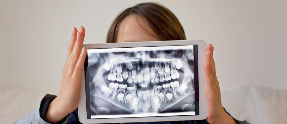
Panoramic X-Ray
Panoramic X-rays, also known as panoramic radiographs or orthopantomograms (OPGs), are a type of dental X-ray that provides a broad and comprehensive view of the entire oral cavity. Unlike bitewing or periapical X-rays, which focus on individual teeth, panoramic X-rays capture a panoramic or panoramic view of the upper and lower jaws, including the teeth, jawbones, temporomandibular joints (TMJ), and surrounding structures.
Panoramic X-rays are taken using a specialized machine called a panoramic X-ray unit. The patient stands or sits in an upright position while the machine rotates around their head, capturing a single continuous image. This rotating motion allows the X-ray machine to create a panoramic image by capturing the X-rays that pass through the patient’s mouth and jaw from multiple angles.
The resulting panoramic image shows a detailed overview of the entire oral cavity, displaying the teeth, their roots, the jawbones, sinuses, and other structures. The image also provides valuable information about the relationship between the upper and lower jaws, the positioning of teeth, and any abnormalities or irregularities.
Panoramic X-rays offer several benefits in dental diagnostics and treatment planning:
- Comprehensive View
- Impacted Teeth
- Orthodontic Evaluations
- TMJ Evaluation
- Sinus Examination
Comprehensive View
Panoramic X-rays provide a comprehensive and holistic view of the oral cavity, making them useful for evaluating overall dental health and identifying a wide range of dental conditions.
Impacted Teeth
Panoramic X-rays can detect impacted teeth, such as wisdom teeth, and evaluate their position and orientation relative to neighboring teeth and structures. This information is crucial for determining if extraction is necessary.
Orthodontic Evaluations
Panoramic X-rays are commonly used in orthodontics to assess the alignment of teeth, evaluate the eruption patterns of permanent teeth in children, and plan orthodontic treatments.
TMJ Evaluation
These X-rays allow dental professionals to evaluate the temporomandibular joints (TMJ), which connect the jawbone to the skull. They can reveal any joint abnormalities or disorders that may be causing jaw pain or dysfunction.
Sinus Examination
Panoramic X-rays capture images of the maxillary sinuses, providing valuable information about sinus infections, cysts, or other sinus-related conditions.
Panoramic X-rays are relatively quick and comfortable for patients, as they do not require the insertion of X-ray film or sensors inside the mouth. However, it is important to note that panoramic X-rays have some limitations. They may not provide the same level of detail as other types of X-rays, such as bitewing or periapical X-rays, for detecting early-stage cavities or assessing specific areas of concern.
In summary, panoramic X-rays offer a broad view of the entire oral cavity, assisting dental professionals in diagnosing dental conditions, planning treatments, and evaluating overall oral health. They are especially valuable for assessing impacted teeth, orthodontic evaluations, TMJ examinations, and sinus evaluations. By providing a comprehensive perspective, panoramic X-rays contribute to more accurate diagnoses and effective treatment planning in dentistry.
Cone Beam Computed Tomography
Cone Beam Computed Tomography (CBCT) is an advanced imaging technique used in dentistry to provide three-dimensional (3D) images of the oral and maxillofacial structures. CBCT scanners utilize a cone-shaped X-ray beam and a specialized detector to capture a series of images from various angles. These images are then reconstructed using computer algorithms to create a detailed 3D representation of the scanned area.
CBCT offers several advantages over traditional two-dimensional X-rays and panoramic radiographs:
- Enhanced Visualization
- 3D Imaging
- Precise Treatment Planning
- Pathology and Trauma Assessment
- Minimal Radiation Exposure
Enhanced Visualization
CBCT provides a more comprehensive view of the oral and maxillofacial structures, including teeth, jaws, TMJ, sinuses, nerves, and bone. It allows for precise evaluation of anatomical relationships and a more accurate assessment of complex dental conditions.
3D Imaging
Unlike traditional X-rays, which offer a two-dimensional view, CBCT provides a 3D representation of the scanned area. This enables dental professionals to examine structures from different angles, rotate the image, and view cross-sectional slices for a more thorough evaluation.
Precise Treatment Planning
CBCT scans are invaluable for treatment planning in various dental procedures. They aid in implant placement by determining the ideal location, size, and orientation of implants, and assessing bone quality and quantity. CBCT is also used in orthodontics to evaluate tooth position, root morphology, and airway analysis for improved treatment outcomes.
Pathology and Trauma Assessment
CBCT allows for detailed assessment of dental pathologies, such as cysts, tumors, infections, and fractures. It provides valuable information for accurate diagnosis and treatment planning in complex cases.
Minimal Radiation Exposure
While CBCT uses a higher radiation dose compared to traditional dental X-rays, advancements in technology have reduced radiation exposure significantly. The cone-shaped beam focuses on the specific area of interest, limiting radiation exposure to the surrounding tissues and reducing potential risks.
CBCT scans are typically performed in a specialized dental imaging center or dental office equipped with a CBCT machine. During the scan, the patient is positioned in a standing or sitting position, and the machine rotates around the head, capturing the necessary images. The scanning process is quick, usually taking less than a minute, and is painless for the patient.
It is important to note that CBCT scans are typically reserved for specific cases that require detailed 3D imaging. The increased complexity and higher cost associated with CBCT imaging may limit its routine use for general dental examinations.
In summary, Cone Beam Computed Tomography (CBCT) is a state-of-the-art imaging technique in dentistry that provides detailed 3D images of the oral and maxillofacial structures. Its advanced capabilities offer enhanced visualization, precise treatment planning, and accurate assessment of complex dental conditions. CBCT plays a crucial role in various dental specialties, contributing to improved patient care and treatment outcomes.
Benefits of Dental X-Ray
- Early Detection and Diagnosis
- Treatment Planning
- Monitoring Development and Progression
Early Detection and Diagnosis
Dental X-rays allow dental professionals to detect and diagnose oral health conditions at their early stages when they are often invisible to the naked eye. This enables timely intervention and treatment, preventing the progression of diseases such as cavities, gum disease, and oral infections.
Treatment Planning
Dental X-ray offers a deeper understanding of the patient’s oral health, aiding in treatment planning. Dentists can use these images to determine the best course of action, including the need for fillings, root canal treatment, tooth extraction, or orthodontic procedures.
Monitoring Development and Progression
For orthodontic treatments and monitoring the development of wisdom teeth, dental X-rays are essential. These images provide dentists with insights into the growth and alignment of teeth, ensuring optimal treatment planning and preventing potential complications.
Dental X-Ray Procedures
- Preparing for Dental X-Ray
- The X-Ray Process
- Safety Measures
Preparing for Dental X-Ray
Before undergoing a dental X-ray, patients may be asked to remove any jewelry, eyeglasses, or metal objects that could interfere with the images. The dental professional will provide a lead apron to protect the patient’s body from unnecessary radiation exposure.
The X-Ray Process
During the X-ray procedure, the dental professional will position the X-ray machine next to the patient’s mouth or face. The patient may be asked to bite down on a specialized film or sensor while the X-ray machine is positioned to capture the necessary images. The process is quick and painless.
Safety Measures
Dental X-rays are performed using low levels of radiation. However, dental professionals take several precautions to minimize radiation exposure further. These measures include using lead aprons, collars, thyroid shields, and high-speed films or digital sensors that require less exposure time.
Dental X-Ray Safety and Precautions
- Radiation Exposure and Risk
- Protective Measures
- Limitations for Special Groups
- Regular Evaluation of X-ray Equipment
Radiation Exposure and Risk
Although dental X-rays involve radiation, the exposure levels are minimal, and the risks associated with modern dental X-rays are extremely low. The equipment and techniques used in dental practices adhere to strict guidelines and regulations to ensure patient safety.
Protective Measures
Dental professionals prioritize patient safety by implementing protective measures during X-ray procedures. Lead aprons shield the body from radiation, and thyroid collars protect the thyroid gland. Additionally, dental offices employ digital radiography systems that significantly reduce radiation exposure compared to traditional film-based X-rays.
Limitations for Special Groups
Certain patient groups require additional considerations regarding X-ray usage. Pregnant women, for instance, are advised to postpone routine X-rays, unless absolutely necessary, to minimize fetal exposure. However, in emergency situations, dental X-rays may be necessary, and appropriate shielding is provided.
Regular Evaluation of X-ray Equipment
Dental offices are required to regularly evaluate and maintain their X-ray equipment to ensure optimal functionality and safety. This includes periodic inspections, calibration, and adherence to regulatory standards to minimize any potential risks.
The Role of Dental X-Rays in Oral Health
- Dental X-Rays for Comprehensive Examination
- Monitoring Progress and Treatment Success
- Preventive Dentistry
Dental X-Rays for Comprehensive Examination
Dental X-rays are an integral part of a comprehensive oral examination. They provide vital information that may not be visible during a visual inspection alone. X-rays reveal hidden decay, abscesses, impacted teeth, bone loss, and other abnormalities, enabling accurate diagnosis and treatment planning.
Monitoring Progress and Treatment Success
After dental procedures, X-rays play a crucial role in monitoring the progress and success of the treatment. They allow dental professionals to evaluate the healing process, the integration of dental implants, and the effectiveness of orthodontic treatment.
Preventive Dentistry
Dental X-rays aid in preventive dentistry by detecting early signs of dental problems. They enable dentists to identify areas of decay, cavities, or gum disease that may not be visible to the naked eye. Early intervention based on X-ray findings can prevent further damage and save teeth from extraction.
Conclusion
In the ever-evolving field of dentistry, dental X-rays have emerged as an invaluable tool for oral health professionals. Their ability to reveal hidden conditions, aid in accurate diagnoses, and assist in treatment planning has revolutionized patient care. Through minimal radiation exposure and advanced safety measures, dental X-rays offer significant benefits while prioritizing patient safety. By embracing dental X-rays as an essential component of comprehensive oral care, patients and dental professionals can work together to achieve and maintain optimal oral health throughout a lifetime.