
Radiation plays a crucial role in dentistry, aiding in diagnostics, treatment, and oral health management. With its pivotal role comes concerns about safety, exposure levels, and its impact on patients and dental professionals. This comprehensive guide aims to delve into the various facets of radiation in dentistry, encompassing its types, safety protocols, advancements, and impact on oral health.
Table of Contents
ToggleRadiation in Dentistry
Radiation usage in dentistry primarily revolves around imaging techniques and therapeutic interventions. Dental radiographs, commonly known as X-rays, remain a cornerstone in diagnosing various oral conditions. These imaging techniques aid in identifying cavities, examining tooth roots, assessing bone health, and planning orthodontic treatments.
Types of Dental Radiation
- Intraoral X-rays
- Extraoral X-rays
- Cone Beam Computed Tomography (CBCT)
Intraoral X-rays
These are the most common type, involving placing a sensor inside the mouth to capture images of individual teeth and their supporting structures.
Extraoral X-rays
These encompass panoramic radiographs that capture a broad view of the entire mouth, providing a comprehensive look at the teeth, jaws, and surrounding structures.
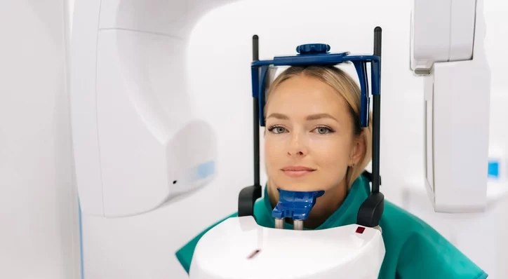
Cone Beam Computed Tomography (CBCT)
This technology offers three-dimensional imaging, beneficial in complex dental procedures like dental implant placements and orthodontic evaluations.
Safety Measures and Regulations
Dental professionals adhere to stringent safety measures to minimize radiation exposure for both patients and staff. Lead aprons, thyroid collars, and high-speed film are among the protective tools used during X-rays. Additionally, dental offices follow guidelines established by organizations like the American Dental Association (ADA) and the Food and Drug Administration (FDA) to ensure proper equipment maintenance, radiation dosage regulation, and staff training.
The Amount of Radiation Exposure in Dentistry
The amount of radiation exposure from dental imaging is measured in millisieverts (mSv), which quantifies the absorbed dose of ionizing radiation. Here are approximate radiation doses for common dental imaging procedures:
Intraoral X-rays
Each intraoral X-ray exposes a patient to a very low dose, typically ranging from about 0.005 to 0.01 mSv per X-ray. This is equivalent to a few hours of natural background radiation or even less.
Panoramic X-rays (Extraoral X-rays)
Panoramic radiographs generally result in a higher radiation dose compared to intraoral X-rays due to their broader coverage. On average, a panoramic X-ray might expose a patient to around 0.01 to 0.02 mSv per image.
Cone Beam Computed Tomography (CBCT)
CBCT, offering three-dimensional images, involves a higher radiation dose compared to intraoral and panoramic X-rays due to its detailed and comprehensive imaging. The dose for a typical CBCT scan can range from approximately 0.05 to 0.2 mSv or more, depending on the size and parameters of the scan.
It’s important to understand these figures are approximate values and can vary based on factors such as the specific equipment used, imaging settings, and the individual patient’s anatomy. The doses mentioned are generally considered low in comparison to other medical imaging procedures like full-body CT scans, which can expose a patient to higher doses ranging from 5 to 10 mSv or more.
Dental professionals are trained to follow ALARA principles (As Low As Reasonably Achievable) to ensure that radiation exposure is kept to the minimum necessary for obtaining the required diagnostic information while prioritizing patient safety. Ongoing advancements in technology and imaging techniques continually strive to reduce radiation exposure further while maintaining diagnostic accuracy.
How Much is The Safe Dose?
When discussing radiation exposure, the concept of a “safe dose” can be challenging because it depends on various factors, including the type of radiation, the individual’s age, health status, and the purpose of exposure. In general, for medical procedures involving ionizing radiation, including dental imaging, the goal is to keep the radiation exposure “as low as reasonably achievable” (ALARA principle).
The International Commission on Radiological Protection (ICRP) and regulatory bodies like the US Nuclear Regulatory Commission (NRC) and the Food and Drug Administration (FDA) provide guidance on radiation safety and permissible exposure levels.
For context, the average annual background radiation exposure from natural sources is around 2 to 3 millisieverts (mSv) globally. However, it’s crucial to understand that this background exposure varies depending on geographic location and other environmental factors.
In terms of radiation exposure from medical imaging, including dental X-rays:
- Intraoral X-rays: The average dose per X-ray is very low, usually ranging from 0.005 to 0.01 mSv, which is significantly lower than background radiation exposure.
- Panoramic X-rays (Extraoral X-rays): These may result in slightly higher doses, typically ranging from 0.01 to 0.02 mSv per image.
- Cone Beam Computed Tomography (CBCT): CBCT scans involve higher doses compared to intraoral or panoramic X-rays. A typical CBCT scan might range from approximately 0.05 to 0.2 mSv or more, depending on various factors.
The concept of a “safe dose” in radiation is often approached with the understanding that any exposure carries some level of risk. The goal in medical procedures is to keep radiation exposure as low as reasonably achievable while balancing the benefits of the procedure in diagnosing or treating health conditions.
Medical professionals and regulatory bodies continuously review and update guidelines to ensure that the radiation exposure from imaging procedures remains within safe limits and that the benefits of the procedure outweigh the associated risks.
For individuals receiving dental X-rays, the exposure levels from these procedures are generally considered safe. Dentists follow strict protocols and use equipment designed to minimize radiation exposure while ensuring accurate diagnostics and effective treatment planning. If you have specific concerns about radiation exposure, discussing them with your dentist can provide reassurance and further information about the safety measures in place during dental procedures.
Impact on Oral Health and Diagnosis
Despite concerns, the benefits of dental radiographs outweigh the associated minimal risks. These images aid in early detection of oral issues, facilitating timely treatment and preventing the progression of dental problems. From identifying hidden cavities to evaluating the progression of gum disease, these radiographs are indispensable tools in a dentist’s diagnostic arsenal.
Additional Considerations Regarding Radiation
Certain groups of patients require additional considerations regarding radiation exposure during dental procedures due to various factors affecting their sensitivity or susceptibility to radiation. These groups include:
Children
- Higher Sensitivity
- Longer Lifespan
- Risk-Benefit Analysis
Higher Sensitivity
Children are more sensitive to radiation’s potential effects due to their developing bodies and increased cell division rates.
Longer Lifespan
The longer lifespan of children means a longer period for potential radiation-induced effects to manifest.
Risk-Benefit Analysis
Dentists carefully consider the necessity of X-rays for children, opting for the lowest radiation exposure while ensuring adequate diagnostic information.
Pregnant Women
- Fetal Development
- Shielding and Timing
Fetal Development
Radiation exposure can potentially affect fetal development, especially during the first trimester.
Shielding and Timing
When X-rays are unavoidable during pregnancy, shielding with lead aprons and collars is crucial. Dentists often postpone non-emergency imaging procedures to the second trimester or later, minimizing potential risks.
Patients with Compromised Health
- Immunocompromised Patients
- Patients Undergoing Multiple Procedures
Immunocompromised Patients
Individuals with weakened immune systems might be more susceptible to the effects of radiation.
Patients Undergoing Multiple Procedures
Those requiring frequent or multiple imaging procedures might accumulate higher cumulative radiation doses, warranting careful monitoring and optimization of imaging frequency.
Individuals with Specific Conditions
- Previous Radiation Exposure
- Radiosensitive Conditions
Previous Radiation Exposure
Patients with a history of high radiation exposure from medical treatments or other sources might need cautious consideration to avoid unnecessary additional exposure.
Radiosensitive Conditions
Some rare genetic conditions make individuals more susceptible to the effects of radiation, necessitating careful dosage considerations.
Patients with Special Needs
- Communication Challenges
- Increased Sensitivity
Communication Challenges
Patients with cognitive or communication difficulties may require additional effort to ensure understanding and consent regarding dental procedures, including X-rays.
Increased Sensitivity
Some patients with sensory issues or anxiety may be more sensitive to the imaging environment, requiring additional measures for comfort and cooperation during the procedure.
Advances in Radiation Technology
Technological advancements continue to revolutionize dental radiology. Digital radiography has replaced conventional film-based methods, offering quicker image acquisition, lower radiation doses, and enhanced image quality. Moreover, the introduction of CBCT technology has transformed treatment planning, especially in implant dentistry and oral surgeries, by providing detailed 3D images.
Managing Patient Concerns
Patients often express apprehension regarding radiation exposure during dental procedures. Dentists address these concerns by educating patients about the necessity and safety of these procedures. Clear communication regarding the benefits and minimal risks associated with dental radiographs helps alleviate patient anxiety.
Conclusion
Radiation in dentistry is an invaluable tool for diagnosing and treating oral health issues. With stringent safety measures, technological advancements, and adherence to regulatory guidelines, the dental community ensures that radiation exposure remains minimal while maximizing diagnostic accuracy and treatment efficacy. The ongoing evolution of imaging technologies promises even safer and more efficient methods for enhancing oral health care.