
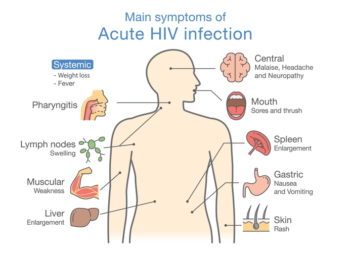
Acquired Immunodeficiency Syndrome (AIDS) represents the advanced stage of infection caused by the Human Immunodeficiency Virus (HIV). HIV is a retrovirus that primarily targets CD4+ T-helper lymphocytes, progressively weakening the immune system. As immune function deteriorates, patients become increasingly susceptible to opportunistic infections, unusual malignancies, and systemic complications. Because the oral cavity contains a complex microbial ecosystem and is constantly exposed to environmental pathogens, it is one of the earliest and most frequently affected sites in HIV infection.
Approximately 70–75% of individuals with HIV infection develop oral manifestations during the course of their disease. In many cases, oral lesions are among the earliest clinical signs of immune suppression and may serve as important diagnostic and prognostic indicators. Although most oral manifestations are not pathognomonic (absolutely diagnostic) of HIV infection, their presence—especially in young or otherwise healthy individuals—should raise suspicion of underlying immunodeficiency.
Oral manifestations of HIV infection are commonly classified into three groups:
- Group I — Strongly associated with HIV
- Group II — Less strongly associated with HIV
- Group III — Possibly associated with HIV
Table of Contents
ToggleGroup I — Strongly Associated with HIV
These lesions are closely linked to HIV infection and often correlate with declining CD4 counts and disease progression.
1. Candidosis (Oral Candidiasis)
Oral candidosis is the most common oral manifestation of HIV infection, occurring in approximately 70% of patients. It may appear early in the disease and is frequently the first clinical sign of immune compromise.
Candida species, particularly Candida albicans, are commensal organisms in the oral cavity. However, when cell-mediated immunity declines, overgrowth occurs.
Clinical forms include:
- Erythematous (atrophic) candidosis – Often an early presentation; appears as red, depapillated areas on the dorsum of the tongue or palate.
- Pseudomembranous candidosis (thrush) – White plaques that can be wiped away, leaving a red or bleeding surface beneath.
- Hyperplastic candidosis – White plaques that cannot be scraped off.
- Angular cheilitis – Fissuring and inflammation at the corners of the mouth.
HIV-associated candidosis has prognostic significance. Approximately 50% of patients with HIV-related thrush may progress to AIDS within five years if untreated.
Management includes topical antifungals (nystatin, clotrimazole) in mild cases and systemic agents (fluconazole, itraconazole) in severe or recurrent cases.
2. Hairy Leukoplakia
Oral hairy leukoplakia presents as bilateral, white, corrugated, non-removable plaques most commonly affecting the lateral borders of the tongue. Unlike candidosis, these lesions do not respond to antifungal therapy.
Hairy leukoplakia is strongly associated with Epstein–Barr virus (EBV) infection and occurs almost exclusively in immunocompromised individuals. It may resolve with antiviral therapy such as aciclovir or valaciclovir but typically recurs when treatment is discontinued.
Its presence is a marker of immune suppression and may indicate worsening disease. It is also associated with increased risk of lymphoma.
3. HIV Gingivitis (Linear Gingival Erythema)
HIV gingivitis is characterized by an intense red band along the marginal gingiva, often disproportionate to the amount of plaque present. Unlike conventional plaque-induced gingivitis, it does not respond well to routine oral hygiene measures alone.
Features include:
- Persistent erythema
- Spontaneous bleeding
- Mild discomfort
The condition reflects altered host immune response and may require antimicrobial mouth rinses such as chlorhexidine in addition to improved oral hygiene.
4. Necrotizing Ulcerative Gingivitis (NUG)
Necrotizing ulcerative gingivitis in HIV-positive patients may occur in individuals who otherwise appear healthy. It presents with:
- Rapid onset pain
- Ulceration of interdental papillae
- Pseudomembrane formation
- Spontaneous bleeding
- Halitosis
In HIV patients, NUG may be more aggressive and may progress to necrotizing ulcerative periodontitis (NUP) with rapid attachment loss and bone destruction.
Management involves debridement, antimicrobial therapy (e.g., metronidazole), chlorhexidine mouth rinses, and pain control.
5. HIV Periodontitis
HIV-associated periodontitis is characterized by severe, localized periodontal destruction that appears disproportionate to local factors. It may progress rapidly with:
- Deep periodontal pockets
- Severe pain
- Gingival necrosis
- Rapid bone loss
The aggressive nature is attributed to immune dysregulation and altered microbial flora. Early intervention is critical to prevent tooth loss.
6. Kaposi’s Sarcoma (KS)
Kaposi’s sarcoma is the most common malignancy associated with HIV infection. It is linked to Human Herpesvirus-8 (HHV-8).
Clinically, it appears as:
- Red, purple, or brown macules
- Nodular swellings
- Frequently located on the palate, gingiva, or tongue
Approximately 50% of cases involve intraoral or perioral sites. In young males not receiving immunosuppressive therapy, oral Kaposi’s sarcoma is considered highly suggestive of HIV infection.
Treatment options include:
- Antiretroviral therapy (often results in regression)
- Radiotherapy
- Intralesional vincristine
- Surgical excision
7. Non-Hodgkin’s Lymphoma
Non-Hodgkin’s lymphoma is another AIDS-defining malignancy. Burkitt’s lymphoma is significantly more common in HIV-positive individuals.
Oral features may include:
- Rapidly enlarging mass
- Ulceration
- Tooth mobility
- Jaw swelling
Prompt biopsy and oncologic referral are essential.
Group II — Less Strongly Associated with HIV
These conditions are seen in HIV patients but are not exclusive to HIV infection.
1. Atypical Oropharyngeal Ulceration
HIV patients may develop persistent, large, painful ulcers that do not resemble typical aphthous ulcers. These ulcers may be:
- Deep
- Necrotic
- Slow healing
Management may require corticosteroids or systemic therapy.
2. Immune Thrombocytopenic Purpura (ITP)
ITP may manifest orally as:
- Petechiae
- Ecchymosis
- Spontaneous gingival bleeding
It results from immune-mediated platelet destruction.
3. HIV-Associated Salivary Gland Disease
This includes:
- Lymphoepithelial cysts
- Focal lymphocytic sialadenitis
- Parotid gland enlargement
It is more common in children with HIV. Xerostomia may result, increasing risk of caries and candidosis.
4. Common Viral Infections
Due to immunosuppression, patients are prone to:
- Herpes simplex virus (HSV)
- Varicella-zoster virus (VZV)
- Human papillomavirus (HPV)
- Cytomegalovirus (CMV)
These infections may be more severe, recurrent, or persistent.
Group III — Possible Association with HIV
These conditions have been reported in HIV patients but lack strong evidence of a direct association.
- Rare bacterial and fungal infections
- Oral hyperpigmentation (may also be drug-induced)
- Cat scratch disease
- Neurological abnormalities affecting oral function
- Squamous cell carcinoma (SCC) of the oropharynx
- Persistent generalized lymphadenopathy (PGL)
Persistent generalized lymphadenopathy is defined as lymph node enlargement greater than 1 cm, persisting for more than 3 months in two or more extra-inguinal sites. Cervical lymph nodes are commonly involved. It may represent early HIV infection or progression to AIDS.
Management (Rx)
Antiretroviral Therapy (ART)
The introduction of Highly Active Antiretroviral Therapy (HAART) revolutionized HIV management. Modern antiretroviral therapy:
- Suppresses viral replication
- Restores immune function
- Reduces opportunistic infections
- Improves life expectancy to near normal
Effective ART has dramatically reduced the incidence of many oral manifestations.
However, ART may have adverse effects such as:
- Facial lipoatrophy
- Xerostomia
- Drug interactions
Early diagnosis and management of oral lesions significantly improve quality of life.
Dental Management of Patients with AIDS
Dental professionals play a crucial role in early detection and management of HIV-related oral conditions.
Risks to Dental Personnel
The risk of HIV transmission through needlestick injury from an infected patient is approximately 0.3–0.45%.
Strict adherence to universal precautions is mandatory:
- Gloves
- Masks
- Eye protection
- Safe sharps handling
- Proper sterilization
Considerations in Immunocompromised Patients
Because HIV patients may be immunocompromised:
- Procedures with infection risk (e.g., extractions) may require antimicrobial prophylaxis.
- Surgery should be minimally traumatic.
- Healing may be delayed.
- There may be increased bleeding tendency.
Local haemostatic measures should be available when needed.
Post-Exposure Prophylaxis (PEP)
If a contaminated needlestick injury occurs, especially from a hollow-bore needle:
- Immediate washing of the area
- Reporting and risk assessment
- Initiation of post-exposure prophylaxis within 24 hours (preferably sooner)
Antiretroviral therapy offers the best chance of preventing seroconversion. Follow-up testing is required.
Prognostic Significance of Oral Lesions
Oral manifestations often correlate with declining CD4 counts:
- Candidosis and hairy leukoplakia indicate moderate immunosuppression.
- Kaposi’s sarcoma and lymphoma indicate advanced disease.
Thus, oral examination is a valuable, non-invasive tool in monitoring disease progression.
Conclusion
Oral manifestations are common, clinically significant features of HIV infection and AIDS. They may serve as early diagnostic indicators, markers of disease progression, and predictors of prognosis. With the advent of modern antiretroviral therapy, the prevalence and severity of many oral conditions have declined, but they remain important clinical findings.
Dentists and healthcare professionals must be able to recognize these manifestations, implement appropriate infection control measures, and contribute to multidisciplinary management. Early identification and treatment not only improve oral health but also enhance overall quality of life in individuals living with HIV.