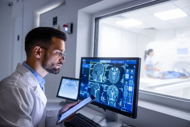
Magnetic Resonance Imaging (MRI) has long been a cornerstone in medical diagnostics, offering unparalleled imaging capabilities for soft tissues, organs, and other intricate anatomical structures. In recent years, its application has expanded into the field of dentistry, where it holds promise for revolutionizing diagnostic precision and patient care. This article explores the principles of MRI, its unique advantages, and its growing uses in dentistry, emphasizing its potential to reshape oral health management.
Table of Contents
TogglePrinciples of MRI
MRI operates on the principle of nuclear magnetic resonance, using powerful magnetic fields and radiofrequency (RF) waves to create detailed images of internal structures. Unlike X-rays or computed tomography (CT), MRI does not rely on ionizing radiation, making it a safer option for repeated imaging.
The process involves aligning hydrogen protons in the body within a strong magnetic field. These protons are then stimulated using RF pulses, causing them to emit signals that are captured and converted into detailed cross-sectional images. The contrast in MRI images arises from differences in proton density and relaxation times, providing exceptional clarity for soft tissue structures.
Unique Advantages of MRI in Dentistry
- Non-ionizing Radiation
- Superior Soft Tissue Imaging
- Multiplanar Imaging
- Functional Imaging
Non-ionizing Radiation
One of the most significant advantages of MRI is its reliance on non-ionizing radiation. Dental imaging techniques like X-rays and CT scans expose patients to varying degrees of ionizing radiation, which, while low, carries cumulative risks over time. MRI eliminates this concern, making it a preferable option for patients requiring frequent imaging, such as those with chronic conditions or children.
Superior Soft Tissue Imaging
MRI excels in visualizing soft tissues, which is crucial in dentistry for assessing the temporomandibular joint (TMJ), salivary glands, and peri-implant tissues. Conventional dental imaging techniques primarily focus on hard tissues like bones and teeth, whereas MRI complements these modalities by offering a detailed view of surrounding soft tissue anatomy.
Multiplanar Imaging
MRI provides the capability for multiplanar imaging, allowing dentists to view anatomical structures from various angles without repositioning the patient. This feature enhances diagnostic accuracy and aids in treatment planning for complex dental cases.
Functional Imaging
Functional MRI (fMRI) can assess physiological processes such as blood flow and neural activity. Although still in its nascent stages in dentistry, fMRI holds potential for applications such as evaluating neural responses to oral sensory stimuli or studying pain pathways in temporomandibular disorders (TMD).
Applications of MRI in Dentistry
- Temporomandibular Joint Disorders (TMD)
- Assessment of Salivary Gland Pathologies
- Oral Cancer and Tumor Imaging
- Dental Implantology
- Orthodontics
- Endodontics
- Pediatric Dentistry
Temporomandibular Joint Disorders (TMD)
One of the most established uses of MRI in dentistry is the diagnosis and management of temporomandibular joint disorders. The TMJ is a complex structure comprising bones, cartilage, ligaments, and an articular disc. MRI is considered the gold standard for imaging the TMJ due to its ability to visualize both hard and soft tissues with high resolution.
MRI can detect:
- Disc displacement and deformity
- Joint effusion
- Inflammatory changes
- Degenerative conditions like osteoarthritis
These insights are invaluable for tailoring treatment plans, whether through conservative therapies or surgical interventions.
Assessment of Salivary Gland Pathologies
MRI is highly effective in evaluating salivary gland disorders, including:
- Sialadenitis (inflammation of the salivary glands)
- Sialolithiasis (salivary gland stones)
- Tumors or cysts
- Sjogren’s syndrome
MRI can provide a clear distinction between benign and malignant lesions, guide biopsy procedures, and monitor treatment response.
Oral Cancer and Tumor Imaging
MRI plays a pivotal role in the diagnosis, staging, and monitoring of oral cancers and benign tumors. Its superior soft tissue contrast helps delineate tumor margins, assess invasion into adjacent structures, and identify lymph node involvement. MRI can also evaluate perineural invasion, a critical factor in oral cancer prognosis.
Dental Implantology
The success of dental implants relies on precise placement and osseointegration with the surrounding bone. While CT scans are commonly used for preoperative planning, MRI offers a radiation-free alternative for assessing:
- Bone quality and volume
- Soft tissue integration
- Peri-implant inflammation or infection
MRI’s ability to detect early inflammatory changes can aid in the timely management of peri-implantitis, a significant cause of implant failure.
Orthodontics
Orthodontic treatment planning often requires detailed imaging of craniofacial structures. MRI can provide 3D reconstructions of the jaw, teeth, and surrounding tissues without exposing patients to radiation. Additionally, it can help evaluate:
- Airway dimensions in cases of obstructive sleep apnea
- The impact of orthodontic forces on TMJ structures
Endodontics
MRI’s capability to visualize soft tissues makes it an emerging tool in endodontics. It can identify:
- Periapical lesions
- Cystic versus solid masses
- Pulpal inflammation
Although traditional radiographs and cone-beam computed tomography (CBCT) remain the primary imaging modalities in endodontics, MRI offers complementary benefits, particularly in cases with ambiguous findings.
Pediatric Dentistry
MRI’s safety profile makes it an attractive option for imaging in pediatric dentistry. It can be used to evaluate:
- Congenital anomalies such as cleft lip and palate
- Pediatric TMJ disorders
- Trauma-related injuries to soft tissues and developing teeth
Challenges and Limitations of MRI in Dentistry
- Cost and Accessibility
- Image Artifacts
- Longer Imaging Time
- Learning Curve
Cost and Accessibility
MRI systems are expensive to purchase and maintain, limiting their availability in dental practices. The cost of MRI scans is also higher than conventional imaging modalities, potentially posing a financial burden on patients.
Image Artifacts
Dental restorations, such as metal crowns and braces, can cause artifacts that degrade image quality. Advances in MRI-compatible materials and artifact reduction techniques are addressing this issue, but it remains a challenge.
Longer Imaging Time
MRI scans take significantly longer than X-rays or CT scans, which can be uncomfortable for patients, particularly those with anxiety or claustrophobia. Open MRI systems and faster imaging sequences are helping mitigate this limitation.
Learning Curve
Interpreting MRI images requires specialized training, and many dental professionals are not yet proficient in this area. Collaboration with radiologists is often necessary to leverage MRI’s full potential.
Future Directions for MRI in Dentistry
- Development of Dental MRI Systems
- Integration with Artificial Intelligence (AI)
- Functional Imaging in Dentistry
- Hybrid Imaging Techniques
Development of Dental MRI Systems
Dedicated dental MRI systems are being developed to address the unique requirements of oral imaging. These systems feature smaller, open designs for patient comfort and optimized imaging protocols for dental applications.
Integration with Artificial Intelligence (AI)
AI has the potential to enhance MRI’s diagnostic capabilities by automating image analysis, detecting subtle abnormalities, and predicting disease progression. AI-driven MRI tools could streamline workflows and improve diagnostic accuracy.
Functional Imaging in Dentistry
As fMRI technology advances, its application in dentistry could expand to include studies on:
- Pain pathways in chronic orofacial pain conditions
- Neuroplasticity in response to dental treatments
- Muscle activity during mastication and speech
Hybrid Imaging Techniques
Combining MRI with other imaging modalities, such as PET-MRI or MRI-ultrasound, could provide comprehensive diagnostic information. These hybrid techniques are particularly promising for complex cases, such as oral cancer staging or TMJ disorders.
Conclusion
MRI is emerging as a transformative tool in dentistry, offering unparalleled insights into soft tissue anatomy and pathology. Its applications span a wide range of dental specialties, from diagnosing TMJ disorders and salivary gland diseases to enhancing implantology and orthodontics. While challenges such as cost and accessibility persist, ongoing technological advancements and interdisciplinary collaboration are paving the way for broader adoption of MRI in dental practice.
As the field continues to evolve, MRI has the potential to become a standard diagnostic tool, elevating the precision and safety of dental care. For practitioners and researchers alike, embracing MRI represents a step forward in the quest to deliver comprehensive and patient centered oral health solutions.

