
The maxilla, a crucial component of the human craniofacial complex, plays a central role in various biological and clinical aspects. It is a paired bone that forms the upper jaw and the majority of the midface. The maxilla serves essential functions in speech, mastication, and facial aesthetics, making it a subject of great interest in various medical and dental fields. In this comprehensive article, we will delve into the anatomy, function, and clinical significance of the maxilla, exploring its role in dental health, craniofacial development, and surgical interventions.
Table of Contents
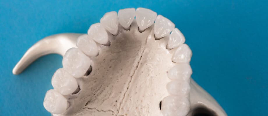
ToggleAnatomy of the Maxilla
Location and Structure
The maxilla is a paired bone that comprises the upper jaw. It is located in the midface, forming the central portion of the facial skeleton. The maxilla consists of two main parts: the right and left maxillae. These bones are fused at the midline to create the upper jaw, which houses the upper teeth and contributes significantly to the facial skeleton’s structural integrity.
Bony Features
The maxilla presents various bony features, each with its own specific function and anatomical significance:
- Alveolar Process
- Palatine Process
- Nasal Process
- Zygomatic Process
- Frontal Process
- Infraorbital Rim
Alveolar Process
The alveolar process of the maxilla contains sockets known as alveoli, which house the upper teeth. These sockets support and anchor the teeth in place.
Palatine Process
The palatine process of the maxilla forms the anterior part of the hard palate, separating the oral and nasal cavities.
Nasal Process
The nasal process extends superiorly, forming the bridge of the nose and connecting with the nasal bones.
Zygomatic Process
The zygomatic process articulates with the zygomatic bone (cheekbone), contributing to the formation of the zygomatic arch.
Frontal Process
The frontal process of the maxilla forms part of the medial orbital wall, contributing to the eye socket’s structure.
Infraorbital Rim
The infraorbital rim, located below the orbit, plays a role in the support and protection of the eye.
Articulations
The maxilla articulates with several neighboring bones in the craniofacial complex:
- Palatine Bones
- Nasal Bones
- Zygomatic Bones
- Ethmoid Bone
Palatine Bones
The palatine process of the maxilla articulates with the horizontal plates of the palatine bones to form the posterior part of the hard palate.
Nasal Bones
The nasal process of the maxilla articulates with the nasal bones, contributing to the formation of the bridge of the nose.
Zygomatic Bones
The zygomatic process of the maxilla articulates with the zygomatic bones, creating the zygomatic arch.
Ethmoid Bone
The frontal process of the maxilla articulates with the ethmoid bone, which is part of the nasal cavity and the eye socket.
Functions of the Maxilla
The maxilla performs various essential functions that contribute to an individual’s overall well-being and quality of life. These functions can be broadly categorized into the following areas:
- Mastication
- Speech and Articulation
- Facial Aesthetics
- Support for the Eye and Nasal Structures
Mastication
The alveolar process of the maxilla contains sockets that support and anchor the upper teeth. These teeth are vital for the process of mastication, or chewing, which is the first step in digestion. Proper mastication helps break down food into smaller particles, facilitating digestion and nutrient absorption.
Speech and Articulation
The maxilla plays a crucial role in speech and articulation. The shape and positioning of the maxilla affect the resonance and articulation of speech sounds. Conditions that impact the maxilla, such as cleft palate, can lead to speech difficulties and require surgical correction.
Facial Aesthetics
The maxilla significantly contributes to an individual’s facial aesthetics. Its position and structure influence the overall shape and contour of the face. A well-aligned maxilla is essential for a symmetrical and harmonious facial appearance.
Support for the Eye and Nasal Structures
The maxilla’s frontal and nasal processes provide structural support to the eye and nasal cavities. These processes help maintain the integrity of the eye sockets and the shape of the nose.
Development of the Maxilla
The development of the maxilla is a complex and highly regulated process that occurs during embryonic and postnatal growth. Understanding maxillary development is essential for diagnosing and treating various craniofacial anomalies and malformations.
Embryonic Development
The maxilla begins to form during the early stages of embryonic development. It originates from the first branchial arch, also known as the mandibular arch. During embryogenesis, the maxilla undergoes a process of ossification, where cartilaginous structures are gradually replaced by bone tissue. This process involves the coordination of various genes and signaling pathways.
Growth and Fusion
The maxillae develop on both sides of the midline and gradually move medially toward each other. They eventually fuse at the midline, forming the upper jaw. Proper fusion and growth of the maxilla are critical for facial symmetry and function. Abnormalities in maxillary development can lead to conditions such as cleft lip and cleft palate.
Postnatal Growth
Maxillary growth continues after birth and throughout childhood. The maxilla grows both vertically and horizontally to accommodate the development of the permanent dentition. This growth is influenced by genetic and environmental factors and may require orthodontic interventions to ensure proper alignment of the teeth and facial structures.
Clinical Significance of the Maxilla
The maxilla holds great clinical significance in various medical and dental disciplines. Its anatomical and functional role impacts both health and aesthetics, making it a focus of research and treatment in the following areas:
Dentistry
- Orthodontics
- Prosthodontics
- Periodontics
Orthodontics
Proper alignment of the maxilla is essential for orthodontic treatment. Orthodontists use various appliances and techniques to correct malocclusions, which are misalignments of the upper and lower teeth.
Prosthodontics
Prosthodontists may design and place prosthetic devices in the maxilla to replace missing teeth, improve function, and restore aesthetics.
Periodontics
Periodontists focus on the health of the tissues surrounding the teeth, including the maxillary alveolar process. Periodontal disease can lead to tooth loss and affect the maxilla’s supporting structures.
Oral and Maxillofacial Surgery
- Cleft Lip and Palate Repair
- Orthognathic Surgery
- Dental Implant Surgery
Cleft Lip and Palate Repair
Cleft lip and palate are congenital conditions that require surgical intervention to correct the separation of tissues in the maxilla and surrounding structures.
Orthognathic Surgery
Orthognathic surgery is performed to correct severe malocclusions and facial asymmetry by repositioning the maxilla and/or mandible.
Dental Implant Surgery
The maxilla serves as the foundation for dental implants, which are surgically placed to replace missing teeth. Proper evaluation and surgical techniques are crucial for implant success.
Otolaryngology
Rhinoplasty
Surgeons may perform rhinoplasty to reshape the nasal structures, which can involve modifying the maxilla’s nasal process to enhance facial aesthetics and breathing function.
Ophthalmology
Orbital Fracture Repair
Trauma to the face can lead to orbital fractures that involve the maxilla’s infraorbital rim. Ophthalmologists or oculoplastic surgeons may be involved in the repair of such fractures to ensure the proper functioning and aesthetics of the eye.
Speech and Language Pathology
Cleft Palate Speech Therapy
Individuals born with cleft palate often require speech therapy to address speech and articulation difficulties caused by the malformation. Speech-language pathologists work closely with patients to improve communication.
Radiology
Imaging and Diagnosis
Maxillary disorders, such as fractures, tumors, and infections, are often diagnosed through various imaging modalities, including X-rays, CT scans, and MRIs. Radiologists play a key role in the accurate diagnosis and assessment of maxillary conditions.
Research and Advancements
- Maxillary Distraction Osteogenesis
- 3D Printing and Maxillofacial Surgery
Maxillary Distraction Osteogenesis
This surgical technique involves the gradual separation of the maxillary bones to stimulate new bone formation. It is used to treat severe maxillary deficiencies and has contributed to advancements in craniofacial surgery.
3D Printing and Maxillofacial Surgery
Three-dimensional printing technology has revolutionized the planning and execution of complex maxillofacial surgeries. Surgeons can create accurate anatomical models and custom implants to enhance surgical precision and outcomes.
Common Maxillary Disorders and Conditions
The maxilla can be affected by various disorders and conditions, both congenital and acquired. Understanding these conditions is essential for early diagnosis and appropriate management. Some common maxillary disorders and conditions include:
- Cleft Lip and Palate
- Maxillary Fractures
- Maxillary Sinusitis
- Maxillary Tumors
- Maxillary Hypoplasia
- Maxillary Dentofacial Deformities
- Maxillary Dental Issues
- Temporomandibular Joint Disorders (TMD)
Cleft Lip and Palate
Cleft lip and palate are congenital conditions characterized by openings or gaps in the upper lip and/or palate. Surgical repair is typically required to restore proper function and appearance.
Maxillary Fractures
Trauma or accidents can result in fractures of the maxilla. Treatment may involve surgical fixation to restore the bone’s alignment and function.
Maxillary Sinusitis
The maxillary sinuses, located within the maxilla, can become inflamed due to infections or allergies, leading to sinusitis. Proper diagnosis and treatment are essential to alleviate symptoms and prevent complications.
Maxillary Tumors
Benign or malignant tumors can develop in the maxilla, requiring surgical excision and, in some cases, radiation or chemotherapy.
Maxillary Hypoplasia
Maxillary hypoplasia refers to an underdeveloped upper jaw. It can lead to malocclusion, aesthetic concerns, and impaired function. Orthodontic treatment and orthognathic surgery are often required.
Maxillary Dentofacial Deformities
Conditions that affect the alignment and positioning of the maxilla relative to the mandible can result in dentofacial deformities. Orthodontic and surgical interventions are used to correct these abnormalities.
Maxillary Dental Issues
Dental problems such as tooth decay, gum disease, and missing teeth can impact the maxilla’s health and function. Dental professionals provide treatments to address these issues.
Temporomandibular Joint Disorders (TMD)
The temporomandibular joints, which are closely related to the maxilla, can develop disorders that cause jaw pain, clicking, and difficulty with jaw movement. Treatment options may include physical therapy and oral appliances.
Surgical Interventions Involving the Maxilla
The maxilla is a common focus of surgical interventions in various medical and dental specialties. These procedures are performed to correct congenital and acquired conditions, enhance facial aesthetics, and improve overall function. Some of the most notable surgical interventions involving the maxilla include:
- Orthognathic Surgery
- Cleft Lip and Palate Repair
- Maxillary Sinus Surgery
- Maxillofacial Trauma Surgery
- Dental Implant Surgery
- Maxillary Distraction Osteogenesis
- Rhinoplasty
Orthognathic Surgery
Orthognathic surgery, also known as jaw surgery, is performed to correct severe malocclusions, dentofacial deformities, and facial asymmetry. Surgeons reposition the maxilla and/or mandible to achieve proper alignment and balance.
Cleft Lip and Palate Repair
Surgical correction of cleft lip and palate involves the reconstruction of the maxillary structures to close gaps and restore normal function. This procedure is typically performed in multiple stages over a period of several years.
Maxillary Sinus Surgery
When the maxillary sinuses are affected by chronic sinusitis, polyps, or other conditions, endoscopic sinus surgery may be necessary to alleviate symptoms and improve sinus drainage.
Maxillofacial Trauma Surgery
In cases of facial trauma or fractures involving the maxilla, surgical repair may be required to restore bone alignment and facial aesthetics.
Dental Implant Surgery
Dental implants, which replace missing teeth, are often anchored in the maxilla’s alveolar process. Oral and maxillofacial surgeons perform implant placement procedures to restore function and aesthetics.
Maxillary Distraction Osteogenesis
This surgical technique involves the gradual separation of the maxillary bones to stimulate new bone formation. It is used to treat severe maxillary deficiencies and has become a valuable option in craniofacial surgery.
Rhinoplasty
Rhinoplasty, or nose surgery, may involve modifications to the maxilla’s nasal process to improve the aesthetics and function of the nose. This procedure is often performed by plastic and reconstructive surgeons.
Arteries Associated with the Maxilla
The major arteries associated with the maxilla include:
Maxillary Artery
The maxillary artery is the primary arterial supply to the maxilla and its surrounding structures. It is a branch of the external carotid artery, one of the two main divisions of the common carotid artery.
The maxillary artery has a complex course and multiple branches, some of which provide blood supply to the maxilla directly. The maxillary artery’s branches include the following:
- Deep Auricular Artery
- Middle Meningeal Artery
- Inferior Alveolar Artery
- Posterior Superior Alveolar Artery
- Infraorbital Artery
- Greater Palatine Artery
- Lesser Palatine Arteries
Deep Auricular Artery
This branch provides blood to the external ear and the temporomandibular joint (TMJ), which is closely associated with the maxilla.
Middle Meningeal Artery
The middle meningeal artery supplies blood to the meninges of the brain. It is located in the middle cranial fossa, which is adjacent to the superior part of the maxilla.
Inferior Alveolar Artery
The inferior alveolar artery travels through the mandibular foramen and canal in the mandible to provide blood supply to the lower teeth and alveolar processes. While this artery primarily serves the mandible, it is relevant to the maxilla as it contributes to the overall blood supply of the dental arch.
Posterior Superior Alveolar Artery
This artery is responsible for supplying blood to the maxillary molar and premolar teeth, the maxillary sinus, and the periodontal tissues in the maxilla’s posterior region.
Infraorbital Artery
The infraorbital artery is a major branch of the maxillary artery. It runs through the infraorbital canal and emerges on the face to supply blood to the maxillary teeth, as well as the upper lip and cheek.
Greater Palatine Artery
The greater palatine artery travels through the greater palatine foramen and supplies blood to the hard palate, which is part of the maxilla.
Lesser Palatine Arteries
These arteries provide blood to the soft palate and tonsil region, which are also anatomically related to the maxilla.
Facial Artery
The facial artery is another branch of the external carotid artery, and it provides blood supply to the soft tissues of the face. While the facial artery itself doesn’t directly supply the maxilla, its branches, particularly the superior labial artery, contribute to the vascularization of the upper lip, which is closely associated with the maxillary region.
Angular Artery
The angular artery is a terminal branch of the facial artery that anastomoses with the dorsal nasal artery. It supplies blood to the medial canthus of the eye and the upper part of the nose. This artery’s contributions are important for the maxilla’s support and maintenance of the nasal structures.
Nerves Associated with the Maxilla
The major nerves associated with the maxilla include:
- Trigeminal Nerve (Cranial Nerve V)
- Facial Nerve (Cranial Nerve VII)
- Greater and Lesser Palatine Nerves
- Sphenopalatine Ganglion
Trigeminal Nerve (Cranial Nerve V)
The trigeminal nerve is the fifth cranial nerve and is responsible for the innervation of the maxilla, as well as the entire face. It has three main branches, two of which are closely associated with the maxilla:
Ophthalmic Nerve (V1)
The ophthalmic nerve is the first branch of the trigeminal nerve and provides sensory innervation to the upper face, including the forehead, upper eyelid, and part of the nose. In the context of the maxilla, it supplies the skin of the forehead and upper part of the nasal region, including the root of the nose.
Maxillary Nerve (V2)
The maxillary nerve is the second branch of the trigeminal nerve and is specifically associated with the maxilla. It provides sensory innervation to various structures within and around the maxilla, including the upper teeth, upper lip, palate, nasal mucosa, and maxillary sinus. The maxillary nerve gives rise to several significant branches, such as:
Infraorbital Nerve
This branch travels through the infraorbital canal and exits onto the face through the infraorbital foramen, supplying sensory innervation to the upper lip, cheek, and the lower eyelid.
Greater Palatine Nerve
The greater palatine nerve travels through the greater palatine foramen and provides sensory innervation to the hard palate and adjacent oral structures.
Lesser Palatine Nerves
These nerves supply sensory innervation to the soft palate and tonsil region.
Posterior Superior Alveolar Nerves
These nerves provide sensation to the maxillary molars and premolars, including the dental pulp and periodontal tissues.
Facial Nerve (Cranial Nerve VII)
The facial nerve is the seventh cranial nerve and is responsible for motor function and the control of facial expressions. While the facial nerve does not directly innervate the maxilla, it does control the muscles of facial expression, including those in the upper lip and cheek, which are closely associated with the maxillary region. Damage or dysfunction of the facial nerve can lead to conditions like facial droop or paralysis.
Greater and Lesser Palatine Nerves
These nerves originate from the maxillary nerve (V2) and specifically innervate the palatal region of the maxilla. The greater palatine nerve supplies sensory innervation to the hard palate, while the lesser palatine nerves provide sensory input to the soft palate and tonsil area.
Sphenopalatine Ganglion
The sphenopalatine ganglion, also known as the pterygopalatine ganglion, is a collection of nerve cell bodies located in the pterygopalatine fossa. It is associated with the maxilla because it is connected to various branches of the maxillary nerve, including the greater and lesser palatine nerves, and contributes to autonomic functions like controlling blood flow to the nasal mucosa.
Muscles Associated with the Maxilla
Muscles associated with the maxilla play a critical role in facial expression, mastication (chewing), and speech. These muscles are responsible for various movements and functions within the craniofacial complex. Here are some of the main muscles associated with the maxilla:
- Levator Labii Superioris
- Levator Anguli Oris
- Zygomaticus Major and Zygomaticus Minor
- Buccinator
- Risorius
- Masseter
- Temporalis
- Medial Pterygoid
- Lateral Pterygoid
- Depressor Labii Inferioris
Levator Labii Superioris
The levator labii superioris is a facial muscle that originates from the infraorbital rim and inserts into the upper lip.
It is responsible for elevating the upper lip, contributing to actions like smiling and exposing the upper teeth. It also helps in wrinkling the nose.
Levator Anguli Oris
The levator anguli oris, also known as the levator labii superioris alaeque nasi, is a facial muscle located around the upper lip.
It elevates the angle of the mouth, contributing to smiling and other facial expressions that involve the upper lip.
Zygomaticus Major and Zygomaticus Minor
These muscles are responsible for raising the corners of the mouth and are key muscles involved in smiling.
The zygomaticus major originates from the zygomatic bone (cheekbone), while the zygomaticus minor arises from the infraorbital rim of the maxilla.
Buccinator
The buccinator muscle is located in the cheek and is responsible for compressing the cheeks during activities like chewing, blowing, and sucking.
It helps to keep food or liquids within the oral cavity during mastication.
Risorius
The risorius is a thin muscle that extends from the fascia superficial to the masseter muscle to the corner of the mouth.
It assists in retracting the corner of the mouth, especially during grinning.
Masseter
The masseter muscle is one of the primary muscles of mastication. It originates from the zygomatic arch and inserts onto the mandibular ramus.
It plays a significant role in elevating and closing the jaw during chewing.
Temporalis
The temporalis muscle is another key muscle of mastication. It originates from the temporal fossa of the skull and inserts onto the coronoid process of the mandible.
It functions in closing the jaw and assisting in side-to-side movements during chewing.
Medial Pterygoid
The medial pterygoid muscle is located within the pterygoid space and plays a role in elevating the mandible, as well as contributing to side-to-side movements during mastication.
It is closely associated with the maxillary region due to its location in the maxillofacial complex.
Lateral Pterygoid
The lateral pterygoid muscle is another important muscle of mastication. It has two heads (superior and inferior) and is involved in the depression of the mandible and jaw opening.
The lateral pterygoid muscle is also responsible for moving the mandible forward, which is essential for functions like speaking and swallowing.
Depressor Labii Inferioris
The depressor labii inferioris is a muscle located in the lower lip. It helps to depress the lower lip and contributes to various facial expressions and lip movements.
Conclusion
The maxilla, a fundamental component of the craniofacial complex, plays a pivotal role in various aspects of human life, from mastication and speech to facial aesthetics. Its intricate anatomy and function have led to extensive research and clinical interventions in fields such as dentistry, oral and maxillofacial surgery, orthodontics, and more.
Understanding the development, anatomy, and clinical significance of the maxilla is crucial for healthcare professionals, researchers, and patients alike. Ongoing advancements in surgical techniques, regenerative medicine, and 3D technologies continue to shape the future of maxillary research and treatment, offering hope for improved outcomes and enhanced quality of life for those affected by maxillary conditions.