
Dental implants have revolutionized restorative dentistry by offering a long-term, functional, and aesthetic solution for missing teeth. Among the many advancements in implantology, guided implant surgery stands out as a significant leap forward. This technology-driven approach enhances precision, improves outcomes, and reduces both surgery time and patient discomfort.
Table of Contents
ToggleWhat is Guided Implant Surgery?
Guided implant surgery is a technique that uses digital imaging and computer-assisted planning to place dental implants with a high degree of accuracy. It involves creating a 3D virtual model of the patient’s oral structures using Cone Beam Computed Tomography (CBCT) and intraoral scans. Using this data, dentists or oral surgeons plan the optimal dental implant position, angle, and depth—before even making an incision.
This virtual plan is then translated into a surgical guide, typically a 3D-printed template that fits over the patient’s teeth or gums. The guide contains precise channels or sleeves to direct the surgical instruments and implant placement.
Advantages of Guided Implant Surgery
1. Improved Accuracy
Traditional implant placement relies heavily on the clinician’s experience and intraoperative judgment. Guided surgery, by contrast, is based on detailed anatomical data, ensuring implants are placed exactly where planned. This reduces the risk of damaging nerves, sinuses, or adjacent teeth.
2. Minimally Invasive Procedures
In many cases, guided implant surgery allows for flapless surgery, meaning the gum tissue doesn’t need to be cut and flapped open. This results in less bleeding, reduced postoperative pain, and faster healing.
3. Reduced Surgery Time
The detailed preoperative planning and the use of surgical guides streamline the procedure, significantly reducing time in the chair for both the patient and the clinician.
4. Predictable Outcomes
With fewer variables left to chance, guided implant surgery offers greater predictability, especially in complex cases or for patients with limited bone availability.
5. Enhanced Patient Communication
The digital planning process allows clinicians to show patients a visual representation of the procedure and expected results, improving understanding and trust.
Step-by-Step Overview
1. Diagnostic Imaging and Data Collection
The first step is gathering high-resolution, three-dimensional data of the patient’s oral anatomy. This typically involves:
- CBCT Scan (Cone Beam Computed Tomography): This scan provides a detailed 3D image of the patient’s jawbone, showing bone quality, density, location of anatomical structures (such as nerves and sinuses), and any pathology.
- Digital Intraoral Scanning or Impressions: These capture the soft tissue and existing dentition in detail. If intraoral scanners are not available, traditional impressions can be digitized using a desktop scanner.
These datasets are then merged in specialized planning software, aligning the CBCT data with the surface scan to create a comprehensive digital model of the patient’s oral environment.
2. Virtual Treatment Planning
Using advanced implant planning software (such as NobelClinician, BlueSkyPlan, or coDiagnostiX), the clinician:
- Selects the optimal implant size, type, and manufacturer based on bone volume and the restorative requirements.
- Determines the ideal angulation, depth, and placement site to support the final prosthesis while avoiding critical structures (e.g., mandibular nerve, maxillary sinus).
- Plans the final restoration first (“restorative-driven planning”)—ensuring that the implant is placed in the best position for function and esthetics.
- Collaborates with a prosthodontist or dental lab if necessary, especially in complex or full-arch cases.
Once the plan is finalized, it is exported for guide design and manufacturing.
3. Surgical Guide Design and Fabrication
The finalized plan is used to design a custom surgical guide, which can be:
- Tooth-supported: Rests on adjacent teeth for high stability (common in partially edentulous cases).
- Mucosa-supported: Rests on soft tissue (common for fully edentulous arches).
- Bone-supported: Used in cases with no teeth and thin mucosa; requires a flap to be opened and the guide positioned directly on bone.
The guide design includes metal sleeves or plastic cylinders that guide the drill and implant placement tools to the exact planned trajectory.
The guide is then fabricated using 3D printing technology, often with biocompatible resin. Some clinics print in-house, while others outsource to dental labs.
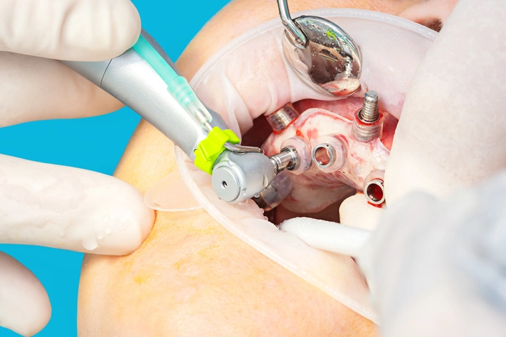
4. Surgical Procedure
On the day of surgery:
- The surgical guide is placed and verified for passive fit and stability.
- If flapless surgery is possible, the guide is used without incising the soft tissue. In other cases, a minimal flap is created.
- A series of guided drills are used in sequence through the guide sleeves to create the osteotomy (implant site) with precise depth and angulation.
- The implant is inserted through the guide or immediately afterward, using guided insertion tools or freehand if required by the system.
- The guide is then removed, and healing abutments or cover screws are placed depending on the treatment protocol (immediate loading vs. delayed).
5. Postoperative Care and Healing
After implant placement:
- A follow-up CBCT or panoramic X-ray may be taken to confirm the position of the implant(s).
- The patient receives instructions on oral hygiene, medications, and healing protocols.
- Healing typically takes 3 to 6 months, depending on bone quality and implant stability.
6. Prosthetic Restoration
Once osseointegration (bone integration) is confirmed:
- Digital or conventional impressions are taken of the implant position.
- The final prosthetic restoration (crown, bridge, overdenture, etc.) is fabricated.
- The prosthesis is then fitted, adjusted, and secured—completing the full implant workflow.
In immediate loading cases, temporary restorations may be placed on the same day as surgery, guided by the same digital planning principles.
Limitations and Considerations
While guided implant surgery offers numerous advantages in terms of precision, safety, and efficiency, it also presents a range of challenges and limitations that clinicians must understand before adopting it into routine practice.
1. Additional Costs
One of the primary concerns with guided implant surgery is cost:
- Initial Investment: Clinics need to invest in CBCT machines, intraoral scanners, planning software, and potentially 3D printers.
- Case-Specific Expenses: Each case typically requires the fabrication of a unique surgical guide, which may cost several hundred dollars, especially if outsourced to a lab.
- Software and Licensing Fees: Many planning platforms operate on subscription-based models, adding ongoing expenses.
- Patient Costs: These expenses may be passed on to the patient, which could limit acceptance, especially in cost-sensitive markets.
2. Learning Curve and Technical Expertise
Implementing guided surgery demands a steep learning curve:
- Software Proficiency: Clinicians must be comfortable using digital planning software to make precise implant decisions.
- 3D Imaging Interpretation: Accurate diagnosis and virtual treatment planning require advanced understanding of CBCT anatomy.
- Guide Usage: Knowing how to seat and secure surgical guides properly is critical to prevent errors in implant trajectory.
Mistakes during the digital planning stage can carry through to the surgical outcome, so clinician training is essential.
3. Guide Fit and Stability
The accuracy of guided surgery relies heavily on the fit of the guide:
- Poor Fit: If the surgical guide does not seat precisely, it can cause implant misalignment or unintended anatomical complications.
- Intraoral Conditions: Saliva, limited mouth opening, or soft-tissue movement can affect how securely the guide stays in place.
- Tooth-Supported vs. Tissue-Supported Guides: Mucosa-supported guides (for edentulous patients) are more prone to movement or flex compared to tooth-supported ones.
Proper scanning protocols, guide design, and sometimes the use of fixation pins are required to ensure guide stability.
4. Limited Intraoperative Flexibility
Unlike freehand surgery, guided implant surgery offers little room for intraoperative adjustments:
- If unexpected anatomical findings occur (e.g., poor bone quality, hidden pathology), the pre-planned guide may no longer be appropriate.
- Modifying implant positioning on the spot is challenging without abandoning the guide altogether.
- Hybrid workflows (partially guided) can help retain some flexibility but reduce the control offered by fully guided approaches.
5. Accuracy Can Be Affected by Workflow Errors
Several points in the workflow can introduce inaccuracies:
- Inaccurate Impressions or Scans: Any distortion in the digital scan or physical impression will compromise guide fit.
- CBCT Resolution and Artifacts: Low-quality scans or metal artifacts can distort anatomical landmarks, leading to planning errors.
- Improper Data Merging: Mismatching CBCT and scan data in the planning software can lead to discrepancies in virtual versus actual anatomy.
Rigorous protocols must be followed throughout the digital workflow to maintain surgical precision.
6. Not Always Necessary for Simple Cases
In straightforward cases involving single implants with adequate bone volume and few anatomical challenges, conventional freehand surgery may be just as effective, especially in the hands of an experienced surgeon. In such scenarios:
- Guided surgery may not significantly improve outcomes.
- The additional time and cost may outweigh the benefits.
Careful case selection is essential to ensure guided surgery is used when it offers a real clinical advantage.
7. Dependence on External Partners
In many practices, especially smaller ones:
- Planning and guide fabrication may be outsourced to third-party labs or planning centers.
- This can cause delays in case turnaround time, and less control over the surgical plan if communication is lacking.
- Errors can occur if the information provided to the lab (e.g., CBCT scan orientation or soft tissue scans) is incomplete or inconsistent.
Internalizing the full digital workflow (planning, design, and printing) can mitigate this but requires more investment and training.
Summary of Key Considerations
| Limitation | Impact |
|---|---|
| Additional cost | May deter patients and increase clinic overhead |
| Learning curve | Requires investment in training and experience |
| Fit and stability of surgical guide | Can compromise accuracy if improperly seated |
| Workflow errors | Risk of compound inaccuracies through digital steps |
| Limited flexibility | Less adaptable to intraoperative surprises |
| Not always necessary | May not justify cost/complexity in simple cases |
| External dependencies | Possible delays and quality-control challenges |