
Digital radiography has revolutionized diagnostic procedures in modern dentistry. It involves the use of digital X-ray sensors instead of traditional photographic film. Since its introduction in the 1980s, digital radiography has become increasingly prevalent due to its numerous advantages, including reduced radiation exposure, instantaneous image viewing, and enhanced image manipulation capabilities. This article explores the history, technology, types, applications, advantages, limitations, and future trends of digital radiography in the dental field.
Table of Contents
ToggleHistorical Background
The journey of radiography in dentistry began shortly after the discovery of X-rays by Wilhelm Conrad Roentgen in 1895. Traditional film-based radiography remained the standard for nearly a century. The advent of digital technology in the 1980s marked a turning point. The first digital radiography system was introduced by Dr. Frances Mouyen, who developed the RadioVisioGraphy (RVG) system. This innovation laid the groundwork for the digital imaging systems we use today.
Technological Framework
Digital radiography systems in dentistry comprise three main components: the X-ray machine, the digital sensor, and the computer software. These components work together to capture, process, and display high-quality diagnostic images.
- X-ray Machine
- Digital Sensors
- Computer Software
- Networking and Data Management
X-ray Machine
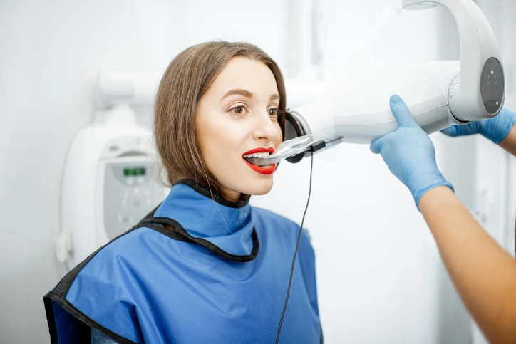
Modern X-ray units used in digital radiography are often equipped with adjustable settings to optimize image quality while minimizing radiation exposure. The control console allows the operator to set exposure time, tube current, and voltage based on the diagnostic need and patient size. These machines may be handheld or wall-mounted, with configurations tailored for intraoral, panoramic, or CBCT imaging.
Digital Sensors
These devices replace traditional photographic film and capture X-ray photons, converting them into digital signals. There are three primary types of sensors used:
- Charged-Coupled Device (CCD): CCD sensors have been widely used since the inception of digital radiography. They consist of an array of light-sensitive elements that generate electronic signals when exposed to radiation. These signals are processed to form a digital image. CCDs are known for their excellent image resolution and diagnostic reliability. However, they require a wired connection and are relatively more expensive.
- Complementary Metal-Oxide Semiconductor (CMOS): CMOS sensors are a newer alternative to CCDs. They offer several advantages, including lower power consumption, faster image processing, and greater affordability. CMOS sensors use active pixel technology, allowing each pixel to amplify and process signals individually, resulting in faster image acquisition.
- Photostimulable Phosphor (PSP) Plates: PSP plates resemble traditional film in flexibility and size, making them more comfortable for patients. They store X-ray energy and must be scanned using a specialized device to convert the stored energy into a digital image. While not as immediate as CCD or CMOS sensors, PSP systems are cost-effective and widely used in practices transitioning from film to digital.
Computer Software
Once the digital sensor captures an image, it is transmitted to computer software for display, storage, and manipulation. Software platforms allow for real-time image viewing, enhancing clinical workflow. Features typically include brightness and contrast adjustment, zoom, measurement tools, annotations, and filters that aid in diagnosis. Many software solutions also support image archiving, integration with electronic health records (EHR), and compatibility with 3D imaging and printing technologies.
Networking and Data Management
Digital radiography systems often integrate with a clinic’s IT infrastructure, enabling images to be accessed from multiple workstations and shared with other professionals. This interoperability is critical for multidisciplinary collaboration, referrals, and maintaining comprehensive patient records. Additionally, cloud-based storage solutions are increasingly popular for secure, remote access to radiographic data.
Types of Digital Radiographic Techniques in Dentistry
Digital radiography in dentistry encompasses several imaging modalities, each suited for different diagnostic purposes:
- Intraoral Radiography
- Extraoral Radiography
- Advanced Imaging Techniques
Intraoral Radiography
- Bitewing Radiographs: Commonly used to detect interproximal caries between teeth and to evaluate alveolar bone levels. These images are essential for routine checkups and early caries detection.
- Periapical Radiographs: Provide a detailed view of the entire tooth, including the crown, root, and surrounding bone. These are useful for diagnosing root infections, periapical lesions, and bone loss.
- Occlusal Radiographs: Capture larger sections of the jaw and are used to view the arch of teeth in either the upper or lower jaw. These are helpful in identifying supernumerary teeth, cysts, and other abnormalities in pediatric and adult patients.
Extraoral Radiography
- Panoramic Radiographs (Orthopantomograms): Offer a comprehensive view of the upper and lower jaws, teeth, temporomandibular joints, nasal area, and sinuses. These are widely used for general assessments, surgical planning, and identifying impacted teeth or jaw disorders.
- Cephalometric Radiographs: Typically used in orthodontics, these lateral skull X-rays help assess the relationship between the teeth, jaw, and skull. Cephalometric Radiographs are invaluable for planning orthodontic treatment and evaluating growth changes.
- Lateral Oblique Views and Skull Radiographs: Occasionally used in maxillofacial diagnostics to evaluate trauma, pathologies, and developmental anomalies.
Advanced Imaging Techniques
- Cone Beam Computed Tomography (CBCT): Delivers 3D imaging with high spatial resolution, allowing for accurate visualization of hard tissues. CBCT is indispensable for implant placement, endodontic diagnosis, assessment of jaw pathologies, and temporomandibular joint evaluation. It offers volumetric data, cross-sectional views, and precise anatomical measurements.
- Digital Subtraction Radiography (DSR): Primarily used in periodontal and endodontic research, DSR allows for the comparison of sequential radiographs by subtracting one image from another to highlight subtle changes over time.
- Digital Tomosynthesis: A limited-angle tomography technique used to reconstruct layered images, helping to reduce the effects of overlapping structures in dental imaging.
Clinical Applications
Digital radiography serves multiple clinical applications in dentistry:
- Caries Detection: Enhanced image contrast and diagnostic tools enable early identification of incipient carious lesions, especially in interproximal and occlusal surfaces, which might be missed during a visual examination.
- Periodontal Assessment: Radiographs assist in evaluating alveolar bone loss, the presence of periodontal pockets, furcation involvements, and calculus deposits. Digital imaging supports longitudinal comparisons in periodontal therapy.
- Endodontics: Provides accurate visualization of root canal morphology, periapical pathology, and helps in determining working length during root canal treatments. Post-operative assessments and retreatment evaluations also rely heavily on radiographs.
- Orthodontics: Aids in cephalometric analysis, assessment of tooth angulation, eruption patterns, and bone growth. It helps in creating treatment plans and tracking changes over time.
- Oral and Maxillofacial Surgery: Essential for pre-operative planning and post-operative evaluation of procedures like tooth extractions, cyst removal, trauma management, and corrective jaw surgeries. CBCT, in particular, provides three-dimensional insights.
- Implant Planning and Placement: Digital radiographs and CBCT imaging help assess bone volume, proximity to vital anatomical structures, and suitability of the implant site. They also assist in designing surgical guides for accurate implant placement.
- Detection of Pathologies: Enables identification of benign and malignant tumors, cysts, impacted teeth, and other developmental anomalies.
- Temporomandibular Joint (TMJ) Evaluation: TMJ disorders can be evaluated through panoramic and CBCT images, identifying joint space, condylar position, and signs of degeneration.
- Forensic Dentistry: Provides essential evidence for human identification, bite mark analysis, and age estimation. Digital records can be archived indefinitely and shared easily.
- Pediatric Dentistry: Facilitates the monitoring of developing dentition, assessment of trauma, detection of caries, and evaluation of congenital anomalies.
Advantages of Digital Radiography
The transition to digital radiography offers numerous benefits:
- Reduced Radiation Exposure: Digital radiography significantly decreases radiation dose by up to 80% compared to conventional film-based methods, enhancing patient safety.
- Immediate Image Availability: Digital systems allow for the instantaneous viewing of images, improving workflow efficiency and reducing patient chair time.
- Superior Image Enhancement: Digital images can be manipulated post-capture for improved diagnostic accuracy. Features like zoom, color inversion, contrast and brightness adjustment, and measurement tools help clinicians detect subtle abnormalities.
- Efficient Data Storage and Sharing: Digital radiographs can be stored electronically, eliminating the need for physical storage space. They can be easily shared via secure networks or cloud platforms, facilitating interdisciplinary collaboration and consultations.
- Environmentally Friendly: The absence of film processing chemicals and lead foils makes digital radiography more sustainable and reduces environmental impact.
- Patient Education and Engagement: Real-time display of digital images allows clinicians to visually explain conditions and treatment plans to patients, enhancing understanding and trust.
- Cost-Effectiveness Over Time: While initial investment is high, digital radiography reduces recurring costs associated with film, chemicals, and storage, offering financial benefits in the long run.
- Integration with Modern Dental Systems: Digital radiographs can be seamlessly integrated with practice management software, EHRs, and digital charting tools, streamlining administrative tasks and improving patient care continuity.
Limitations and Challenges
Despite its many advantages, digital radiography is not without drawbacks:
- Initial Cost: The cost of acquiring digital radiography systems, including sensors, software, and compatible X-ray machines, can be prohibitively high, particularly for smaller or newly established dental practices.
- Sensor Discomfort: Intraoral digital sensors are often more rigid and bulkier than traditional film, which can cause discomfort or gagging in sensitive patients, especially children and individuals with smaller oral cavities.
- Learning Curve and Training Requirements: Dental staff and clinicians must undergo specialized training to correctly use digital systems, interpret images, and make the most of advanced software features. Mistakes during this learning phase can impact image quality and diagnostic accuracy.
- Technical Malfunctions: Like any technology, digital radiography is prone to hardware failures, software glitches, and connectivity issues. These can disrupt workflow, delay diagnosis, and result in data loss if adequate backup systems are not in place.
- Image Quality Variability: Improper sensor placement, exposure settings, or software calibration can result in poor-quality images that hinder diagnosis. Additionally, digital sensors may be more sensitive to movement, leading to blurred images if patient cooperation is not optimal.
- Data Security and Privacy Concerns: The storage and sharing of digital radiographs require robust cybersecurity measures. Clinics must ensure compliance with data protection laws such as HIPAA to safeguard patient information.
- Maintenance and Upgrades: Digital systems require regular maintenance and software updates to ensure optimal performance. Over time, hardware components may become obsolete, necessitating reinvestment in newer technologies.
Infection Control Considerations
Infection control is a critical component of digital radiography, especially due to the reuse of sensors and imaging plates in clinical settings. Because digital sensors often come into direct contact with mucous membranes, there is a risk of cross-contamination between patients if proper protocols are not followed.
To mitigate this risk:
- Barrier Protection: Single-use plastic barriers should be placed over digital sensors and PSP plates during every use. These barriers must be replaced for each patient to ensure hygiene.
- Disinfection Protocols: After removing the barrier, sensors should be wiped and disinfected using approved intermediate-level disinfectants. Care must be taken to avoid damage to sensitive electronic components during cleaning.
- Sterilizable Holders: Sensor holders and accessories must be sterilized between uses, ideally using an autoclave or chemical sterilant in accordance with the manufacturer’s instructions.
- Hand Hygiene: Operators should perform hand hygiene before and after handling sensors, equipment, or barriers. Gloves must be changed between patients.
- Staff Training: Dental professionals and assistants must be regularly trained in infection control measures, including the proper handling and cleaning of digital radiographic equipment.
- Surface Protection: Work surfaces in radiographic areas should be disinfected regularly and covered with disposable barriers when appropriate.
- Regulatory Compliance: Clinics must follow guidelines issued by national and international health organizations (e.g., CDC, WHO) regarding infection control in dental imaging to maintain safety standards.
By implementing comprehensive infection control measures, dental practices can ensure both patient safety and the longevity of expensive radiographic equipment.
Regulatory and Ethical Aspects
Digital radiography must adhere to strict regulatory and ethical standards. Radiation safety is governed by national regulatory bodies, requiring that dental professionals follow ALARA (As Low As Reasonably Achievable) principles to minimize patient exposure. Clinics must ensure radiographic equipment is routinely calibrated and maintained, and personnel must undergo radiation safety training.
Patient privacy is equally vital. Digital images are part of a patient’s health record and must be protected under data protection laws such as HIPAA in the United States or GDPR in the European Union. Clinics must implement secure data storage and transmission systems to prevent unauthorized access, hacking, or breaches.
Ethically, dental professionals must obtain informed consent before performing any radiographic procedure, explaining the purpose, risks, and benefits. Radiographs should only be taken when clinically justified to avoid unnecessary exposure. Practitioners are also responsible for maintaining transparency in image interpretation, storing records securely for the legally required duration, and referring to specialists when advanced diagnostics are needed.