
Gingival cysts are relatively rare, benign lesions found within the soft tissues of the mouth, particularly the gingiva, or gums. Although typically asymptomatic, they can cause concern among patients due to their visible presentation. These cysts are usually small, slow growing, and non-invasive, but they play a significant role in the differential diagnosis of oral soft tissue lesions. The accurate identification and management of gingival cysts are crucial for dental professionals to avoid unnecessary treatment and reassure affected individuals.
This article provides an in-depth examination of gingival cysts, covering their types, etiology, clinical presentation, diagnostic approaches, histopathology, treatment options, and prognosis. We will also discuss their significance within the broader spectrum of oral pathology and their differentiation from other similar lesions.
Table of Contents
ToggleWhat Is a Gingival Cyst?
A gingival cyst is a type of developmental odontogenic cyst that occurs within the soft tissues of the gingiva, typically without involvement of the underlying alveolar bone. It originates from the rests of dental lamina epithelium, remnants from the embryological development of teeth. Gingival cysts are classified as non-neoplastic, meaning they are not tumors and do not exhibit aggressive or infiltrative growth.
They are most often asymptomatic, present as small localized swellings on the gingiva, and are discovered incidentally during routine dental examinations. Despite their benign nature and small size, accurate diagnosis is important due to the similarity of their clinical presentation to other more serious gingival or periodontal lesions.
Gingival cysts are part of the wider family of odontogenic cysts, which are cystic lesions derived from tooth-forming tissues. However, unlike other odontogenic cysts like the radicular cyst or dentigerous cyst, gingival cysts are extraosseous, meaning they are located outside the bone and confined to the soft tissue.
Historical Context
The gingival cyst of the adult was not well-documented until the mid-20th century. It was first clearly differentiated from the lateral periodontal cyst in the 1960s, when it became evident that a similar lesion could arise in the soft tissue without radiographic bone involvement.
In contrast, the gingival cyst of the newborn has long been observed and often referred to using other names such as Epstein pearls, Bohn’s nodules, and dental lamina cysts. These terms were initially used interchangeably, but later histological and anatomical distinctions clarified their individual identities.
Classification of Gingival Cysts
Gingival cysts can be classified based on age of onset, origin, and location. The two major categories are:
1. Gingival Cyst of the Adult
A gingival cyst of the adult is a rare, developmental odontogenic cyst that occurs in the gingiva of adults, arising from remnants of the dental lamina. It is the soft tissue counterpart to the lateral periodontal cyst, which involves alveolar bone.
Characteristics:
- Age group: Typically presents in middle-aged adults (40–60 years).
- Size: Small, usually less than 5 mm in diameter.
- Color: Clear, bluish, or similar to surrounding mucosa.
- Consistency: Soft and fluctuant.
- Symptoms: Usually asymptomatic; may occasionally cause mild discomfort or cosmetic concern.
- Common location: Facial gingiva of the mandibular canine-premolar region, followed by the anterior maxilla.
Etiopathogenesis:
The adult gingival cyst arises from epithelial remnants of the dental lamina, also known as the rests of Serres. These rests are residual embryonic tissue left after tooth development and can persist in the gingival connective tissue.
In some individuals, these epithelial remnants may proliferate and undergo cystic degeneration due to minor trauma, inflammation, or idiopathic stimuli, resulting in the formation of a gingival cyst.
Relationship to Lateral Periodontal Cyst:
The gingival cyst of the adult is considered the soft tissue analog of the lateral periodontal cyst, which presents similarly but occurs within bone, producing a radiolucency adjacent to the roots of vital teeth. Both cysts likely share a common histogenetic origin, but their locations (soft tissue vs. intraosseous) distinguish them clinically and radiographically.
2. Gingival Cyst of the Newborn
Also referred to as dental lamina cysts, gingival cysts of the newborn are small, keratin-filled cysts found on the alveolar ridges of infants. They are benign, developmental lesions resulting from remnants of the dental lamina.
Characteristics:
- Age group: Present at birth or within the first few weeks of life.
- Size: Typically 1–3 mm in diameter.
- Color: White or yellowish.
- Location: Crest of alveolar ridges (buccal, lingual, or midline).
- Symptoms: Asymptomatic; do not cause discomfort or interfere with feeding.
- Resolution: Self-limiting; usually rupture and disappear spontaneously within the first few months of life.
Etiopathogenesis:
During tooth development, the dental lamina is responsible for giving rise to tooth buds. After tooth formation, the lamina disintegrates, but small fragments can remain embedded within the alveolar mucosa. In newborns, these remnants may keratinize and form small cysts.
These cysts are often confused with:
- Epstein pearls: Located on the midline of the palate; arise from entrapped epithelium during palatal fusion.
- Bohn’s nodules: Located on the buccal or lingual aspects of the dental ridges; believed to originate from minor salivary glands.
Only the dental lamina cysts located on the alveolar ridges are true gingival cysts of the newborn.
Summary Table: Gingival Cyst Classification
| Feature | Gingival Cyst of the Adult | Gingival Cyst of the Newborn |
|---|---|---|
| Age | 40–60 years | Neonates (birth–3 months) |
| Origin | Rests of dental lamina | Rests of dental lamina |
| Size | <5 mm | 1–3 mm |
| Location | Facial gingiva (canine/premolar) | Alveolar ridge (crest) |
| Symptoms | Asymptomatic | Asymptomatic |
| Resolution | Requires excision | Spontaneous resolution |
| Bone involvement | None | None |
| Recurrence | Rare | Not applicable |
Rare Variants and Related Lesions
In addition to the two main types, some unusual presentations and closely related lesions include:
Multiple Gingival Cysts in Adults:
Though rare, multiple gingival cysts may appear in adults, potentially mimicking vesiculobullous diseases like mucous membrane pemphigoid. Careful histopathological evaluation is required.
Peripheral Odontogenic Cysts:
Occasionally, other peripheral (soft tissue) odontogenic cysts can mimic gingival cysts, such as:
- Peripheral calcifying odontogenic cyst
- Peripheral dentigerous cyst (rare)
Eruption Cyst:
Sometimes confused with gingival cysts, eruption cysts are soft tissue analogs of dentigerous cysts that form over erupting teeth in children. They involve partially erupted teeth and are often bluish due to hemorrhagic contents. These are not true gingival cysts as they are associated with developing teeth and not remnants of dental lamina in soft tissue.
Epidemiology
Prevalence
- Gingival cysts of the adult are uncommon, accounting for less than 0.5% of all odontogenic cysts.
- Gingival cysts of the newborn are more prevalent, seen in up to 60-85% of neonates, although they often go unnoticed due to their asymptomatic nature.
Age and Gender Distribution
- Adults: Most commonly seen in individuals aged 40-60 years. There is no significant gender predilection.
- Newborns: Present at birth or within the first few weeks of life, with equal occurrence among genders.
Common Locations
- Gingival cysts of the adult most frequently occur in the mandibular canine and premolar region, followed by the anterior maxilla.
- Newborn cysts are often seen along the alveolar ridges, both maxillary and mandibular.
Etiology and Pathogenesis
Understanding the etiology and pathogenesis of gingival cysts provides essential insight into their formation, clinical presentation, and management. Both the adult and newborn variants of gingival cysts are developmental in origin, arising from residual embryological structures related to tooth formation. However, the specific pathways, triggers, and tissue behaviors differ between the two types.
General Etiology: Developmental Origin
Gingival cysts are categorized as odontogenic cysts—that is, they arise from tissues involved in the formation of teeth. These tissues include the dental lamina, enamel organ, and other epithelial structures derived from the ectoderm. During normal odontogenesis (tooth development), these structures typically regress or transform. However, residual epithelial cells may persist within the gingival connective tissue and become the source of cystic change.
The dental lamina plays a central role in the development of both forms of gingival cysts. This band of epithelial tissue, which forms during early embryogenesis, gives rise to the enamel organs of developing teeth. After tooth bud formation, parts of the dental lamina disintegrate, but scattered fragments—called the rests of Serres—may remain embedded in the surrounding soft tissue.
These epithelial remnants can later undergo proliferation, keratinization, or degeneration, resulting in the formation of a cystic cavity lined with epithelium and often filled with keratin or fluid.
A. Etiology and Pathogenesis of Gingival Cyst of the Adult
Origin: Rests of Serres
The gingival cyst of the adult arises specifically from the rests of Serres, which are epithelial remnants of the dental lamina that persist in the gingival soft tissue after the tooth development process is complete. These remnants are quiescent under normal circumstances but may be reactivated by various stimuli.
Pathogenesis
The proposed mechanism for cyst formation involves:
Epithelial Proliferation
For reasons not completely understood, one or more epithelial rests begin to proliferate. This may be a spontaneous event or triggered by:Minor trauma (e.g., food impaction, brushing)
Chronic low-grade inflammation
Hormonal or age-related changes in tissue
Genetic or molecular signaling abnormalities
Cystic Degeneration
As epithelial proliferation progresses, the central cells may undergo degeneration due to lack of vascular supply. This creates a cavity that expands and becomes filled with fluid or keratin.Encapsulation
Surrounding connective tissue forms a fibrous capsule around the proliferating epithelium, resulting in a well-circumscribed, true cyst.Epithelial Differentiation
The epithelial lining often remains thin, typically 1–3 layers thick, and composed of cuboidal to squamous cells. In many cases, clear cells may be observed—these are glycogen-rich epithelial cells, a characteristic histological feature.Lack of Bone Involvement
Importantly, this entire process occurs in the soft tissue only, with no extension into the alveolar bone. This is a key distinguishing feature from other odontogenic cysts.
Possible Contributing Factors
- Genetic predisposition: While not conclusively proven, some individuals may possess a greater tendency for odontogenic epithelial remnants to proliferate.
- Environmental triggers: Trauma, irritation, or smoking could hypothetically contribute by inducing tissue changes.
- Aging process: Age-related changes in connective tissue metabolism or vascularity may create an environment that promotes cyst formation.
B. Etiology and Pathogenesis of Gingival Cyst of the Newborn
Origin: Dental Lamina Remnants in Neonates
Gingival cysts of the newborn result from dental lamina fragments that remain within the alveolar mucosa after the formation of the primary tooth buds. During embryogenesis, the dental lamina invaginates from the oral epithelium and forms a series of swellings that develop into the deciduous dentition. After tooth bud formation, the majority of the lamina breaks down.
However, remnants may persist within the alveolar ridge mucosa and form cysts during late fetal or early neonatal life.
Pathogenesis
The pathogenesis of gingival cysts in newborns follows a simpler and more self-limiting pathway:
- Keratinization of Epithelial Rests
Epithelial remnants undergo keratinization as part of a normal degenerative process. - Cyst Formation
Keratin accumulates within the lumen of the epithelial structures, resulting in a small keratin-filled cyst. - Eruption or Rupture
The cyst gradually enlarges but remains superficial. Eventually, the overlying mucosa thins and ruptures, releasing the keratin contents. In most cases, this occurs spontaneously within a few weeks after birth. - Resorption
After rupture, the cyst resolves completely without scarring or any long-term consequences.
Key Features of Pathogenesis in Newborns
- No inflammation: The cysts are not associated with any inflammatory response unless secondarily infected (which is very rare).
- No clinical symptoms: Because of their superficial location and painless nature, they do not interfere with feeding or cause discomfort.
- High prevalence: Their presence in up to 85% of newborns suggests that they may be considered a normal developmental phenomenon rather than a pathological condition.
Comparison of Pathogenesis Between Adults and Newborns
| Feature | Adult Gingival Cyst | Newborn Gingival Cyst |
|---|---|---|
| Origin | Rests of Serres (dental lamina remnants) | Dental lamina remnants in neonates |
| Trigger | Possibly trauma, irritation, aging | Natural degeneration of epithelial rests |
| Cellular Activity | Proliferation, degeneration, cystic change | Keratinization and superficial cysting |
| Histology | Thin epithelial lining, glycogen-rich cells | Parakeratinized epithelium, keratin lumen |
| Resolution | Requires surgical removal | Spontaneous rupture |
| Inflammatory Response | Minimal to none | None (unless secondary infection) |
Clinical Features
Gingival cysts, whether in adults or newborns, exhibit distinct clinical characteristics that reflect their developmental origin and soft tissue location. A clear understanding of their presentation is crucial not only for diagnosis but also for distinguishing them from other more serious or symptomatic oral lesions. These cysts are typically asymptomatic, but their visibility and physical presence on the gingiva may prompt concern or curiosity from patients, parents, or clinicians.
This section outlines the clinical features of both types of gingival cysts—adult and newborn—with attention to their appearance, location, symptoms, behavior over time, and clinical course.
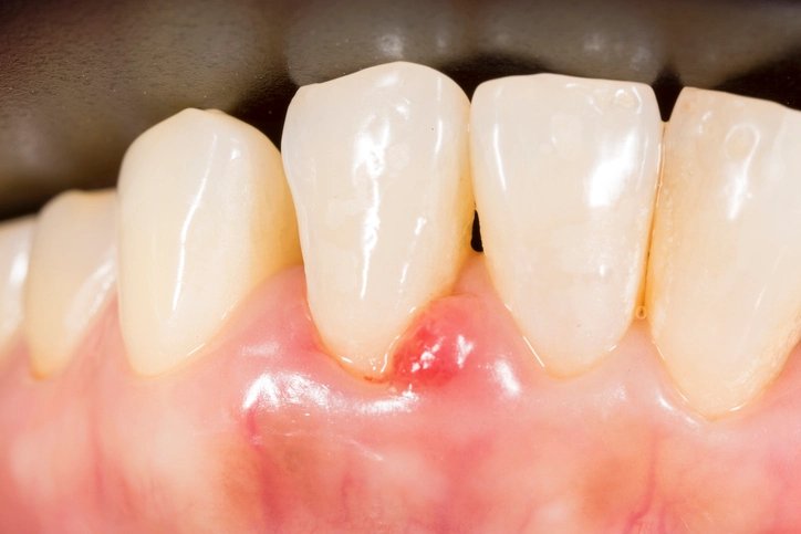
A. Clinical Features of Gingival Cyst of the Adult
General Characteristics
Asymptomatic: The majority of adult gingival cysts do not produce pain, tenderness, or inflammation. Patients may not be aware of their presence until noticed by a dentist during a routine examination.
Small Size: Typically less than 5 mm in diameter, often in the range of 2–4 mm.
Color and Appearance:
Often translucent, pale, or bluish due to the fluid contents or thin overlying mucosa.
Occasionally, they may match the color of the surrounding gingiva and appear as a subtle swelling or nodule.
Shape: Round, ovoid, or dome-shaped.
Location and Distribution
- Facial (labial/buccal) gingiva is the most common site.
- Strong predilection for the mandibular canine and premolar region (also called the “mandibular anterior sextant”).
- Less commonly seen in the maxillary anterior region.
- Usually unilateral and solitary, but rare cases of multiple or bilateral cysts have been reported.
Consistency and Palpation
- Soft and fluctuant to touch, indicating fluid-filled nature.
- Non-tender, unless secondarily traumatized or inflamed.
- Surface mucosa is typically intact and non-ulcerated.
Progression and Behavior
- Slow-growing lesion, often stable in size for long periods.
- Do not invade surrounding tissues or bone.
- May persist for months to years unless treated.
- No spontaneous resolution—surgical excision is typically required.
Tooth Vitality and Periodontal Health
- Teeth adjacent to the cyst are typically vital and show no signs of pathology.
- No periodontal pocketing or mobility associated with the lesion.
- Radiographic examination reveals no underlying bone changes, which helps distinguish it from lateral periodontal cysts or periapical pathology.
Patient Complaints
- Most patients are unaware of the lesion and present only after it is discovered incidentally.
- In cases where the lesion becomes cosmetically concerning or interferes with oral hygiene, patients may request removal.
B. Clinical Features of Gingival Cyst of the Newborn
Also known as dental lamina cysts, these lesions are a common finding in neonates and are part of the normal developmental process in many cases. While often asymptomatic and self-limiting, they can be mistaken for more serious congenital anomalies by concerned parents or caregivers.
General Characteristics
- Asymptomatic: No pain or discomfort to the infant.
- Small Nodules: Usually 1 to 3 mm in diameter.
- White or Yellowish: Due to the keratin-filled lumen, giving a pearly or creamy appearance.
- Smooth Surface: Dome-shaped with intact overlying mucosa.
Location and Pattern
- Found along the alveolar ridges, especially on the crest of the maxillary or mandibular alveolar ridges.
- May appear unilaterally or bilaterally.
- Occur on both anterior and posterior segments but most commonly in the incisor and canine areas.
Number of Lesions
- May occur as single or multiple nodules.
- Frequently appear in clusters, sometimes involving several sites on the alveolar ridge.
- In contrast to adult gingival cysts, multiple lesions are the norm, not the exception.
Duration and Natural Course
- These cysts appear at birth or within the first few weeks of life.
- They rupture spontaneously within 2 to 12 weeks and typically disappear without intervention.
- No scarring or recurrence is observed after resolution.
Parental Concerns
Despite their benign nature, these cysts can cause anxiety among new parents who may mistake them for:
Natal or neonatal teeth
Thrush (oral candidiasis)
Herpes or other infections
Abscesses or tumors
It is critical for healthcare providers to educate and reassure caregivers that the lesions are harmless, painless, and will resolve on their own.
Associated Conditions
Gingival cysts of the newborn are sometimes mistaken for Epstein pearls (located on the palatal midline) or Bohn’s nodules (located on the buccal or lingual parts of the alveolar ridge), although the histogenesis differs.
C. Differential Features at a Glance
| Feature | Gingival Cyst of Adult | Gingival Cyst of Newborn |
|---|---|---|
| Age of Onset | 40–60 years | Birth to a few weeks old |
| Symptoms | Asymptomatic | Asymptomatic |
| Size | 2–5 mm | 1–3 mm |
| Number | Usually solitary | Often multiple |
| Color | Translucent, bluish, mucosa-colored | White or yellowish |
| Location | Facial gingiva (mandibular canine/premolar) | Alveolar ridges (crest) |
| Resolution | Persistent without treatment | Spontaneous within weeks |
| Radiographic Findings | None | None |
When to Be Cautious
Although gingival cysts are benign, clinicians should remain vigilant when evaluating oral lesions. Certain features may suggest an alternative or more serious diagnosis:
- Rapid increase in size
- Ulceration or bleeding
- Pain or tenderness
- Involvement of underlying bone (visible on radiographs)
- Tooth mobility or non-vitality
- Foul discharge or signs of infection
If such features are present, a biopsy and histopathological examination are warranted to rule out other odontogenic or soft tissue pathologies.
Diagnosis
Clinical Examination
Diagnosis is primarily clinical, particularly in the case of newborns. For adult gingival cysts, a differential diagnosis is important due to their resemblance to other soft tissue lesions.
Differential Diagnosis
- Mucocele
- Fibroma
- Peripheral ossifying fibroma
- Pyogenic granuloma
- Peripheral giant cell granuloma
- Lateral periodontal cyst (if bone involvement is suspected)
Radiographic Examination
- Gingival cysts of the adult do not show radiographic findings because they do not affect bone.
- In contrast, lateral periodontal cysts show radiolucency adjacent to the roots of teeth.
Histopathology
Microscopic examination is essential for definitive diagnosis, especially if the lesion is excised:
- Epithelial lining: Thin (1-3 cell layers), cuboidal to squamous epithelium.
- Cyst wall: Fibrous connective tissue with minimal inflammation.
- Clear cells: May be present in the epithelial lining, representing glycogen-rich epithelial cells.
Management and Treatment
The management of gingival cysts varies significantly based on the type—adult vs. newborn—because of differences in their clinical behavior, resolution patterns, and patient age. In general, treatment is simple and definitive for adult lesions and non-interventional for neonatal cysts due to their spontaneous resolution.
A comprehensive understanding of the appropriate management approach is essential for dentists, oral surgeons, pediatricians, and general practitioners to avoid unnecessary procedures, address cosmetic or functional concerns, and reassure patients and caregivers.
A. Management of Gingival Cyst of the Adult
Gingival cysts of the adult are non-neoplastic and benign but do not spontaneously resolve. Therefore, once identified and confirmed, the standard treatment is surgical excision. The goal is to:
- Confirm the diagnosis histologically.
- Remove the cyst entirely to prevent persistence or rare recurrence.
- Restore normal gingival contour and function.
1. Surgical Excision
This is the treatment of choice for adult gingival cysts.
Procedure Overview
Anesthesia: Local infiltration anesthesia is administered at the site.
Incision and Exposure: A small linear or elliptical incision is made to expose the lesion.
Cyst Enucleation:
The cyst is carefully dissected and enucleated in its entirety to avoid rupture.
Care is taken to avoid damaging adjacent gingival or periodontal structures.
Hemostasis and Closure:
Hemostasis is achieved using gauze compression or electrocautery, if necessary.
The site may be closed with resorbable or non-resorbable sutures, depending on size and location.
Histopathologic Confirmation
All excised tissue should be submitted for histopathologic examination to:
Confirm the diagnosis.
Rule out other odontogenic or soft tissue lesions with similar clinical appearance.
Detect rare dysplastic changes (although extremely uncommon in gingival cysts).
2. Postoperative Care
- Analgesics: Mild pain can be managed with over-the-counter NSAIDs (e.g., ibuprofen).
- Antibiotics: Not typically required unless signs of infection or compromised immunity.
- Oral hygiene instructions: Patients should avoid brushing the area directly for a few days and use warm saline rinses.
- Suture removal: If non-resorbable sutures are used, they are typically removed after 7–10 days.
3. Follow-Up and Prognosis
- Follow-up visits are recommended to monitor healing and ensure no recurrence.
- Recurrence is exceedingly rare when complete excision is achieved.
- Cosmetic results are generally excellent, especially in esthetically sensitive areas like the anterior gingiva.
B. Management of Gingival Cyst of the Newborn
No treatment is required for gingival cysts of the newborn. These lesions are self-limiting and harmless, representing a normal developmental phenomenon. Intervention is unnecessary and may cause undue trauma or distress to the infant.
1. Observation
The optimal management strategy is clinical observation with parental education and reassurance.
- Most cysts rupture spontaneously within 2 weeks to 3 months after birth.
- The keratin content is released into the oral cavity and swallowed without complication.
- The lesion leaves no residual defect or scarring.
2. Parental Counseling
A vital aspect of management is to inform and reassure caregivers, who may be anxious about the presence of nodules in their infant’s mouth. Counseling should include:
- An explanation that the cysts are benign and common.
- A description of their natural course and resolution.
- Assurance that they do not interfere with feeding, speech, or teething.
- Avoidance of any attempts to manually rupture or remove the lesions at home.
3. When to Reconsider Management
Although extremely rare, intervention or referral may be warranted if:
- Lesions become infected (e.g., erythema, warmth, discharge).
- Lesions persist beyond 6 months without signs of resolution.
- Atypical features are present (e.g., ulceration, rapid growth, associated systemic signs).
- Differential diagnosis includes other neonatal oral lesions (e.g., candidiasis, neonatal teeth, congenital epulis).
In such cases, referral to a pediatric dentist or oral pathologist may be appropriate.
C. Considerations for Differential Management
| Feature | Adult Gingival Cyst | Newborn Gingival Cyst |
|---|---|---|
| Management Approach | Surgical excision | No treatment (observation only) |
| Anesthesia Required | Local | None |
| Histopathology | Recommended (to confirm diagnosis) | Not required unless lesion persists |
| Recurrence Risk | Extremely low if completely excised | None |
| Resolution Timeframe | Persists indefinitely until removed | Resolves in 2–12 weeks |
| Parental Education | Not typically needed | Crucial to reduce unnecessary anxiety |
| Follow-Up | One or two post-op visits recommended | Usually none needed unless atypical |
D. Other Management Scenarios
1. Misdiagnosed Lesions
If a gingival cyst is misdiagnosed as another soft tissue lesion, unnecessary or incorrect treatment (e.g., incisional biopsy, pharmacotherapy) may be initiated. Clear identification avoids this.
2. Cosmetic Concerns
In some adults, even small cysts in esthetic zones (e.g., anterior maxilla) may become a concern due to visibility when smiling or talking. In these cases, elective excision may be pursued for cosmetic reasons alone, even if the cyst is asymptomatic.
3. Lesions with Unusual Features
- Rapid enlargement
- Ulceration
- Pain or drainage
Such signs warrant further investigation and possibly more extensive excision and biopsy, as they deviate from the benign course typical of gingival cysts.
E. Prognosis
Adult Cysts
- Excellent prognosis following complete surgical removal.
- Minimal complications, such as minor post-operative discomfort or temporary gingival contour changes.
- Recurrence is almost nonexistent if the cyst is fully enucleated.
Newborn Cysts
- Near-universal resolution without intervention.
- No impact on teething or oral development.
- No recurrence or long-term sequelae.
F. Clinical Pearls for Practitioners
- In adults, always differentiate gingival cysts from lateral periodontal cysts, fibromas, and mucocele-like lesions using clinical exam and radiography.
- In infants, reassurance is treatment—do not overmanage or refer unnecessarily.
- Submit all excised adult cysts for histopathologic confirmation, even if the clinical diagnosis appears clear.
- Educate dental and medical teams (including pediatricians and family physicians) about neonatal cysts to reduce unnecessary referrals or concerns.
Conclusion
Gingival cysts, though uncommon, represent an important category of odontogenic cysts, particularly due to their benign and self-limiting nature. Proper clinical evaluation, accurate diagnosis, and appropriate management are key to ensuring optimal patient outcomes. For clinicians, distinguishing gingival cysts from other similar oral lesions is crucial to avoid overtreatment. For patients, especially parents of affected newborns, understanding the non-threatening nature of these cysts helps in alleviating anxiety.
While most gingival cysts require minimal to no intervention, their recognition underscores the broader importance of comprehensive oral examinations in both pediatric and adult populations. Continued awareness and documentation of such lesions also enhance our understanding of developmental anomalies within the oral cavity.