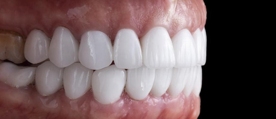
Dental occlusion is a critical aspect of oral health that refers to the way teeth come together when the jaws close. It involves the alignment, contact, and function of the upper and lower teeth during various movements, such as biting, chewing, and speaking. A harmonious dental occlusion is essential for proper mastication, speech, and overall oral function. This comprehensive article explores the concepts and significance of dental occlusion, its role in oral health, common occlusal disorders, diagnostic techniques, and treatment options.
Table of Contents
ToggleUnderstanding Dental Occlusion
Dental occlusion involves the relationship between the upper and lower teeth and how they meet when the jaws are closed. The occlusion can be influenced by various factors, including the shape and size of the teeth, jaw alignment, and muscular coordination. The primary components of dental occlusion are:
- Centric Occlusion (CO)
- Centric Relation (CR)
- Occlusal Schemes
Centric Occlusion (CO)
It refers to the position of the teeth when the jaws are closed in maximum intercuspal contact, with the condyles of the mandible in their most superior and retruded position.
Centric Relation (CR)
It is the most retruded, repeatable, and stable position of the mandible in relation to the maxilla. CR is independent of tooth contact and is determined by the position of the temporomandibular joints.
Occlusal Schemes
Various occlusal schemes, such as Angle’s classification, describe the relationship between the upper and lower teeth in different types of malocclusion.
Types of Dental Occlusion
When discussing dental occlusion, three primary types are commonly referenced: Class I, Class II, and Class III occlusion. These classifications describe the relationship between the upper and lower teeth when the jaws are closed. Here’s a closer look at each type:
- Class I Occlusion (Neutrocclusion)
- Class II Occlusion (Distocclusion)
- Class III Occlusion (Mesiocclusion)
Class I Occlusion (Neutrocclusion)
Class I occlusion is considered the ideal occlusal relationship, where the upper teeth slightly overlap the lower teeth. In this type of occlusion, the molars fit together in a manner where the upper first molar sits just in front of the lower first molar. The canines of the upper and lower arches are aligned, allowing for proper function during biting and chewing. Class I occlusion is typically characterized by good dental alignment, with minimal crowding or spacing issues.
However, it’s important to note that even in Class I occlusion, variations and individual differences in tooth alignment and jaw relationships can exist. It is crucial to evaluate other factors such as tooth angulations, midline discrepancies, and occlusal plane discrepancies to fully assess occlusion.
Class II Occlusion (Distocclusion)
Class II occlusion, also known as distocclusion or retrognathic occlusion, refers to a condition where the upper first molar is positioned significantly further forward compared to the lower first molar. This results in a prominent overbite and a significant horizontal overlap of the upper teeth over the lower teeth. Class II occlusion can be further classified into two divisions:
Class II Division 1
In this type, the upper incisors are protruded, causing a significant overjet (horizontal distance between the upper and lower incisors). The upper incisors may be inclined forward, and the lower jaw is typically retruded in relation to the upper jaw.
Class II Division 2
Here, the upper incisors are retruded, and there is a deep overbite. The lower central incisors may be inclined towards the tongue, and the lower jaw may appear more vertically positioned in relation to the upper jaw.
Class II occlusion can be associated with various functional and aesthetic issues, including difficulty in proper tooth alignment, increased risk of tooth wear, speech difficulties, and potential temporomandibular joint (TMJ) problems.
Class III Occlusion (Mesiocclusion)
Class III occlusion, also referred to as mesiocclusion or prognathic occlusion, is characterized by an anterior crossbite, where the lower teeth are positioned ahead of the upper teeth when the jaws are closed. This results in a negative overjet or an underbite. Class III occlusion can also be divided into two divisions:
Class III Division 1
In this type, the lower incisors are protruded, causing the anterior crossbite and a negative overjet. The lower jaw may be positioned forward in relation to the upper jaw.
Class III Division 2
Here, the lower incisors are retruded, and the anterior crossbite is present. The upper central incisors are typically inclined towards the tongue, and the upper jaw may be retruded compared to the lower jaw.
Class III occlusion can be associated with challenges in mastication, speech difficulties, and facial aesthetics. TMJ issues may also arise in severe cases.
It’s important to note that while Class I, II, and III occlusions provide a general framework for understanding dental relationships, individual variations exist, and occlusion can be influenced by factors such as tooth alignment, skeletal relationships, and muscular coordination. A thorough examination by a dental professional is necessary to evaluate and diagnose occlusal conditions accurately.
Significance of Dental Occlusion in Oral Health
Proper dental occlusion plays a crucial role in maintaining oral health and overall well-being. The significance of dental occlusion can be understood through the following aspects:
- Mastication and Digestion
- Speech and Articulation
- Temporomandibular Joint (TMJ) Health
- Tooth Wear and Trauma
Mastication and Digestion
Optimal occlusion allows for efficient biting and chewing, promoting proper food breakdown and digestion. Malocclusion or occlusal imbalances can lead to difficulties in chewing, impacting the overall nutritional intake.
Speech and Articulation
Dental occlusion affects speech production and clarity. The position and contact of teeth during speech sounds are essential for proper articulation, and any occlusal abnormalities can result in speech impairments.
Temporomandibular Joint (TMJ) Health
Dental occlusion influences the function and health of the TMJ. Imbalances in occlusion can lead to TMJ disorders, causing symptoms such as jaw pain, clicking or popping sounds, and restricted jaw movement.
Tooth Wear and Trauma
Malocclusion and occlusal interferences can cause excessive tooth wear, chipping, or fractures. Uneven distribution of biting forces may also result in tooth mobility and periodontal problems.
Common Occlusal Disorders
Several occlusal disorders can affect dental occlusion and oral health. This section highlights some common occlusal disorders:
- Malocclusion
- Temporomandibular Joint Disorders (TMD)
- Bruxism
Malocclusion
Malocclusion refers to the misalignment or incorrect positioning of teeth when the jaws are closed. It can involve issues such as crowded teeth, overbite, underbite, crossbite, and open bite.
Temporomandibular Joint Disorders (TMD)
TMD encompasses a range of conditions affecting the TMJ and surrounding structures. Occlusal factors, such as uneven contact or bite discrepancies, can contribute to TMD symptoms like jaw pain, headaches, and muscle stiffness.
Bruxism
Bruxism is the habitual grinding or clenching of teeth, often associated with occlusal imbalances and stress. It can lead to tooth wear, TMJ problems, and muscle discomfort.
Diagnostic Techniques for Assessing Dental Occlusion
Accurate assessment of dental occlusion is crucial for proper diagnosis and treatment planning. Various diagnostic techniques are employed to evaluate dental occlusion. Here are some commonly used methods:
- Clinical Examination
- Dental Models and Study Casts
- Radiographic Evaluation
- Occlusal Analysis
- Digital Technologies
Clinical Examination
A comprehensive clinical examination involves visually inspecting the teeth, evaluating the bite relationship, and assessing any signs of malocclusion or occlusal abnormalities. Dentists may use instruments like articulating paper or occlusal indicators to identify areas of tooth contact and occlusal interferences.
Dental Models and Study Casts
Dental models or study casts are replicas of a patient’s teeth, which provide a three-dimensional representation of the occlusion. These models are used to analyze tooth alignment, occlusal contacts, and the relationship between the upper and lower dental arches.
Radiographic Evaluation
Radiographic techniques, such as panoramic radiographs or cephalometric radiographs, can provide valuable information about the dental and skeletal relationships. They help in assessing the jaw position, tooth angulations, and any abnormalities that may affect dental occlusion.
Occlusal Analysis
Occlusal analysis involves evaluating the patient’s bite relationship using various methods. This may include assessing the distribution of biting forces, identifying areas of premature contacts or interferences, and analyzing the occlusal plane and curve of occlusion.
Digital Technologies
Advancements in digital dentistry have introduced innovative tools for assessing dental occlusion. Intraoral scanners, cone-beam computed tomography (CBCT), and computerized occlusal analysis systems provide detailed digital information about the occlusion, aiding in accurate diagnosis and treatment planning.
Treatment Options for Occlusal Disorders
Treating occlusal disorders depends on the specific condition and its underlying causes. Here are some common treatment options:
- Orthodontic Treatment
- Occlusal Adjustments
- Restorative Dentistry
- Temporomandibular Joint Therapy
- Behavior Modification
Orthodontic Treatment
Orthodontics involves the use of braces, aligners, or other orthodontic appliances to correct malocclusion and align the teeth properly. This treatment aims to achieve an optimal dental occlusion and improve both aesthetics and functionality.
Occlusal Adjustments
Occlusal adjustments involve reshaping the tooth surfaces to achieve a harmonious bite relationship. This may include selective grinding of specific areas to eliminate interferences and create proper tooth contacts.
Restorative Dentistry
In cases of severe tooth wear or tooth damage due to occlusal issues, restorative dentistry procedures like dental crowns, veneers, or dental implants may be required to restore proper tooth alignment and function.
Temporomandibular Joint Therapy
For occlusal disorders associated with TMJ dysfunction, treatments such as physical therapy, occlusal splints, or medication may be recommended to alleviate symptoms and improve joint function.
Behavior Modification
In cases of bruxism or parafunctional habits, behavior modification techniques, stress management, and the use of occlusal splints can help reduce excessive tooth grinding and clenching.
Conclusion
Dental occlusion plays a vital role in oral health, affecting mastication, speech, TMJ function, and overall dental well-being. Understanding the concepts and implications of dental occlusion is crucial for dentists and patients alike. By recognizing occlusal disorders and employing appropriate diagnostic techniques and treatment options, dental professionals can help restore proper occlusal function, improve oral health, and enhance patients’ quality of life. Regular dental check-ups and early intervention can contribute to the prevention and timely management of occlusal issues, ensuring long-term oral health and well-being.

