
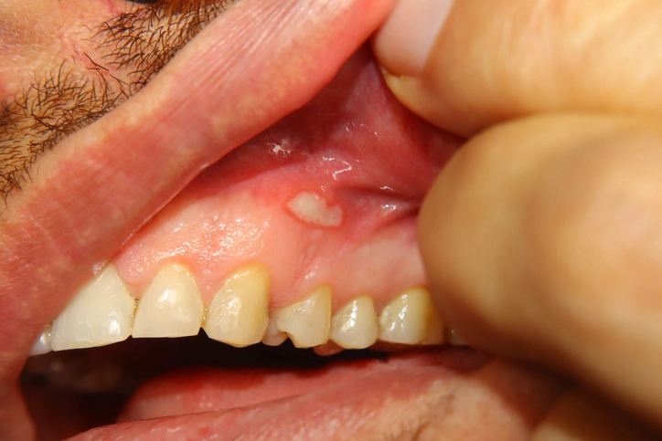
Oral ulceration is one of the most common conditions affecting the oral mucosa encountered in clinical practice. While many oral ulcers are benign and self-limiting, some may represent serious underlying systemic disease or malignancy. Therefore, a structured and methodical approach to assessment is essential. A clinician must be able to distinguish between harmless recurrent conditions and lesions that require urgent investigation or referral.
A systematic evaluation of oral ulcers involves careful consideration of duration, onset, presence of blistering, distribution, recurrence pattern, pain characteristics, associated systemic features, and risk factors. Each of these elements contributes to narrowing the differential diagnosis and guiding management.
1. Duration: How Long Has the Ulcer Been Present?
Duration is one of the most critical diagnostic clues in evaluating an oral ulcer.
Ulcers Present for More Than Three Weeks
Any oral ulcer persisting for more than three weeks must be regarded with suspicion. Such lesions require urgent referral for specialist evaluation, typically to an oral medicine specialist or oral and maxillofacial surgeon. Investigation often includes biopsy to exclude malignancy, particularly oral squamous cell carcinoma (SCC).
Persistent ulcers may represent:
- Oral SCC
- Chronic infections (e.g., tuberculosis, deep fungal infections)
- Chronic traumatic ulcer
- Immune-mediated disease
- Drug-induced ulceration
A non-healing ulcer is a red flag. Delayed diagnosis of oral cancer significantly worsens prognosis. Therefore, the “three-week rule” is a cornerstone principle in oral medicine.
Ulcers of Recent Onset
If the ulcer has appeared recently, further clarification is needed:
- Was it preceded by blistering?
- Is the ulcer single or multiple?
- Are other parts of the body involved?
- Has the patient experienced similar ulcers before?
- Is there an identifiable trigger (e.g., trauma, stress, medication)?
Short-duration ulcers are commonly caused by:
- Recurrent aphthous stomatitis (RAS)
- Viral infections
- Traumatic injury
- Acute necrotizing ulcerative gingivitis (NUG)
Understanding the temporal pattern helps differentiate acute self-limiting conditions from chronic or malignant processes.
2. Blistering: Was There a Preceding Vesicle?
The presence or history of blistering prior to ulcer formation is a vital diagnostic clue.
Vesiculo-Bullous Disorders
If an ulcer develops following rupture of a blister, this suggests a vesiculo-bullous condition. These may be intraepithelial or subepithelial and include:
- Pemphigus vulgaris
- Mucous membrane pemphigoid
- Bullous pemphigoid
- Herpes simplex infection
- Herpes zoster
Intraepithelial blistering disorders typically produce fragile bullae that rupture quickly, leaving painful erosions. Subepithelial blistering may result in more intact bullae before rupture.
Blistering with Systemic Involvement
If blistering or ulceration occurs both in the oral cavity and elsewhere on the body, systemic conditions should be considered. Examples include:
- Erythema multiforme
- Hand, foot, and mouth disease
- Stevens–Johnson syndrome
Erythema multiforme often presents with target lesions on the skin and acute oral ulceration. Identifying this condition early allows avoidance of the precipitating antigen (often medication or infection).
Thus, asking about preceding blisters and examining other body areas is essential.
3. Distribution: Where Are the Ulcers Located?
The anatomical distribution of oral ulcers provides strong diagnostic direction.
Gingival Ulcers
If ulceration is limited to the gingivae, especially with necrosis and bleeding, acute necrotizing ulcerative gingivitis (NUG) should be suspected. NUG is characterized by:
- Painful, bleeding gums
- “Punched-out” interdental papillae
- Halitosis
- Possible systemic symptoms (fever, malaise)
This condition is often associated with stress, smoking, immunosuppression, and poor oral hygiene.
Unilateral Distribution
Ulcers confined to one side of the mouth suggest a viral etiology, particularly herpes zoster. Herpes zoster affects a single dermatome and does not cross the midline. Oral lesions may occur along the distribution of the trigeminal nerve.
Early diagnosis is important because prompt treatment with antiviral therapy (e.g., aciclovir) may reduce post-herpetic neuralgia.
Under Dentures or Appliances
If ulceration is located beneath a denture or orthodontic appliance, trauma should be considered. Ill-fitting prostheses or sharp components may cause chronic irritation.
Traumatic ulcers typically:
- Correspond to a mechanical source
- Improve when the source is removed
- Heal within 7–14 days
Failure to heal despite removal of trauma warrants biopsy to exclude malignancy.
Specific Site Patterns
- Non-keratinized mucosa (labial, buccal mucosa, floor of mouth): common site for aphthous ulcers.
- Hard palate and attached gingiva: common sites for recurrent herpes simplex infection.
- Lateral tongue and floor of mouth: high-risk sites for oral cancer.
Thus, precise documentation of ulcer location is crucial.
4. Recurrence: Has This Happened Before?
A history of repeated episodes strongly suggests recurrent aphthous stomatitis (RAS).
Recurrent Aphthous Stomatitis (RAS)
RAS is one of the most common causes of oral ulceration. It typically presents as:
- Small, round or oval ulcers
- Yellow or grey base
- Erythematous halo
- Painful
- Occurring on non-keratinized mucosa
Episodes often resolve within 7–14 days, but recur periodically.
Three types exist:
- Minor aphthae (most common)
- Major aphthae (larger, may scar)
- Herpetiform aphthae (multiple small ulcers)
Triggers may include stress, trauma, hormonal changes, nutritional deficiencies (iron, folate, B12), and systemic conditions such as coeliac disease or inflammatory bowel disease.
Recurrence after apparent complete healing is characteristic of RAS and helps distinguish it from malignancy.
5. Pain: Is the Ulcer Painful?
Pain is often less diagnostically useful than clinicians assume.
Painful Ulcers
Common causes of painful ulcers include:
- Aphthous ulcers
- Herpes simplex infection
- Traumatic ulcers
- NUG
Pain may vary in intensity depending on size, depth, and location.
Painless Ulcers
A painless ulcer is particularly concerning if it persists beyond three weeks. Oral squamous cell carcinoma may initially be painless. Pain often develops later due to nerve involvement or secondary infection.
Therefore:
- Pain does not confirm benignity.
- Absence of pain does not exclude malignancy.
Character of pain may help:
- Burning sensation: viral or erosive conditions
- Severe neuralgic pain: herpes zoster
- Dull persistent pain: possible malignancy
6. Trial of Therapy
For many ulcers of recent onset, especially where malignancy is unlikely, a short therapeutic trial may assist diagnosis.
Examples include:
- Symptomatic management for viral ulcers
- Topical corticosteroids for aphthous ulcers
- Removal of traumatic source
If the ulcer resolves appropriately, the presumptive diagnosis is supported. However, failure to respond or persistence beyond expected healing time mandates referral.
Importantly, a therapeutic trial must not delay biopsy in suspicious cases.
7. Ulcers Requiring Early Diagnosis
Some ulcerative conditions require urgent recognition due to potential complications.
1. Herpes Zoster
Early antiviral treatment (e.g., aciclovir) may reduce severity and risk of post-herpetic neuralgia. Delay can result in chronic neuropathic pain.
2. Erythema Multiforme
Identifying and eliminating the triggering factor (often a drug or infection) prevents recurrence and progression to more severe forms such as Stevens–Johnson syndrome.
3. Erosive Lichen Planus
This chronic inflammatory condition may require systemic therapy and long-term specialist follow-up. There is also a small but real risk of malignant transformation.
4. Traumatic Ulcerative Granuloma with Stromal Eosinophilia (TUGSE)
TUGSE can clinically resemble squamous cell carcinoma. It may persist for several weeks but typically resolves spontaneously. Because of its similarity to malignancy, biopsy is often required.
5. Oral Squamous Cell Carcinoma (SCC)
SCC is the most serious cause of persistent oral ulceration. Risk factors include:
- Tobacco use
- Alcohol consumption
- Betel quid chewing
- Human papillomavirus (HPV) infection
Features suggestive of SCC:
- Persistent ulcer (>3 weeks)
- Indurated margins
- Rolled borders
- Fixation to underlying tissues
- Unexplained bleeding
- Associated neck lymphadenopathy
Early detection significantly improves survival.
8. Additional Considerations
Systemic Associations
Oral ulcers may be manifestations of systemic disease, including:
- Behçet’s disease
- Crohn’s disease
- Ulcerative colitis
- HIV infection
- Hematological disorders (e.g., leukemia, neutropenia)
A comprehensive medical history is essential.
Drug-Induced Ulceration
Certain medications may cause ulceration directly or through immune mechanisms, including:
- NSAIDs
- Nicorandil
- Chemotherapy agents
Nutritional Deficiencies
Iron, folate, and vitamin B12 deficiencies may predispose to recurrent ulceration.
Conclusion
A structured approach to oral ulcers is essential for safe and effective clinical practice. The key components include:
- Assess duration (three-week rule).
- Determine whether blistering preceded ulceration.
- Evaluate distribution and site.
- Identify recurrence pattern.
- Assess pain characteristics.
- Consider systemic involvement.
- Recognize red flags requiring urgent referral.
Most oral ulcers are benign and self-limiting. However, a small but significant proportion represent serious disease, including malignancy. A systematic, thoughtful approach ensures timely diagnosis, appropriate management, and improved patient outcomes.
Early recognition of potentially serious conditions, combined with judicious use of referral and biopsy, forms the foundation of competent oral medicine practice.