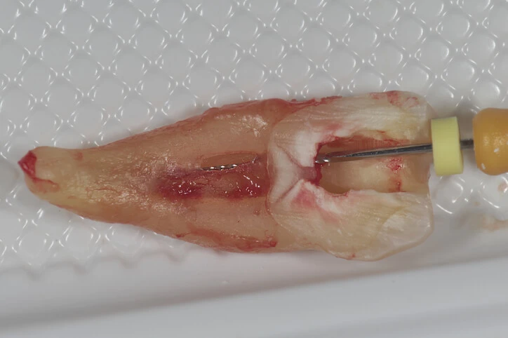
Tooth root perforation is a significant complication in dentistry, particularly in endodontics, that occurs when the integrity of the tooth’s root is compromised due to a mechanical or pathological process. This condition involves the unintended creation of a hole or communication between the pulp cavity and the surrounding periodontal tissues. Such perforations can significantly impact the prognosis of endodontic treatments and are associated with a range of challenges, from pain and infection to tooth loss.
This article delves into the causes, diagnosis, treatment options, and prognosis of tooth root perforations. Additionally, it highlights the importance of timely diagnosis and appropriate management to ensure the best possible outcomes for patients.
Table of Contents
ToggleCauses of Tooth Root Perforation
Tooth root perforations can be classified into two broad categories: iatrogenic and pathological.
Iatrogenic Causes
- Endodontic Procedures
- Post Placement and Restoration
- Access Cavity Preparation
Endodontic Procedures
One of the most common causes of root perforations is errors during root canal treatment. In particular, the use of rotary instruments and endodontic files can result in perforation if the clinician misjudges the anatomy of the root canal or if excessive force is applied during the procedure. Over-preparation of the canal or misdirected files can inadvertently breach the root structure.
Post Placement and Restoration
In cases where posts are inserted for the stabilization of crowns or bridges, excessive drilling or misaligned post placement can result in perforation of the root. This occurs when the practitioner extends the preparation too deeply or in an incorrect direction, penetrating the root structure or into the surrounding periodontal ligament space.
Access Cavity Preparation
Improper location or extension of access cavity preparation can also result in perforation, especially when working in teeth with complex root canal anatomy or calcified canals.
Pathological Causes
- Caries
- Resorption
- Trauma
Caries
Advanced dental caries (decay) can extend into the root structure, causing perforation. In cases where the decay progresses unnoticed, it can create a communication between the pulp and the surrounding tissues, compromising the integrity of the root.
Resorption
Root resorption, either external or internal, can lead to perforations. External resorption typically results from trauma or infection, while internal resorption occurs within the pulp chamber or root canal. Both can weaken the structure of the root to the point of perforation.
Trauma
Direct trauma to a tooth, such as from an accident or injury, can lead to root fractures and eventual perforation if not properly managed.
Clinical Presentation and Symptoms
The clinical symptoms of tooth root perforation can vary depending on the location and extent of the perforation. The perforation may be asymptomatic initially, particularly if it occurs in an area without direct communication with the oral environment. However, as the perforation progresses or becomes infected, several symptoms can arise:
- Pain
- Swelling
- Fistula Formation
- Increased Tooth Mobility
- Periodontal Pocketing
- Persistent Infection
- Discoloration of the Tooth
Pain
Patients may experience sharp, localized pain, particularly when chewing or applying pressure to the affected tooth.
Swelling
Perforations that result in infection can lead to localized swelling of the surrounding gingival or facial tissues.
Fistula Formation
In some cases, a fistula (sinus tract) may develop as a result of chronic infection. This can cause a small, pus-filled pimple to form on the gum near the affected tooth.
Increased Tooth Mobility
Severe perforations, particularly those affecting the periodontal ligament, may result in increased mobility of the tooth.
Periodontal Pocketing
A deep periodontal pocket can develop in the area of the perforation due to the inflammation and breakdown of the surrounding periodontal tissues.
Persistent Infection
A perforated root may result in a persistent periapical or periodontal infection that does not resolve with standard root canal therapy.
Discoloration of the Tooth
The tooth may show signs of discoloration if the pulp becomes necrotic (dies) as a result of the perforation.
Diagnosis of Tooth Root Perforation
Accurate diagnosis of a tooth root perforation is critical for successful treatment. There are several diagnostic tools and techniques that clinicians can use to identify the presence and location of a perforation:
- Clinical Examination
- Radiographs
- Electronic Apex Locators
- Periodontal Probing
- Dye Staining
Clinical Examination
A thorough clinical examination is essential. Dentists will look for signs of swelling, fistulas, and periodontal pocketing that may indicate a perforation. Percussion tests and palpation can also reveal areas of tenderness that suggest a perforation.
Radiographs
Periapical radiographs are a common diagnostic tool used to assess the anatomy of the root and identify perforations. However, radiographs may not always reveal the exact location of the perforation, particularly if it occurs on the buccal or lingual aspect of the root. Cone-beam computed tomography (CBCT) is a more advanced imaging technique that can provide three-dimensional views of the root and surrounding structures, allowing for more precise localization of perforations.
Electronic Apex Locators
These devices can help determine the working length of a root canal during endodontic treatment. A sudden reading that indicates the canal is shorter than expected can suggest that a perforation has occurred, especially when the apex locator signals a communication with the periodontal ligament.
Periodontal Probing
In cases where the perforation occurs at or near the cervical region of the tooth, probing the sulcus with a periodontal probe may reveal an unusually deep pocket, indicating the presence of a perforation.
Dye Staining
In some cases, perforations may be difficult to detect visually. Applying a dye, such as methylene blue, to the suspected area can help reveal the perforation as the dye will seep into the defect.
Treatment Options for Tooth Root Perforation
The treatment of tooth root perforations depends on several factors, including the location and size of the perforation, the degree of periodontal involvement, and the overall prognosis of the affected tooth. The primary goal of treatment is to seal the perforation, prevent infection, and preserve the tooth. Treatment options can be divided into nonsurgical and surgical approaches.
Nonsurgical Management
- Direct Sealing with MTA (Mineral Trioxide Aggregate)
- Calcium Hydroxide
- Gutta-Percha
- Internal Resorption and Perforation Repair
Direct Sealing with MTA (Mineral Trioxide Aggregate)
MTA is a biocompatible material that has become the gold standard for sealing root perforations. It can be placed directly into the perforation site to form a seal and promote healing. MTA has excellent sealing properties, is highly biocompatible, and can set in the presence of moisture, making it ideal for use in perforation repairs.
Calcium Hydroxide
In cases where the perforation is small and located in an area that allows for direct access, calcium hydroxide may be used as a temporary filling material to promote healing before final restoration with a more permanent material, such as MTA.
Gutta-Percha
In some instances, gutta-percha, the traditional root canal filling material, can be used to seal a small perforation if it occurs within the canal system itself. However, gutta-percha is generally not as effective for sealing larger or more complex perforations.
Internal Resorption and Perforation Repair
For perforations caused by internal resorption, MTA or similar materials can be used to fill the resorbed area and restore the structural integrity of the tooth. However, success depends on early detection and treatment of the resorption before it progresses too far.
Surgical Management
- Root-End Resection (Apicoectomy)
- Intentional Replantation
- Crown Lengthening or Hemisection
Root-End Resection (Apicoectomy)
If the perforation is located near the apex of the root and cannot be effectively sealed through nonsurgical means, an apicoectomy may be performed. This involves the removal of the apical portion of the root, followed by sealing of the perforation with MTA or another suitable material.
Intentional Replantation
In rare cases, intentional replantation of the tooth may be considered. This involves extracting the tooth, repairing the perforation outside the mouth, and then replanting the tooth back into its socket. While this procedure carries risks, it may be the only option for saving a tooth with a complex perforation.
Crown Lengthening or Hemisection
For perforations that occur near the cervical region of the tooth, crown lengthening or hemisection may be necessary. Crown lengthening involves removing some of the surrounding bone and gum tissue to expose more of the tooth structure, allowing for repair of the perforation and placement of a crown. Hemisection involves removing one root of a multi-rooted tooth, such as a molar, in cases where the perforation affects only one root.
Prognosis and Factors Influencing Success
The prognosis of a tooth with a root perforation depends on several factors:
- Location of the Perforation
- Size of the Perforation
- Time to Treatment
- Skill of the Clinician
- Patient Factors
Location of the Perforation
Perforations located in the coronal third of the root or at the furcation of multi-rooted teeth tend to have a poorer prognosis compared to perforations that occur in the apical third of the root. This is due to the greater difficulty in sealing perforations located closer to the gingival tissues and the increased risk of periodontal breakdown.
Size of the Perforation
Smaller perforations have a better prognosis because they are easier to seal and less likely to cause significant damage to the surrounding tissues. Larger perforations, on the other hand, may be more challenging to manage and are more likely to lead to infection or failure of the repair.
Time to Treatment
Early detection and prompt treatment are critical for a good prognosis. If a perforation is left untreated for an extended period, bacteria can infiltrate the area, leading to infection and further destruction of the surrounding bone and periodontal ligament.
Skill of the Clinician
The success of perforation repair largely depends on the skill and experience of the dentist or endodontist performing the procedure. Proper technique and the use of appropriate materials, such as MTA, are essential for achieving a good outcome.
Patient Factors
The overall health of the patient, including their periodontal status, immune function, and oral hygiene habits, can influence the success of treatment. Patients with compromised immune systems or advanced periodontal disease may be at greater risk of complications following perforation repair.
Conclusion
Tooth root perforation is a complex and challenging condition that can arise from a variety of causes, both iatrogenic and pathological. While perforations can significantly impact the prognosis of endodontic treatments, advances in diagnostic tools and materials, such as MTA, have improved the ability to manage these complications effectively.
Early diagnosis and timely intervention are critical for the successful treatment of root perforations. By utilizing appropriate nonsurgical or surgical techniques, clinicians can often preserve the affected tooth and prevent the need for extraction. However, the prognosis ultimately depends on factors such as the size and location of the perforation, the time to treatment, and the overall health of the patient.
In summary, root perforation is a condition that requires careful management, but with proper treatment and follow-up care, it is often possible to achieve favorable outcomes.

