
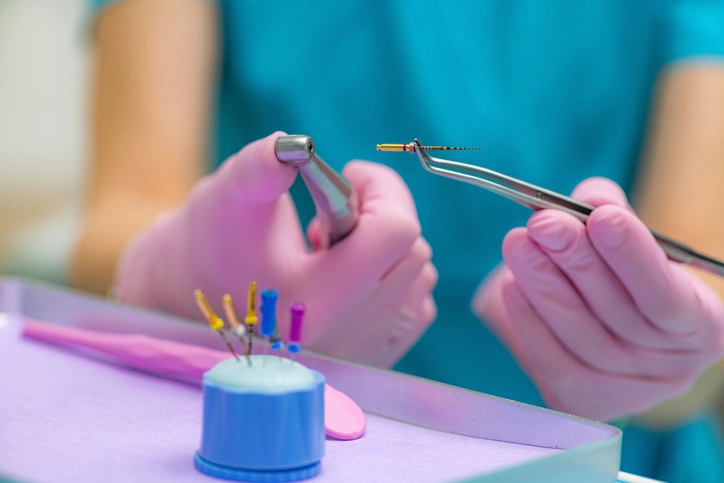
Root canal treatment (RCT), also known as endodontic therapy, remains one of the most common and essential procedures in restorative dentistry. Its primary purpose is the elimination of infection within the root canal system and the prevention of future microbial invasion. Although modern techniques and materials have evolved significantly, the fundamental biological principles underpinning RCT have remained consistent: remove bacteria, destroy their substrate, shape the canal to facilitate disinfection, and obturate the canal to prevent recontamination.
Table of Contents
ToggleRationale for Root Canal Treatment
Conventional root canal therapy is an evidence-based intervention designed to treat or prevent apical periodontitis. Apical periodontitis is primarily a microbial disease arising from bacterial invasion of the pulp tissue and extension into the periradicular tissues.
RCT focuses on the cleaning and shaping of the root canal system to remove:
- Bacteria,
- Bacterial by-products,
- Necrotic or inflamed pulp tissue,
- Smear layer and organic debris.
After this cleaned environment is achieved, the canal is obturated, or sealed, to prevent re-entry of microorganisms and their nutrients. Successful RCT thus disrupts the pathogenic biofilm, destroys its ecological niches, and restores periapical health.
Indications for Root Canal Treatment
RCT is indicated when the dental pulp is irreversibly compromised. Key indications include:
Irreversible Pulp Damage
The irreversible damage may present in two main forms:
- Necrotic pulp
Caused commonly by untreated caries, trauma, or thermal irritation. Necrotic tissue serves as a nutrient-rich environment for bacteria. - Irreversibly inflamed pulp
When the pulp is severely inflamed, healing potential is lost, and vital RCT or extraction are the only options.
Apical Periodontitis
Evidence of apical periodontitis—radiographically or clinically—strongly indicates the need for RCT. In these cases, bacteria have colonized the root canal system, and RCT becomes essential to halt disease progression.
Elective Devitalization
Certain restorative scenarios require intentional pulpal devitalization, such as:
- Teeth prepared for overdentures,
- Abutment teeth requiring extensive prosthodontic work,
- Teeth with strategic restorative value where pulp vitality may complicate treatment.
These indications emphasize preserving strategic teeth wherever possible.
Contraindications for Root Canal Treatment
Although RCT plays a significant role in preserving natural dentition, it is contraindicated in certain circumstances.
Non-functional or Non-restorable Teeth
A tooth that cannot be predictably restored is not suitable for RCT. This includes:
- Severe structural loss,
- Vertical root fractures,
- Unfavorable crown-root ratios.
Insufficient Periodontal Support
If periodontal disease has destroyed the supporting architecture of a tooth (severe bone loss, mobility, or poor prognosis), endodontic treatment becomes futile.
These contraindications reinforce the principle that RCT is only as successful as the restorative and periodontal prognosis of the tooth.
Aims of Root Canal Treatment
Root canal treatment involves three broad objectives:
- Elimination of microorganisms
Bacteria are the primary etiological factor in apical periodontitis; therefore, their removal is essential. - Removal of organic substrate
Any remnants of pulp tissue or necrotic material serve as nutrients for bacteria and must be removed. - Prevention of reinfection
Achieved through hermetic obturation and a well-sealed coronal restoration.
The synergy between cleaning, shaping, and obturation ultimately determines long-term clinical success.
Shaping Aims and Objectives
Shaping the canal is the mechanical aspect of RCT—enabling effective irrigant penetration and ultimately facilitating obturation.
Aims of Shaping
The shaping procedure aims to:
- Remove pulpal debris and microbes,
- Create an ideal internal canal form,
- Generate space for irrigants and medicaments,
- Provide resistance and retention form for the root filling material.
Effective shaping is not about aggressive canal enlargement but about controlled removal of dentine to respect the canal’s original anatomy.
Shaping Objectives
1. Shape
The canal should have a continuously tapering, funnel-shaped form from the coronal portion to the apex. This shape:
- Facilitates irrigant exchange,
- Allows placement of obturation materials,
- Minimizes procedural errors, such as ledging or transportation.
Preserving the apical constriction—the natural barrier at the apex—is critical because it helps maintain the correct working length and creates an ideal environment for the apical seal.
2. Length
Determining the correct working length is essential. The debate about the ideal termination point of instrumentation historically revolved around whether to prepare to the apex or short of it.
The current consensus is:
- Vital teeth: 2–3 mm short of the radiographic apex to avoid unnecessary apical disruption.
- Non-vital or infected teeth: Instrumentation should extend to the full canal length, as bacteria may be present deep into the apical area.
In infected teeth, inadequate preparation of the apical third often leads to persistent infection and treatment failure.
3. Apical Width
Determining the appropriate apical enlargement is a balance between adequate debridement and preservation of tooth structure.
The apical size is typically gauged once the working length is established using hand files. Over-enlargement risks weakening the root and increasing extrusion of debris, while under-preparation may fail to remove infected material.
Cleaning Aims and Objectives
Cleaning refers to the biological reduction of microorganisms and removal of organic and inorganic debris. Mechanical shaping alone cannot adequately debride the canal system because of its complex, irregular anatomy.
Aims of Cleaning
The cleaning phase aims to:
- Remove bacteria and microbial biofilms,
- Eliminate smear layer and dentinal debris,
- Facilitate irrigant penetration,
- Reduce the bacterial load to levels compatible with periapical healing.
A pivotal study by Bystrom and Sundqvist demonstrated that even with meticulous hand instrumentation, only 50% of canals were free of cultivable bacteria. This underscores the importance of chemical disinfection.
Cleaning Objectives
Effective cleaning requires both mechanical and chemical strategies.
Irrigation
1. Functions of Irrigants
Irrigants must:
- Lubricate instruments,
- Flush debris coronally,
- Dissolve both organic and inorganic material (depending on the irrigant),
- Penetrate canal irregularities, fins, and isthmuses,
- Reduce friction and torsional stress on files.
Frequent replenishment and agitation of irrigants significantly enhance their antimicrobial efficacy.
2. Sodium Hypochlorite (NaOCl)
Sodium hypochlorite is considered the gold standard irrigant due to its unique capabilities:
- Broad antimicrobial spectrum,
- Ability to dissolve organic tissue,
- Effectiveness against biofilms,
- Lubrication and flushing action.
Concentration Considerations
Although higher concentrations dissolve tissue more rapidly, they also increase:
- Cytotoxicity,
- Risk of hypochlorite accidents,
- Irritation of periapical tissues.
A commonly accepted concentration is 2.5%, as it balances efficacy with safety.
Delivery and Safety
Key principles include:
- Never extrude NaOCl beyond the apex,
- Keep the needle tip 2–3 mm short of the working length,
- Use side-vented needles to minimize apical pressure.
EDTA (Ethylenediaminetetraacetic Acid)
Instrumentation creates a smear layer composed of organic and inorganic material. NaOCl only dissolves organic components; therefore, a chelating agent such as 17% EDTA is essential for dissolving the inorganic smear layer.
EDTA:
- Opens dentinal tubules,
- Enhances penetration of disinfectants,
- Reduces microleakage risk during obturation.
EDTA is typically used after instrumentation followed by a final NaOCl rinse to remove residual collagen.
Paste or gel-based chelators can negotiate canal blockages but are not recommended with rotary systems due to increased torque.
Aims of Obturation
Once the canal is cleaned and shaped, it must be hermetically sealed to prevent recolonization.
Biological Purposes of Obturation
- Incarceration of residual bacteria
Any remaining microorganisms are entombed, preventing their multiplication. - Apical seal
Prevents inflammatory exudate or microorganisms from entering the root canal system. - Coronal seal
Equally important, as coronal leakage is one of the most common causes of RCT failure.
A well-obturated canal combined with a properly sealed coronal restoration significantly improves long-term prognosis.
Integration of Cleaning, Shaping, and Obturation
The success of RCT depends not on any single step but on the synergy between all three phases.
Cleaning and Shaping
- Shaping facilitates cleaning by enabling irrigant penetration.
- Cleaning enhances shaping by reducing debris and lubricating the canal.
Obturation
The canal is only ready for obturation when it is:
- Adequately cleaned,
- Properly shaped,
- Dried completely,
- Free of symptoms and exudate.
A poorly prepared canal cannot be successfully obturated, even with excellent materials.
Conclusion
Root canal treatment remains a cornerstone of restorative dentistry and endodontics. Its success relies on the clinician’s understanding and execution of biological principles and mechanical strategies.
The rationale for RCT rests on eliminating infection and preventing reinfection through:
- Accurate diagnosis,
- Comprehensive cleaning,
- Precise shaping,
- Effective chemical disinfection,
- Meticulous obturation,
- And a durable coronal restoration.
By adhering to these principles, clinicians can preserve natural dentition, restore function, and maintain long-term oral health.
References
- European Society of Endodontology. Quality guidelines for endodontic treatment: Consensus report of the European Society of Endodontology. International Endodontic Journal. 2006;39:921–930.
- Wu MK, Wesselink PR. The preparation of teeth with necrotic pulps for root canal treatment: A critical review. Oral Surgery, Oral Medicine, Oral Pathology, Oral Radiology, and Endodontology. 2000;89:99–106.
- Byström A, Sundqvist G. Bacteriologic evaluation of the efficacy of mechanical root canal instrumentation in endodontic therapy. Scandinavian Journal of Dental Research. 1981;89:321–328.
- Sjögren U, Figdor D, Persson S, Sundqvist G. Influence of infection at the time of root filling on the outcome of endodontic treatment of teeth with apical periodontitis. International Endodontic Journal. 1997;30(5):297–306.
- Siqueira JF Jr., Rôças IN. Clinical implications and microbiology of bacterial persistence after treatment procedures. Journal of Endodontics. 2008;34(11):1291–1301.
- Peters OA. Current challenges and concepts in the preparation of root canal systems: A review. Journal of Endodontics. 2004;30(8):559–567.
- Haapasalo M, Shen Y, Wang Z, Gao Y. Irrigation in endodontics. British Dental Journal. 2014;216(6):299–303.
- Zehnder M. Root canal irrigants. Journal of Endodontics. 2006;32(5):389–398.
- Schilder H. Cleaning and shaping the root canal. Dental Clinics of North America. 1974;18(2):269–296.
- Ingle JI, Bakland LK, Baumgartner JC (eds). Ingle’s Endodontics. 6th ed. BC Decker; 2008.
- Hargreaves KM, Berman LH (eds). Cohen’s Pathways of the Pulp. 11th ed. Elsevier; 2016.
- Ng YL, Mann V, Rahbaran S, Lewsey J, Gulabivala K. Outcome of primary root canal treatment: Systematic review of the literature – part 1. International Endodontic Journal. 2007;40(12):921–939.