
Dental anatomy is a fascinating field that delves into the intricate structures of our teeth. One of the key components of dental anatomy is the root canal system, which houses the nerves and blood vessels of the tooth. Understanding how many canals each tooth has, as well as exceptions and variations in canal types, is crucial for dental professionals to provide effective treatment. In this article, we will explore the root canal system of teeth, discuss the typical number of canals in each tooth, and examine exceptions and variations in canal types.
Root Canal Anatomy
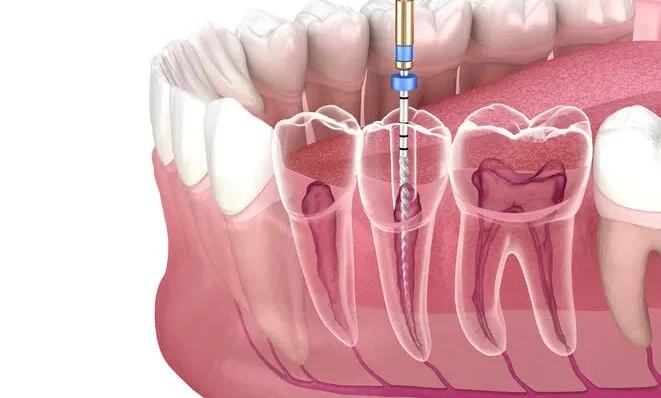
Before delving into the specifics of how many canals each tooth has, it’s essential to understand the anatomy of the root canal system. Each tooth consists of a crown, which is the visible part above the gum line, and one or more roots, which anchor the tooth to the jawbone. Inside the root(s) of the tooth lies the pulp chamber, which houses the dental pulp comprising nerves, blood vessels, and connective tissue.
The root canal system extends from the pulp chamber down to the tip of each root. Within this system, there are intricate networks of canals and tiny branches known as accessory canals. These canals play a crucial role in supplying nutrients to the tooth and transmitting sensory information.
How Many Canals Does Each Tooth Have?
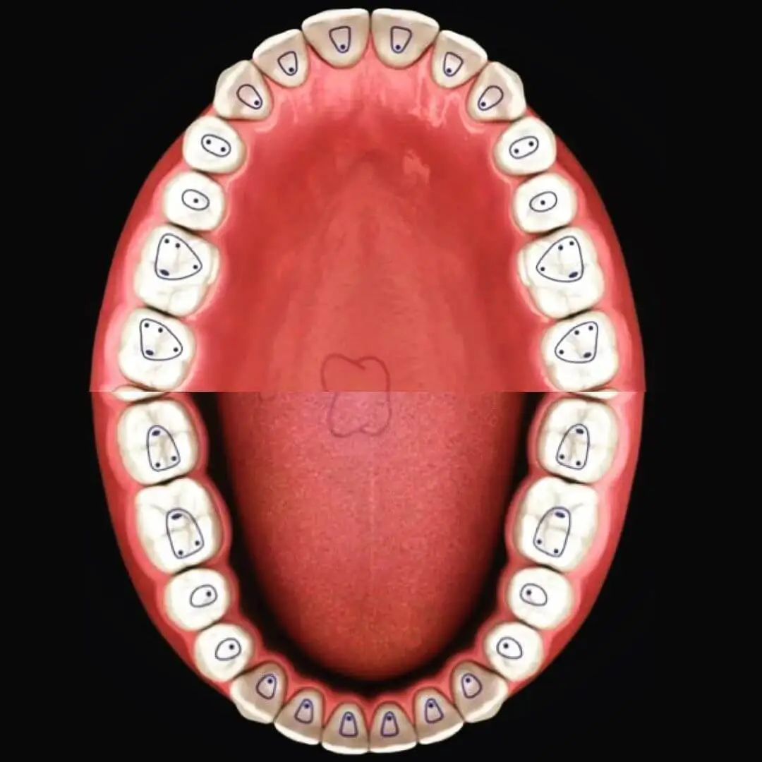
The number of root canals varies depending on the type of tooth. Here’s a general overview of the typical number of canals in each tooth:
- Incisors and Canines: These teeth typically have one root canal.
- Premolars: Most premolars have one or two root canals, although variations can occur.
- Molars: Molars are the most complex teeth when it comes to root canal anatomy. They can have anywhere from one to four root canals, with the lower molars usually having two to three canals and the upper molars having three to four canals.
It’s important to note that while these are the typical numbers of canals, variations and exceptions are common in dental anatomy. Dentists and endodontists must thoroughly examine each tooth to determine the exact number and configuration of canals before performing root canal treatment.
Exceptions and Variations in Canal Types
Despite the general guidelines mentioned above, there are several exceptions and variations in canal types that dental professionals may encounter. Some of these include:
Supernumerary Roots
Occasionally, teeth may have extra roots that contain additional canals. This is more common in certain populations or specific teeth, such as maxillary molars.
C-shaped Canals
In some cases, particularly in mandibular second molars, the root canal system may have a C-shaped configuration instead of the typical oval or round shape. This variation poses challenges during root canal treatment and requires specialized techniques.
Vertucci Classification
The Vertucci classification, named after Dr. Stephen Cohen and Dr. Kenneth Hargreaves, is a system used to categorize the various configurations of root canal anatomy based on their shape and number. This classification system is crucial for endodontists and dental professionals to understand the intricate variations in root canal morphology, which can significantly impact the success of root canal treatment.
The Vertucci classification system consists of eight types, each describing a different configuration of root canals within a tooth. Let’s explore each type in more detail:
Type I:
Type I configuration is the simplest and most common, characterized by a single canal that runs straight from the pulp chamber to the apex (tip) of the root.
Example:
Many anterior teeth (incisors and canines) exhibit Type I canal morphology.
Type II:
Type II configuration features two separate canals that merge into a single canal before reaching the apex.
Example:
Maxillary central incisors are often classified as Type II due to the presence of a second canal (lateral canal) that joins the main canal.
Type III:
Type III configuration consists of one main canal that divides into two separate canals before reaching the apex.
Example:
Mandibular molars commonly exhibit Type III canal morphology, with the main canal splitting into mesial and distal branches.
Type IV:
Type IV configuration involves two separate canals that remain independent throughout the length of the root.
Example:
Maxillary molars may display Type IV canal morphology, where the mesiobuccal and distobuccal canals remain distinct.
Type V:
Type V configuration features a single canal that divides into two separate canals near the apex.
Example:
Mandibular premolars are often classified as Type V, where the main canal divides into buccal and lingual branches.
Type VI:
Type VI configuration consists of two separate canals that merge into a single canal near the apex.
Example:
Maxillary first premolars may exhibit Type VI canal morphology, where the buccal and lingual canals converge into a single canal.
Type VII:
Type VII configuration involves one main canal with an additional accessory canal or lateral canal.
Example:
This type of canal morphology can be found in various teeth, with accessory canals branching off from the main canal.
Type VIII:
Type VIII configuration consists of three separate canals within a single root.
Example:
Maxillary molars are occasionally classified as Type VIII due to the presence of three distinct canals (mesiobuccal, distobuccal, and palatal) within one root.
It’s important to note that while the Vertucci classification provides a framework for understanding root canal morphology, variations and complexities in canal anatomy are common. Therefore, dental professionals must carefully assess each tooth’s anatomy using clinical examination and radiographic imaging to determine the most appropriate treatment approach.
Importance of Accurate Diagnosis and Treatment
Given the complexities and variations in root canal anatomy, accurate diagnosis and treatment are paramount for successful outcomes in endodontic therapy. Dental professionals rely on a combination of clinical examination, radiographic imaging (such as X-rays), and sometimes advanced imaging techniques like cone-beam computed tomography (CBCT) to assess the internal anatomy of teeth accurately.
Once the number and configuration of root canals are determined, the dentist or endodontist can proceed with root canal treatment. This involves removing the infected or damaged pulp, cleaning and shaping the root canal system, and sealing it to prevent reinfection.
Conclusion
In summary, understanding the root canal anatomy of teeth, including how many canals each tooth has and exceptions/variations in canal types, is essential for dental professionals. While there are general guidelines for the typical number of canals in different types of teeth, variations and exceptions are common and must be carefully assessed during diagnosis and treatment.
By employing advanced diagnostic techniques and staying abreast of the latest developments in endodontic therapy, dental professionals can effectively navigate the complexities of root canal anatomy and provide patients with optimal care for their oral health.