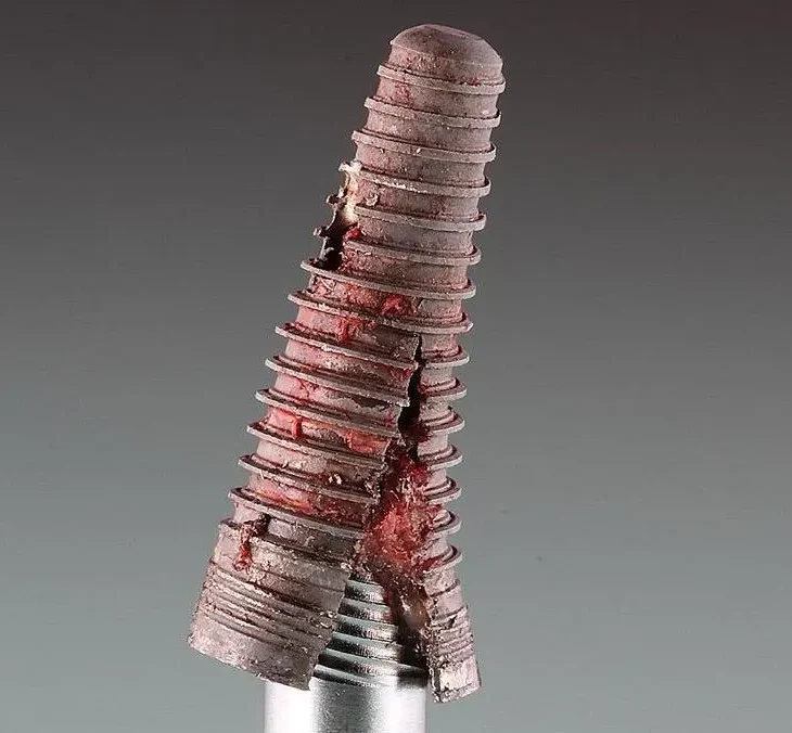
Dental implants have revolutionized modern dentistry, offering durable and aesthetic solutions for tooth replacement. Yet, despite their high success rates, implants can fail even after successful placement and osseointegration due to mechanical complications, including implant fractures. Although relatively rare, understanding their prevalence, causes, management, and prevention is essential for clinicians to ensure long‑term success. Based on the latest literature, this article provides a comprehensive analysis of dental implant fractures.
Table of Contents
ToggleIncidence and Epidemiology
While dental implants generally boast high survival rates, fractures do occur at low frequencies:
- Mishra et al. reviewed 69 studies encompassing 44,521 implants, identifying 827 fractures—an overall incidence of 1.6%.
- A more recent analysis by Goodacre et al. of 12,157 implants reported a 1% fracture rate.
- Others characterize implant fracture as a “rare but possible complication” following prosthetic loading.
Though uncommon, the clinical implications ranging from treatment failure to complex retreatment, underscore the importance of vigilance in prevention and management.
Etiology and Risk Factors
The fracture of a dental implant is rarely due to a single factor; it usually results from a complex interplay of mechanical, biological, and patient-specific influences. Understanding these risk factors in depth allows clinicians to make informed choices in both the surgical and restorative phases.
1. Mechanical Overload & Material Fatigue
Mechanical overload is the primary cause of dental implant fracture. It refers to situations where the forces applied to the implant exceed the material’s fatigue limit over time. While titanium and its alloys have high strength, they are not immune to fatigue failure.
- Cyclic Loading:
Every time a patient chews, the implant experiences a load cycle. In normal function, these loads are well within safe limits. However, when repeated high stresses occur—especially in posterior regions with strong bite forces—microscopic cracks can form within the implant body or abutment screw. These cracks gradually propagate until the remaining cross-section can no longer bear the load, resulting in sudden fracture. - Parafunctional Habits:
Bruxism and clenching dramatically increase cyclic loads. Night-time bruxism is especially destructive because it applies prolonged, uncontrolled force without the protective reflexes present during conscious chewing. - Uneven Occlusal Load:
When prosthetic crowns or bridges are not balanced in occlusion, certain implants may bear disproportionately high forces. For example, if a single implant supports a cantilever bridge without adequate splinting, the bending moments on the implant can skyrocket. - Fatigue Strength Reduction Over Time:
Even with perfect load distribution, repeated stress cycles can weaken the metal over many years. This is why some fractures occur a decade or more after successful function—it’s a slow, cumulative process.
2. Implant Design & Connection Geometry
Implant design heavily influences how forces are transmitted to the implant body and surrounding bone.
- Diameter Considerations:
Narrow and extra-narrow diameter implants (<3.5 mm) have less cross-sectional area to resist bending and shear forces. They may be indicated in limited bone volume cases, but in high-load zones (molars, premolars) they face higher fracture risk. - Length Factors:
Short implants may have a higher bending moment arm because occlusal forces are applied farther from the bone support. However, length alone is less critical than diameter when it comes to pure fracture mechanics. - Implant–Abutment Connection Types: External Hex: Common in older systems, but can be prone to micro-movement and stress concentration at the neck. Internal Hex / Internal Cone (Morse Taper): Distributes loads more evenly, provides better anti-rotational stability, and tends to reduce screw loosening and component fracture.
Clinical studies have suggested that Morse taper connections may offer improved fatigue resistance under dynamic loading. - Thread Design & Platform Switching:
Thread pitch, depth, and shape influence load transfer into bone. Platform switching (using a narrower abutment on a wider implant platform) can reduce crestal bone stress and possibly lower fracture incidence indirectly by preserving bone support.
3. Material Properties and Manufacturing Factors
The choice and quality of implant material is critical.
- Titanium Grades:
Commercially pure titanium (Grades 1–4) and titanium alloys (e.g., Ti-6Al-4V) are most common. CP Titanium has excellent biocompatibility but lower fatigue strength than titanium alloys. Ti-6Al-4V Alloy has superior strength and fatigue resistance, but slightly lower corrosion resistance. - Surface Treatments:
Sandblasting, acid etching, and anodization can influence fatigue performance. While roughened surfaces improve osseointegration, surface micro-defects from manufacturing or finishing can act as stress concentrators. - Manufacturing Defects:
Rarely, microscopic voids, inclusions, or machining marks in the titanium can predispose the implant to early fracture under normal loads.
4. Biological & Bone-Related Factors
The supporting bone plays a crucial role in dissipating forces.
- Bone Density:
Low-density bone (types III and IV) offers less resistance to implant movement, increasing micromotion at the bone–implant interface and concentrating stresses at the crestal region. - Bone Loss Around Implants:
Peri-implantitis or marginal bone resorption can expose more of the implant body to bending forces. The higher the crown-to-implant ratio, the greater the bending moment and fracture risk. - Implant Malpositioning:
Angled placement relative to occlusal forces can create uneven stress distribution and torque on the implant body.
5. Prosthetic Design & Load Distribution
The prosthesis connected to the implant has as much influence on fracture risk as the implant itself.
- Cantilever Extensions:
Unsupported extensions place bending stress on the terminal implant, especially if posterior occlusion is heavy. - Non-Splinted vs. Splinted Restorations:
Single implants in posterior regions are more prone to overload than splinted restorations, which distribute forces across multiple fixtures. - Crown Height Space (CHS):
Excessive CHS acts as a lever arm, magnifying forces at the implant–bone interface.
6. Patient-Related Risk Factors
Beyond bone and prosthetic considerations, patient habits and systemic factors contribute to fracture risk.
- Bruxism:
Possibly the single largest modifiable patient-related factor. The intensity, frequency, and duration of clenching/grinding cycles can exceed the implant’s fatigue threshold. - Dietary Habits:
Patients who frequently chew hard foods (e.g., nuts, ice, hard candy) subject implants to acute high-magnitude loads. - Systemic Conditions:
Osteoporosis, uncontrolled diabetes, and certain medications (like bisphosphonates) can affect bone support and healing capacity. - Oral Hygiene & Compliance:
Poor maintenance can lead to peri-implant disease, bone loss, and mechanical overload due to compromised support.
7. Surgical and Clinical Technique Factors
- Improper Insertion Torque:
Excessive torque during placement can create microcracks in the implant body, which later propagate under function. - Poor Alignment with Antagonist Occlusion:
If the implant axis is significantly misaligned with the opposing tooth’s occlusal forces, lateral loads increase. - Insufficient Healing Before Loading:
Early loading before complete osseointegration can result in micromotion, stress concentration, and eventual material fatigue.
8. Multifactorial Nature
In reality, most fractures are multifactorial—for example:
A narrow-diameter implant placed in soft bone, supporting a long molar crown in a patient with undiagnosed nocturnal bruxism, and restored with a cantilever extension—this combination exponentially increases fracture risk.
Thus, prevention depends on controlling multiple small risks rather than addressing just one factor.
Clinical Presentation & Diagnosis
Implant fracture may present as:
- Sudden mobility of implant components (fixture, abutment, or screw).
- Prosthesis loosening, discomfort, or functional impairment.
- Radiographic evidence of a break in the implant body or connector.
Confirmatory diagnostics include clinical evaluation, radiography, and component disassembly. Diagnostic tools like resonance frequency analysis (RFA) measure implant stability (via ISQ values), with significant drops indicating compromised integrity though not necessarily a fracture.
Management & Treatment Options
The management of a fractured dental implant depends on which part has failed (prosthetic component vs. implant body), the extent of the fracture, and the patient’s clinical circumstances. Treatment decisions should balance mechanical feasibility, biological stability, and patient expectations for function and aesthetics.
1. Clinical Assessment Before Intervention
Before attempting any treatment, a thorough evaluation is critical:
History Taking
When did the patient first notice symptoms?
Was there a sudden “crack” or gradual loss of stability?
Any recent changes in prosthetic work, occlusion, or parafunctional habits?
Clinical Examination
Check for mobility: Is it the crown, abutment, or the entire fixture moving?
Inspect for visible metal fracture lines or missing components.
Assess surrounding gingiva for inflammation, swelling, or exposure of implant threads.
Radiographic Examination
Periapical radiographs can reveal fracture lines in the implant body or abutment screw.
CBCT (cone-beam CT) is invaluable for evaluating bone loss, thread exposure, and exact fracture location.
Decision Point
The clinician must determine whether the fracture is:Prosthetic-only (screw, abutment) → often repairable without fixture removal.
Implant body → typically requires removal and possible site regeneration.
2. Conservative Management: Component Replacement or Repair
If the fracture is limited to the abutment screw or abutment, the fixture can often be salvaged.
Screw Fracture Management
Identify broken fragment location:
If protruding above the implant platform, removal is straightforward using forceps or ultrasonic scalers.
If flush or below the platform, specialized retrieval kits or ultrasonic vibration tips are needed to loosen the fragment.
Replace with a new screw—ideally from the original manufacturer to ensure correct fit and torque values.
Evaluate the cause: If repeated screw fractures occur, occlusion, connection design, and torque protocol must be reassessed.
Abutment Fracture
- Remove remaining abutment parts with manufacturer-specific tools.
- Replace with a stronger abutment material if possible (e.g., from titanium to titanium–zirconium alloy).
- Check that the implant–abutment interface is undamaged before reinstallation.
Advantages of conservative approach:
- Retains original osseointegration.
- Shorter treatment time and cost.
- Less invasive for the patient.
Limitations:
- If the implant platform or internal connection is damaged, full replacement may still be necessary.
- May not address underlying biomechanical overload.
3. Surgical Management: Fixture Removal
If the implant body itself fractures, conservative measures usually cannot restore long-term function.
Indications for Removal
- Visible or radiographic fracture of the implant fixture.
- Severe bone loss with instability.
- Persistent pain, swelling, or infection.
Removal Techniques
- Counter-torque removal: Uses a device to unscrew the remaining fixture from the bone (minimally invasive if osseointegration is weak).
- Trephine burs: Cylindrical burs remove a thin layer of bone around the implant, freeing it for extraction.
- Piezoelectric surgery: Ultrasonic devices can section bone around the implant with reduced trauma to soft tissues.
- Sectioning of the implant: In some cases, splitting the implant vertically allows removal in pieces.
Bone Grafting Considerations
Removal can create bone defects, particularly if a trephine is used. Guided bone regeneration (GBR) or socket preservation is often required before or during future reimplantation.
4. Timing of Reimplantation
After fixture removal, clinicians must decide between immediate or delayed replacement.
Immediate Reimplantation
Feasible if infection is absent, bone damage is minimal, and primary stability can be achieved.
Advantage: Shorter treatment time, maintains soft tissue contours.
Risk: Higher failure if residual biomechanical causes are not addressed.
Delayed Reimplantation
Recommended if bone regeneration is needed or infection is present.
Allows time for graft integration and soft tissue healing.
Typically involves a healing period of 3–6 months before new implant placement.
5. Prosthetic Rehabilitation After Fracture
Whether the implant is repaired or replaced, prosthetic planning should address the cause of the initial fracture.
Key Strategies:
- Load Distribution: Use splinted restorations in posterior regions to share occlusal forces across multiple implants.
- Occlusal Adjustment: Reduce high contact points, particularly in excursive movements.
- Material Selection: In high-load cases, use stronger frameworks (e.g., metal-ceramic instead of all-ceramic in posterior load-bearing zones).
- Crown Height Reduction: Lower lever arm forces when possible.
6. Adjunctive Measures
- Night Guards: Essential for bruxers to minimize nocturnal overload.
- Occlusal Splints: Can be used temporarily during healing phases to stabilize forces.
- Patient Education: Discuss dietary habits (avoiding ice, hard candy, bones), importance of maintenance visits, and early reporting of mobility or noise from prosthetics.
7. Potential Complications of Management
- Incomplete Fragment Retrieval: Residual broken components can prevent new abutment seating.
- Bone Loss During Removal: Overuse of trephine burs can create large defects requiring extensive grafting.
- Recurrent Fracture: If biomechanical or patient-related causes are not addressed, the new implant can fail in the same manner.
- Soft Tissue Recession: More common after surgical fixture removal, potentially affecting aesthetics in anterior cases.
8. Long-Term Follow-Up
Post-treatment monitoring should include:
- Regular Radiographs: Every 6–12 months for the first few years.
- Occlusal Checks: Identify premature contacts or wear facets early.
- Patient Feedback: Encourage patients to report any unusual sensations, mobility, or changes in bite immediately.
Prevention Strategies
1. Pre-operative Planning
- Thorough assessment of bone quality, occlusal forces, and parafunctional habits.
- Selecting appropriate implant diameter, length, and connection design per load expectations and anatomical limitations.
- Favoring internal cone‑type connections when mechanical resilience is key.
2. Prosthetic & Occlusal Management
- Use of splinted prostheses in high-force areas (e.g., molars).
- Balanced occlusion with minimal lateral stress—especially for bruxism patients.
- Designing prosthetics for even force distribution and avoiding cantilever stress.
3. Maintenance & Patient Habits
- Regular checkups for early detection of prosthetic wear, loosening, or micro‑damage.
- Issuing night guards for bruxers or those with parafunctional habits.
- Education on avoiding hard foods, using proper hygiene, and attending routine professional maintenance.
4. Material & Technological Advances
- Selecting high‑quality titanium or titanium alloy fixtures; considering evolving surface treatments and biomaterials for improved fatigue resistance.
- Emerging approaches such as improved surface texturing, nanocoatings, and additive manufacturing may enhance fatigue strength and reduce fracture risk (though such technologies remain largely experimental at present).
Clinical Implications
Though rare—affecting approximately 1–1.6% of implants—dental implant fractures can compromise function, aesthetics, and patient satisfaction. The etiology is multifactorial, involving mechanical overload, material fatigue, design limitations, and patient habits. Successful management depends on prompt diagnosis, appropriate intervention, and restoration planning; prevention hinges on meticulous planning, patient education, and ongoing maintenance.
Clinicians should remain vigilant, strategically selecting implants and prosthetics that match biomechanical demands and adapting treatment protocols based on patient-specific risk factors.

