Cleidocranial Dysplasia (CCD), also known as cleidocranial dysostosis, is a rare autosomal dominant skeletal disorder, affecting one in one million individuals worldwide. It is characterized primarily by hypoplastic or aplastic clavicles, delayed cranial suture ossification, short stature, and numerous dental anomalies. Due to its extensive involvement of craniofacial, clavicular, and dental structures, CCD presents a unique challenge across various fields of medicine, particularly in genetics, orthopedics, and dentistry.
This article provides an in-depth analysis of the genetic etiology, skeletal and dental characteristics, oral manifestations, and multi-disciplinary approaches to managing cleidocranial dysplasia. Special emphasis will be placed on the dental and oral implications of cleidocranial dysplasia as these are among the most common and challenging manifestations requiring intervention.
Table of Contents
ToggleGenetic Etiology and Pathophysiology
Cleidocranial Dysplasia is caused by mutations in the RUNX2 gene, located on chromosome 6p21. RUNX2 is a transcription factor essential for osteoblastic differentiation and bone formation, as well as chondrogenesis and dentition. The RUNX2 gene regulates the maturation of chondrocytes and osteoblasts and controls the differentiation of dental mesenchyme cells into odontoblasts and ameloblasts, which are crucial in the formation of tooth dentin and enamel, respectively. Mutations in RUNX2 disrupt these pathways, leading to defective bone and tooth formation, manifesting as the phenotypic characteristics of CCD.
Skeletal Manifestations of Cleidocranial Dysplasia
- Clavicular Abnormalities
- Craniofacial Dysmorphology
- Dental and Oral Anomalies
Clavicular Abnormalities
One of the hallmark features of cleidocranial dysplasia is clavicular hypoplasia or aplasia, often bilaterally, allowing for increased shoulder mobility, often permitting patients to touch their shoulders together anteriorly. This skeletal abnormality does not usually impact functionality but can lead to cosmetic concerns and occasionally issues with shoulder stability and strength.
Craniofacial Dysmorphology
Craniofacial abnormalities are a defining feature of cleidocranial dysplasia. These include a wide anterior fontanelle, delayed closure of sutures, a large forehead, hypertelorism, and a depressed nasal bridge. The cranial vault abnormalities may persist into adulthood, and while they rarely result in neurological impairment, they contribute significantly to the distinct facial appearance associated with CCD.
Dental and Oral Anomalies in Cleidocranial Dysplasia
The dental manifestations of cleidocranial dysplasia are complex, often requiring multi-phase, interdisciplinary treatment throughout life. Dental anomalies are among the most commonly reported and significant manifestations of CCD, often leading patients to seek treatment initially within a dental setting.
Dental and Oral Issues in Cleidocranial Dysplasia
- Delayed Tooth Eruption
- Supernumerary Teeth
- Malocclusion and Malalignment
- Hypoplastic and Dysplastic Enamel
- Retained Primary Teeth
- Narrow Palate and High Arched Palate
- Gingival Hyperplasia
Delayed Tooth Eruption
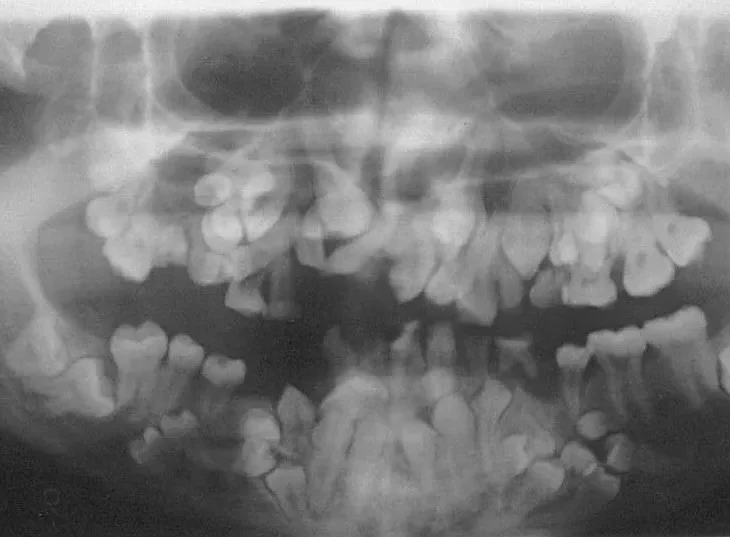
One of the primary dental concerns in cleidocranial dysplasia is delayed or failed eruption of permanent teeth. This is typically due to multiple supernumerary teeth obstructing the eruption pathway of the permanent dentition. The teeth that do manage to erupt often do so in abnormal positions, leading to malocclusion and aesthetic concerns.
Clinical Implications:
- Radiographic Findings: Delayed tooth eruption can be visualized radiographically, showing unerupted permanent teeth below retained primary teeth, often with malformed roots.
- Management: The treatment of delayed eruption often involves the surgical removal of supernumerary teeth followed by orthodontic traction to guide the permanent teeth into the correct positions. This process requires careful planning to prevent further complications and maximize aesthetic and functional outcomes.
Supernumerary Teeth
Supernumerary teeth are commonly present in cleidocranial dysplasia, with multiple extra teeth found in both the maxilla and mandible. These teeth tend to be morphologically abnormal, often resembling premolars or incisors, and can lead to a crowded dental arch, impeding normal eruption.
Clinical Implications:
- Diagnosis: Panoramic and occlusal radiographs are essential for identifying supernumerary teeth. Cone-beam computed tomography (CBCT) provides a 3D visualization, aiding in the assessment of tooth position relative to vital structures.
- Management: Surgical extraction of supernumerary teeth is often indicated, followed by orthodontic therapy to guide normal tooth eruption. However, extraction can be complicated due to the proximity of these teeth to unerupted permanent teeth and other structures.
Malocclusion and Malalignment
Malocclusion, often in the form of anterior open bite and severe crowding, is frequently observed in CCD patients. This malocclusion is compounded by the persistence of primary teeth and the delayed eruption of permanent teeth, which can result in misalignment and functional impairments.
Clinical Implications:
- Orthodontic Intervention: Orthodontic treatment is typically challenging and may be prolonged. Intervention may include the use of fixed appliances to correct malocclusion and align erupted teeth.
- Long-Term Management: Malocclusion in CCD requires ongoing monitoring and sometimes several phases of orthodontic therapy as additional permanent teeth erupt. Multidisciplinary input from orthodontists and oral surgeons is essential for optimal outcomes.
Hypoplastic and Dysplastic Enamel
Cleidocranial Dysplasia can affect the quality of enamel, leading to hypoplastic or dysplastic enamel that is more prone to decay and structural weakness. This can exacerbate dental problems and lead to increased vulnerability to dental caries and other oral health issues.
Clinical Implications:
- Preventive Care: Dental sealants, fluoride treatments, and meticulous oral hygiene practices are essential for reducing the risk of decay in hypoplastic enamel.
- Restorative Treatment: Composite or ceramic restorations may be required to reinforce weak enamel and restore the structural integrity of affected teeth.
Retained Primary Teeth
Due to delayed permanent tooth eruption, cleidocranial dysplasia patients often retain their primary teeth well into adulthood. Retained primary teeth can result in improper dental occlusion and potential aesthetic issues, especially in the anterior region.
Clinical Implications:
- Management Strategy: Retained primary teeth may require extraction once they interfere with permanent teeth eruption. In some cases, retained primary teeth are reshaped or restored to match the adjacent permanent teeth if they remain functional and aesthetically acceptable.
Narrow Palate and High Arched Palate
Many CCD patients present with a narrow or high arched palate, which can further exacerbate malocclusion and impede proper dental arch development. This may lead to crowding of teeth and challenges in achieving a proper bite.
Clinical Implications:
- Orthodontic Expansion: Palatal expanders may be used to address the narrow palate and provide adequate space for tooth alignment. Surgical assistance may be required in some cases.
- Impact on Speech and Function: A narrow or high arched palate may affect speech clarity and chewing efficiency, necessitating early intervention to improve function.
Gingival Hyperplasia
Cleidocranial Dysplasia patients are predisposed to gingival hyperplasia, which can be exacerbated by the presence of multiple unerupted and impacted teeth. Gingival overgrowth can interfere with oral hygiene, increasing the risk of periodontal disease and further complicating orthodontic and surgical interventions.
Clinical Implications:
- Management: Gingivectomy and other periodontal procedures may be necessary to manage hyperplasia. Proper oral hygiene and regular professional cleanings are essential to reduce inflammation and maintain periodontal health.
Diagnosis of Dental and Oral Anomalies in Cleidocranial Dysplasia
Diagnosis of CCD-related dental anomalies involves a thorough clinical examination and the use of various imaging modalities. Panoramic radiographs and CBCT are particularly useful for assessing the number, position, and morphology of supernumerary and impacted teeth, as well as evaluating bone structure abnormalities.
Multidisciplinary Management Approach
Managing CCD-related dental issues requires a comprehensive, multidisciplinary approach that includes coordination among dentists, orthodontists, oral surgeons, and, in some cases, geneticists and craniofacial specialists. The management plan should be tailored to each patient’s specific needs, taking into account the extent of dental anomalies, the presence of functional impairments, and aesthetic considerations.
Phase 1: Early Intervention and Prevention
Early diagnosis and monitoring are critical to managing CCD’s dental manifestations. Pediatric patients should be closely monitored for signs of delayed tooth eruption, and preventive strategies should be implemented to protect the dental enamel.
Phase 2: Surgical and Orthodontic Management
The surgical removal of supernumerary teeth and retained primary teeth is often necessary to facilitate the eruption of permanent teeth. Following surgical intervention, orthodontic treatment is essential to guide the alignment and positioning of teeth. Orthodontic management may require multiple stages due to the extended timeline of permanent tooth eruption in cleidocranial dysplasia.
Phase 3: Long-Term Maintenance and Aesthetic Rehabilitation
Once the permanent teeth have erupted and the alignment is stabilized, long-term maintenance is crucial. Regular dental check-ups, preventive care, and restorative treatments may be required to address hypoplastic enamel and improve aesthetic outcomes. In some cases, dental implants or prosthetic solutions may be considered for patients with missing teeth or poor bone quality.
Advances in Treatment: Bone Regeneration and Gene Therapy
Recent advances in regenerative medicine and gene therapy have opened new possibilities for treating skeletal and dental anomalies in cleidocranial dysplasia. Studies on RUNX2 gene therapy and bone morphogenetic protein (BMP) pathways hold promise for restoring normal bone formation in CCD patients. While these treatments are still in experimental stages, they represent a potential future direction for more definitive management of CCD’s skeletal and dental manifestations.
Frequently Asked Questions (FAQs)
Is Cleidocranial Dysplasia a life-threatening condition?
No, CCD is not a life-threatening condition. However, it can significantly affect skeletal and dental development, leading to functional and aesthetic challenges.
What are the earliest signs of CCD in children?
Common early signs include delayed closure of the fontanelles, absence or underdevelopment of the clavicles, delayed tooth eruption, and a broad forehead.
How is CCD diagnosed?
CCD is diagnosed through clinical examination, radiographic imaging (X-rays, CBCT scans), and genetic testing to confirm RUNX2 mutations.
What are the dental complications associated with CCD?
The most common dental issues include:
- Delayed tooth eruption
- Supernumerary (extra) teeth
- Malocclusion and crowding
- Retained primary teeth
- Enamel hypoplasia
Can CCD be treated?
While there is no cure, surgical, orthodontic, and restorative treatments can significantly improve function and appearance.
Will children with CCD require multiple dental surgeries?
Yes, most patients require multiple dental procedures over time, including extractions, exposure of impacted teeth, and orthodontic interventions.
Can CCD affect speech?
Yes, due to high-arched or narrow palates, some CCD patients experience speech difficulties, which may require therapy.
Are dental implants an option for CCD patients?
Yes, but bone density and delayed eruption of permanent teeth must be considered before planning implant placement.
Can CCD be inherited?
Yes, CCD follows an autosomal dominant inheritance pattern. A parent with CCD has a 50% chance of passing the condition to their child.
What specialists are involved in treating CCD?
Treatment typically involves a multidisciplinary team, including:
- Dentists and orthodontists
- Oral and maxillofacial surgeons
- Geneticists
- Speech therapists (if needed)
Conclusion
Cleidocranial Dysplasia presents complex challenges across multiple domains, particularly in dental and oral care. Dental anomalies, including delayed eruption, supernumerary teeth, malocclusion, and enamel defects, necessitate a structured, interdisciplinary approach to treatment. Early diagnosis, preventive care, and coordinated surgical and orthodontic interventions are essential to achieving optimal functional and aesthetic outcomes.
Advances in genetic and regenerative therapies may provide future options for addressing the underlying causes of CCD. Until then, careful management by a team of specialists will remain the standard approach to improving the quality of life for individuals with this rare but impactful disorder.


kapadokya
2 November 2024Your work is truly inspiring! The content you share is both meticulous and informative, and I look forward to each update with great anticipation.
Danny Sweeney
24 May 2025Looking for a dentist to help me with this condition on my adult mouth. Most do not understand nor do they want to get involved.
Stacie Smith Hamilton
22 October 2025I am looking for a dentist that specializes in C.C.D. I have some gum and bone loss