
Tooth ankylosis occurs when a tooth becomes fused directly to the surrounding alveolar bone, lacking a normal periodontal ligament. While this may sound like a rare anomaly, ankylosed teeth can have serious consequences, especially in growing children, as they may affect both dental alignment and jaw development.
This article aims to provide an in-depth look at ankylosed teeth: what they are, how they develop, how they are diagnosed, and the best approaches for management and treatment.
Table of Contents
ToggleWhat is an Ankylosed Tooth?
Tooth ankylosis is a pathological condition in which there is a direct fusion between the tooth root (specifically, the cementum or dentin) and the alveolar bone, eliminating the periodontal ligament (PDL) that normally separates and cushions the tooth within its socket. This fusion leads to a tooth that is immobile, unresponsive to eruptive or orthodontic forces, and often sunken below the level of adjacent teeth—a condition known as infraocclusion.
To understand the implications of ankylosis, it’s important to first understand the normal dental anatomy and the functions of supporting structures.
Ankylosis Pathophysiology
In ankylosed teeth, the periodontal ligament is lost, either partially or entirely, and the cementum or even dentin becomes directly fused with the alveolar bone. This fusion halts the tooth’s natural ability to erupt or adapt, essentially “freezing” it in place. Over time, especially in growing children, this lack of eruption relative to adjacent teeth results in the tooth appearing submerged or depressed—hence the clinical term infraocclusion.
In advanced cases, this fusion can be so extensive that the tooth root begins to be resorbed and replaced by bone, a process called replacement resorption. Eventually, the root may disappear completely, and only a bony ridge may remain.
Histological Features of Ankylosis
When examined microscopically, ankylosed teeth show distinct characteristics:
- Absence of periodontal ligament fibers in the affected areas
- Direct continuity between cementum/dentin and alveolar bone, without an intervening soft-tissue layer
- In many cases, evidence of replacement resorption, with bone actively replacing tooth root structure
- Loss of Sharpey’s fibers (collagen fibers that connect the PDL to cementum and bone)
These features indicate that ankylosis is not merely a mechanical fusion but a biological remodeling process, where the tooth is gradually absorbed and replaced with osseous tissue.
Types of Tooth Ankylosis
Tooth ankylosis can be classified in various ways depending on extent, timing, and underlying causes:
Based on Dentition Stage
- Primary Ankylosis: Occurs in deciduous (baby) teeth and is more common. It often becomes evident when the tooth fails to exfoliate or becomes infraoccluded.
- Permanent Ankylosis: Less common, typically associated with trauma, reimplantation, or orthodontic complications.
Based on Extent
- Partial Ankylosis: Only a portion of the root is fused to the bone. These cases may present more subtly.
- Complete Ankylosis: The entire root surface is fused. These cases are more resistant to treatment and more likely to lead to significant clinical issues.
Based on Onset or Cause
- Developmental/Idiopathic: Occurs spontaneously without any clear external cause. This is typical in many pediatric cases.
- Post-Traumatic: Follows dental injuries such as luxation, intrusion, or avulsion, especially in reimplanted teeth.
- Inflammatory: Related to chronic infections or severe periodontal disease.
How Ankylosed Teeth Differ from Other Eruption Issues
It is essential to differentiate ankylosed teeth from other conditions that may result in altered eruption or position, such as:
- Impacted Teeth: These fail to erupt due to obstruction, space deficiency, or abnormal angulation. They are not fused to bone.
- Retained Deciduous Teeth: These may simply be late to exfoliate but still have intact PDL and can be extracted or exfoliate naturally.
- Infraerupted Teeth: Can result from hormonal or systemic issues but typically don’t involve bone fusion.
A key distinguishing feature of ankylosis is the absence of mobility, along with the characteristic sound on percussion, which is more metallic or dull due to the rigid connection to bone.
Visual and Functional Signs
Ankylosed teeth are often first suspected due to visual clues:
- A primary tooth that appears sunken below the level of adjacent teeth
- Delayed or absent eruption of the permanent tooth
- Adjacent teeth tipping toward the ankylosed one due to space discrepancies
- Malocclusion, especially in growing children
In older individuals, especially those with ankylosed permanent teeth, signs may also include:
- Changes in occlusion
- Difficulty with orthodontic treatment
- Inability to move the tooth orthodontically
- Progressive root resorption on radiographic evaluation
Clinical Importance
Understanding what an ankylosed tooth is goes far beyond just identifying a non-erupting or infraoccluded tooth. The condition can have broad implications, particularly in children and adolescents:
- It can affect the vertical development of the jaw.
- It may disrupt the eruption sequence and arch alignment.
- It can lead to space loss, crowding, or aesthetic concerns.
- In the permanent dentition, it limits treatment options, especially if orthodontic movement is needed.
Because of these far-reaching effects, early recognition and appropriate treatment planning are essential.
Epidemiology
Tooth ankylosis is relatively uncommon in the general population but is a significant condition due to its impact on dental development, particularly in children. The epidemiology of ankylosed teeth varies depending on several factors, including age, dentition stage (primary vs. permanent), the type of tooth affected, and geographical or ethnic differences.
Prevalence
The reported prevalence of ankylosed teeth varies widely in the literature, primarily due to differences in diagnostic criteria, study populations, and methods of detection (clinical vs. radiographic). The general prevalence rates are:
- Primary Dentition (Children): 1.3% to 14%
- Mixed Dentition: Higher rates observed during this stage due to better visibility of infraocclusion
- Permanent Dentition (Adolescents and Adults): Less common, with prevalence estimated between 0.1% and 2.5%
It is generally accepted that ankylosis is more prevalent in the primary dentition and tends to decrease in occurrence with age. The higher prevalence in children is due to the condition often developing during tooth eruption stages and becoming apparent when adjacent teeth continue to erupt while the ankylosed tooth remains static.
Age and Developmental Considerations
Age plays a significant role in both the presentation and impact of ankylosed teeth:
- In early childhood (2–5 years): Ankylosis of primary molars may first become noticeable.
- During mixed dentition (6–12 years): Ankylosis becomes more clinically significant due to its potential to interfere with the eruption of permanent successors.
- In adolescents and adults: Ankylosis is rarer and usually associated with a history of dental trauma or reimplantation procedures.
Importantly, the consequences of ankylosis are more severe during periods of active growth, as the ankylosed tooth fails to move with the developing alveolar ridge, leading to infraocclusion and potential long term alveolar bone defects.
Teeth Most Commonly Affected
Studies consistently show that posterior teeth are more frequently affected than anterior teeth, particularly in the primary dentition:
Primary mandibular second molars are the most commonly ankylosed teeth.
Primary maxillary second molars follow in frequency.
Ankylosis is much less common in incisors and canines.
In the permanent dentition, ankylosis is rare, but when it occurs, it is often associated with:
Reimplanted anterior teeth (particularly maxillary central incisors after trauma)
Impacted molars that have become ankylosed due to failed eruption or prior infection
Gender Differences
Most epidemiological studies indicate no significant gender predilection, meaning males and females are equally likely to develop tooth ankylosis. However, some localized studies have shown a slightly higher prevalence in boys, potentially due to a higher incidence of traumatic dental injuries.
Ethnic and Geographic Differences
While not extensively studied, some research suggests that genetic and ethnic factors may influence the prevalence of ankylosis:
- Family studies have shown a higher incidence among siblings, suggesting a possible hereditary component.
- Regional studies from parts of Scandinavia, the Middle East, and Asia report varied prevalence rates, indicating environmental, dietary, and genetic influences may play a role.
However, more large-scale, multicenter epidemiological studies are needed to conclusively determine the influence of ethnicity and geography on the prevalence of ankylosed teeth.
Recurrence and Bilateral Occurrence
- Bilateral ankylosis (affecting the same tooth on both sides) occurs in a notable percentage of cases—some studies report up to 50% bilateral involvement for primary second molars.
- Once one ankylosed tooth is diagnosed, it is advisable to examine the contralateral tooth carefully and monitor the eruption of adjacent teeth.
- Recurrence after treatment, particularly in cases of attempted orthodontic extrusion or luxation, is possible due to the tendency of ankylosed teeth to re-fuse with the bone.
Causes and Risk Factors of Tooth Ankylosis
Tooth ankylosis is a complex and multifactorial condition, with a variety of local, systemic, and genetic factors contributing to its development. While the precise pathogenesis is not always clear, current research points to the disruption or degeneration of the periodontal ligament (PDL) as the central initiating event. Once the PDL is compromised, the natural barrier between the root and alveolar bone is lost, allowing for direct fusion and eventual replacement resorption.
Below is a comprehensive look at the most well-documented causes and risk factors associated with ankylosed teeth.
- Dental Trauma
- Reimplantation of Avulsed Teeth
- Genetic Predisposition
- Local Infections and Inflammation
- Developmental Abnormalities
- Iatrogenic Causes (Dentally-Induced)
- Systemic and Syndromic Associations
- Age and Growth Factors
- Mechanical Obstruction or Space Deficiency
Dental Trauma
Dental trauma is one of the most significant and well-documented causes of ankylosis, especially in the permanent dentition.
Types of Trauma Leading to Ankylosis:
- Luxation Injuries (Intrusion, Lateral, or Extrusion): Intrusion, in particular, is highly associated with PDL damage. When a tooth is pushed forcibly into the alveolar bone, the periodontal ligament may be completely crushed or destroyed, leaving no buffer between cementum and bone.
- Avulsion (Complete Tooth Displacement): Avulsed teeth that are reimplanted—especially after a prolonged extra-alveolar period—have a high risk of ankylosis. The success of reimplantation depends on the survival of PDL cells on the root surface. If these cells are necrotic, the healing process may favor bone-to-root fusion rather than ligament regeneration.
- Root Fractures and Alveolar Bone Fractures: Complex traumatic injuries involving both the root and socket walls can disrupt normal healing and promote ankylosis.
Key Risk Factors in Post-Traumatic Ankylosis:
- Delayed reimplantation (>60 minutes)
- Dry storage of avulsed tooth
- Non-physiological storage media (e.g., water instead of milk or saline)
- Poor handling of the tooth root surface
- Lack of splinting or improper splinting technique
- Severe or repeated injury
Reimplantation of Avulsed Teeth
Although technically a subset of trauma, the reimplantation procedure itself deserves special attention due to its high association with ankylosis. Even when reimplantation is done promptly, there’s a significant risk that the root surface may undergo surface resorption, which may later transform into replacement resorption, leading to ankylosis.
Key Factors Influencing Ankylosis After Reimplantation:
- Extra-alveolar dry time: More than 60 minutes almost guarantees PDL necrosis.
- Storage medium: Teeth stored in inappropriate solutions (e.g., water) have higher rates of ankylosis.
- Patient age: Younger patients with open apices may have a better regenerative response.
Genetic Predisposition
Several studies suggest that tooth ankylosis may have a hereditary or genetic component, particularly when it presents bilaterally or in multiple teeth without apparent trauma or infection.
Evidence for Genetic Influence:
- Family clustering of cases has been documented in both primary and permanent dentition.
- Twin studies have shown higher concordance rates of ankylosis among monozygotic twins compared to dizygotic twins.
- Some researchers propose that mutations in genes related to bone metabolism, inflammation, or connective tissue remodeling may predispose individuals to develop ankylosis.
Potential Genetic Mechanisms:
- Abnormal expression of RANK/RANKL/OPG pathways (key regulators of bone remodeling)
- Defects in matrix metalloproteinases (MMPs) that affect collagen breakdown and tissue regeneration
- Irregularities in PDL progenitor cell differentiation, favoring osteoblast-like rather than ligamentous pathways
However, more genetic and molecular research is required to fully elucidate these mechanisms.
Local Infections and Inflammation
Chronic or severe inflammation can degrade the PDL and promote ankylosis. When the periodontal ligament is exposed to prolonged infectious or inflammatory stimuli, the normal repair process may be altered in favor of hard tissue formation (i.e., bone), leading to root-to-bone fusion.
Contributing Conditions:
- Severe periodontitis
- Periapical abscesses or granulomas
- Furcation involvement in primary molars
- Caries-related pulp necrosis in primary teeth
In the case of deciduous teeth, pulpal infections can spread to surrounding periodontal tissues, destroying the PDL and triggering ankylosis, especially when extraction or treatment is delayed.
Developmental Abnormalities
Some children may develop ankylosed primary teeth due to abnormalities in eruption or development:
- Ectopic eruption or impaction may lead to bone remodeling in a way that fuses the developing root with alveolar bone.
- Disturbed exfoliation cycles (e.g., retained primary teeth with missing successors) can cause the roots to become embedded and fused.
These developmental problems are often idiopathic, meaning they occur without a clearly identifiable cause, but they still fall within the broader spectrum of predisposing factors.
Iatrogenic Causes (Dentally-Induced)
Some cases of ankylosis may be the unintended result of dental procedures, particularly those involving trauma to the PDL or root surface:
- Excessive or aggressive tooth movement during orthodontics
- Root surface damage during extraction or apical surgery
- Cementation of posts or crowns with materials that induce hard tissue formation
- Over-instrumentation during root canal therapy
These cases are relatively rare but serve as important reminders of the need for careful handling of teeth during dental procedures.
Systemic and Syndromic Associations
Although less common, some systemic conditions and genetic syndromes have been loosely associated with a higher prevalence of tooth ankylosis, possibly due to altered bone metabolism, connective tissue disorders, or abnormal tooth development.
Examples Include:
- Cleidocranial dysplasia – a disorder known for supernumerary and impacted teeth
- Hypophosphatasia – associated with premature tooth loss and abnormal root-bone interaction
- Osteopetrosis – characterized by dense bone and impaired bone remodeling
However, ankylosis is not a primary feature of these disorders and may occur as a secondary complication.
Age and Growth Factors
Age itself can be a risk modifier rather than a direct cause. In children and adolescents, the dynamic nature of alveolar bone and tooth eruption means that even minor disruptions in eruption timing or path can result in ankylosis. Conversely, in adults, trauma-related ankylosis becomes more prominent, while developmental ankylosis is less commonly identified.
Mechanical Obstruction or Space Deficiency
When a tooth lacks sufficient space to erupt, particularly in crowded arches, the prolonged lack of movement may cause the PDL to degrade, increasing the risk of ankylosis. While this is more often implicated in impaction, it may also lead to localized areas of fusion in rare cases.
Signs and Symptoms of an Ankylosed Tooth
Recognizing the signs and symptoms of an ankylosed tooth is critical for timely diagnosis and intervention. Since the condition can progress silently—especially in early stages—it often goes undetected until it begins to affect eruption patterns, alignment, or occlusion. The presentation can vary based on age, type of tooth, severity of ankylosis, and whether the ankylosed tooth is primary or permanent.
Below is a comprehensive breakdown of the observable and detectable signs and symptoms associated with ankylosed teeth.
- Infraocclusion (Submerged Appearance)
- Lack of Tooth Mobility
- Distinct Percussion Sound
- Impaired Eruption or Delayed Exfoliation
- Tipping or Migration of Adjacent Teeth
- Loss of Interproximal Contact
- Alveolar Ridge Deficiency
- Occlusal and Functional Disturbances
- Esthetic Concerns
- Radiographic Signs
- Orthodontic Resistance
- Pain or Discomfort (Rare)
- Psychological or Behavioral Indicators (in Children)
Infraocclusion (Submerged Appearance)
The most common and characteristic clinical sign of an ankylosed tooth is infraocclusion, where the affected tooth is positioned below the occlusal plane of adjacent teeth. This occurs because:
- The ankylosed tooth does not erupt or migrate with normal alveolar growth.
- In growing children, the surrounding teeth and alveolar ridge continue to develop vertically while the ankylosed tooth remains static.
Grading of Infraocclusion:
- Mild: Occlusal surface is slightly below the plane, often goes unnoticed.
- Moderate: Occlusal surface is visibly below adjacent teeth, but still visible.
- Severe: Tooth is submerged significantly; in some cases, it may appear partially buried in the gums.
Lack of Tooth Mobility
Under normal conditions, teeth have a slight degree of mobility due to the cushioning effect of the periodontal ligament. Ankylosed teeth, however, are completely immobile because they are fused directly to the alveolar bone.
- Even light lateral or vertical pressure during clinical testing reveals no perceptible movement.
- This is a key diagnostic clue, especially in comparison to adjacent teeth.
Distinct Percussion Sound
When an ankylosed tooth is tapped with a dental instrument (percussion test), it produces a sharp, high-pitched, or metallic sound, as opposed to the dull sound heard when percussing a normally suspended tooth.
- This acoustic difference occurs because sound transmits more efficiently through fused bone than through a tooth suspended in soft tissue.
- Percussion testing is especially useful when the infraocclusion is mild and difficult to observe visually.
Impaired Eruption or Delayed Exfoliation
In children, one of the earliest signs of an ankylosed primary tooth is failure to exfoliate on schedule, leading to:
- Delayed eruption of the permanent successor.
- Ectopic eruption, where the permanent tooth erupts out of alignment.
- Occasionally, the permanent tooth may become impacted or deviate from its intended path if blocked by the ankylosed primary tooth.
If the ankylosed tooth is permanent, it may present as a tooth that appears “stuck” and fails to erupt beyond a certain point, despite the eruption of adjacent teeth.
Tipping or Migration of Adjacent Teeth
Because the ankylosed tooth fails to keep pace with the vertical growth of the jaws and adjacent dentition:
- Neighboring teeth may begin to tilt or drift into the infraoccluded space, especially in mixed dentition.
- This can lead to space loss, dental crowding, or rotation of nearby teeth.
In severe cases, arch form may be compromised, affecting not only aesthetics but also the mechanics of occlusion.
Loss of Interproximal Contact
In cases where adjacent teeth tip toward the ankylosed tooth, interproximal contacts (contact points between teeth) can be lost, leading to:
- Food impaction
- Difficulty with flossing
- Potential localized gingival inflammation
These changes can gradually worsen if the ankylosis is not addressed.
Alveolar Ridge Deficiency
Long-standing ankylosis—especially if it involves permanent teeth—can result in a vertical deficiency in the alveolar ridge due to the lack of natural eruption-driven bone growth.
- This may lead to a visible step or dip in the gum line.
- In future prosthetic planning (like implants or bridges), this can complicate ridge contouring and require bone grafting.
Occlusal and Functional Disturbances
In both children and adults, ankylosed teeth can disrupt normal occlusion by:
- Creating an uneven bite plane
- Interfering with mastication (chewing) efficiency
- Inducing malocclusion, especially when multiple teeth are involved
In some cases, the resulting imbalance can cause functional shifting of the jaw, contributing to temporomandibular joint (TMJ) issues or muscle strain.
Esthetic Concerns
For teeth in the esthetic zone (e.g., maxillary anterior region), ankylosis may lead to:
- Asymmetrical smile line
- Visible “sunken” tooth
- Gingival discrepancies (e.g., uneven gum contour)
- Midline shifts due to drifting of adjacent teeth
These cosmetic issues are especially concerning during adolescence when self-esteem is strongly tied to appearance.
Radiographic Signs
Although not “symptoms” per se, radiographic findings are crucial for confirming the diagnosis of ankylosis and may reveal signs not yet evident clinically:
- Absence or partial loss of the periodontal ligament space
- Direct contact between bone and tooth root
- Replacement resorption: areas where root structure is replaced by bone
- Loss of root contour or irregular root surface
In advanced cases, the root may appear mottled or fused with surrounding bone, and normal anatomical landmarks may be lost.
Orthodontic Resistance
One of the most telling signs—often discovered during orthodontic treatment—is that ankylosed teeth:
- Do not move in response to applied forces
- Cause unexpected anchorage loss or abnormal movement patterns in adjacent teeth
- May induce root resorption in neighboring teeth due to force redirection
Orthodontists often suspect ankylosis when treatment stalls or when tooth movement seems asymmetric.
Pain or Discomfort (Rare)
Most ankylosed teeth are not painful, especially in the absence of active infection. However, secondary complications such as:
- Pericoronitis (in partially erupted teeth)
- Gingival inflammation due to food impaction
- Caries in difficult-to-clean areas
…may cause localized discomfort or sensitivity.
Psychological or Behavioral Indicators (in Children)
In pediatric patients, symptoms may manifest behaviorally rather than physically:
- Reluctance to chew on one side
- Avoidance of certain foods
- Self-consciousness about smile appearance
- Difficulty with speech (in cases affecting anterior teeth)
Parents may be the first to notice these subtle behavioral cues, prompting dental evaluation.
Diagnosis
Dentists often detect ankylosed teeth during routine check-ups, especially when noting a tooth that is lower than adjacent ones or fails to erupt in synchrony. Clinical signs like immobility and a characteristic percussive sound can indicate ankylosis.
Radiographic Evaluation
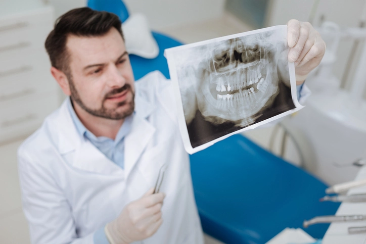
A radiograph (typically a periapical or panoramic X-ray) is the most definitive tool in diagnosing ankylosis. Key radiographic signs include:
- Loss of the periodontal ligament space
- Areas of root resorption
- Direct fusion of the root surface with alveolar bone
- Possible signs of replacement resorption where root structure is replaced with bone
Advanced Imaging
In complex cases, Cone Beam Computed Tomography (CBCT) may be employed to provide a 3D image and assess the extent of bone fusion.
Complications of Ankylosed Teeth
If left untreated, ankylosed teeth can lead to several functional and aesthetic problems:
- Alveolar Bone Deficiency: Since ankylosed teeth do not erupt with normal vertical growth, the surrounding bone may not develop properly, causing a vertical bone defect.
- Malocclusion: Infraocclusion can disrupt the occlusal plane, leading to bite problems.
- Tipping of Adjacent Teeth: Teeth adjacent to an ankylosed tooth may tilt toward it, creating crowding or spacing issues.
- Impaired Eruption of Permanent Teeth: An ankylosed primary tooth may block the path of its permanent successor.
- Facial Asymmetry: Severe infraocclusion in growing children may affect the development of the jaws and facial symmetry.
Treatment Options for Ankylosed Teeth
The treatment of an ankylosed tooth requires a careful, case-by-case approach. There is no single treatment protocol, as the management depends on multiple factors including:
- The patient’s age and growth status
- Whether the tooth is primary or permanent
- Severity of infraocclusion
- Presence or absence of permanent successors
- Location of the tooth (anterior vs. posterior, esthetic vs. functional zone)
- Overall orthodontic and occlusal planning
Because ankylosis affects both hard and soft tissue development—especially in children—early diagnosis and multidisciplinary planning are critical. Left untreated, ankylosed teeth can lead to space loss, malocclusion, esthetic defects, and alveolar bone deformities.
Let’s explore the various treatment options in depth:
- Observation and Monitoring (Conservative Management)
- Extraction of Ankylosed Tooth
- Decoronation
- Orthodontic Management and Space Regaining
- Surgical Luxation and Orthodontic Extrusion
- Prosthetic Replacement (When Successor is Missing)
- Autotransplantation (Rare but Viable Option)
- Ridge Preservation and Bone Grafting
Observation and Monitoring (Conservative Management)
In mild or early cases of ankylosis—particularly in primary teeth—monitoring may be the best initial approach.
When is observation appropriate?
- Mild infraocclusion (tooth is slightly below occlusal plane)
- No effect on adjacent teeth or space development
- Permanent successor is present and developing normally
- Patient is nearing the natural exfoliation age
What does monitoring involve?
- Periodic radiographs to track root resorption and successor eruption
- Clinical checks every 6–12 months for changes in infraocclusion or tipping of adjacent teeth
- Parent/patient education on signs of progression
However, continued infraocclusion or lack of exfoliation may eventually necessitate intervention.
Extraction of Ankylosed Tooth
Extraction is the most common and definitive treatment, especially when the ankylosed tooth interferes with dental development or esthetics.
Indications for extraction:
- Moderate to severe infraocclusion
- Disruption of arch alignment or occlusion
- Lack of permanent successor, risking long-term alveolar defect
- Orthodontic or prosthetic treatment planning
- Ankylosed reimplanted tooth undergoing replacement resorption
Special considerations:
- In children, early extraction may be followed by space maintenance using a band and loop, Nance appliance, or lower lingual holding arch.
- In adults, extraction may require socket preservation techniques to maintain ridge volume for future implants or bridges.
- If the root is severely ankylosed and fused to bone, surgical extraction (corticotomy, bone removal) may be necessary.
Decoronation
Decoronation is a unique, conservative surgical technique used primarily in young patients with an ankylosed permanent tooth, especially when the tooth is undergoing replacement resorption and the goal is to preserve alveolar ridge height.
What is decoronation?
- Removal of the crown and coronal root portion of the ankylosed tooth
- Leaving the apical root in place to allow for natural bone remodeling and ridge preservation
- Typically done when the patient is not yet a candidate for implants due to ongoing growth
Benefits of decoronation:
- Preserves vertical and horizontal alveolar ridge dimensions
- Allows implant placement later without need for major grafting
- Minimally invasive compared to full extraction
Ideal for:
- Children or adolescents with ankylosed anterior teeth
- Reimplanted permanent incisors showing signs of resorption
- Esthetic areas where bone volume must be maintained
Orthodontic Management and Space Regaining
Orthodontic treatment plays a central role in cases where ankylosed teeth affect space or alignment. However, because ankylosed teeth are immobile, treatment focuses on:
Orthodontic Strategies:
- Bypassing the ankylosed tooth: moving other teeth around it
- Extracting the ankylosed tooth and closing or opening space
- Orthodontic extrusion (in rare partial ankylosis cases)
- Using temporary anchorage devices (TADs) or mini-implants to aid in precise space closure or tooth movement
Challenges:
- Orthodontic forces do not work on ankylosed teeth
- Attempting to move an ankylosed tooth can cause root resorption of adjacent teeth
- Careful anchorage control is needed when planning around ankylosed units
A multidisciplinary plan involving the orthodontist and oral surgeon is essential for complex cases.
Surgical Luxation and Orthodontic Extrusion
In select cases where ankylosis is partial and localized, and the fusion is minimal, surgical luxation may allow for orthodontic movement.
What is luxation?
- A minor surgical procedure where the ankylosed tooth is loosened by disrupting the fusion points to the bone
- Followed by application of orthodontic forces to gradually extrude the tooth into position
Limitations:
- High risk of re-ankylosis
- Only effective in early-stage or partial ankylosis
- Usually unsuccessful in severe or complete ankylosis
- Requires careful follow-up and retention phase
This method is considered a salvage option when extraction or decoronation is not preferred.
Prosthetic Replacement (When Successor is Missing)
In cases where the ankylosed tooth is extracted or decoronated and no permanent successor is present, long-term prosthetic planning becomes necessary.
Options include:
- Dental implants (best placed after growth completion to avoid submergence)
- Fixed bridges (less preferred in young patients due to need for adjacent tooth preparation)
- Removable partial dentures (temporary solution during growth)
Timing considerations:
- Implants are typically placed after growth has stabilized (around 18–21 years), especially in the anterior region.
- Bone grafting or ridge augmentation may be required prior to implant placement if bone volume is deficient.
Autotransplantation (Rare but Viable Option)
In select cases, autotransplantation—the surgical movement of a tooth from one site to another within the same patient—may be used to replace an extracted ankylosed tooth, especially in growing patients.
Ideal candidates:
- Young patients with healthy donor teeth (e.g., premolars) and adequate recipient site bone
- Missing anterior teeth due to ankylosis or trauma
This approach requires excellent planning, surgical skill, and strict postoperative monitoring, but can be successful in certain conditions.
Ridge Preservation and Bone Grafting
Following extraction of an ankylosed tooth, particularly in cases of replacement resorption or bone loss, ridge preservation procedures may be needed to prepare for future prosthetic work:
- Socket grafting with bone substitutes or autografts
- Membrane placement (guided bone regeneration)
- Soft tissue augmentation for esthetics
Such procedures ensure long-term stability and esthetics for future implants or bridges.
Treatment by Tooth Type and Dentition Stage
| Tooth Type / Stage | Common Treatment Approach |
|---|---|
| Primary molar (mild) | Observation, monitor exfoliation |
| Primary molar (severe) | Extraction, space maintenance |
| Permanent incisor (traumatized, ankylosed) | Decoronation, possibly extraction and delayed implant |
| Permanent molar (mild) | Surgical luxation (rare), observation |
| Permanent molar (severe or growing child) | Decoronation or extraction, future prosthetic planning |
| Anterior esthetic zone | Decoronation, ridge preservation, implant at skeletal maturity |
Multidisciplinary Approach
Effective management of ankylosed teeth often requires collaboration between:
- Pediatric dentists – for monitoring and early detection
- Orthodontists – for space management and occlusal correction
- Oral surgeons – for extraction, decoronation, grafting, or luxation
- Prosthodontists – for long-term rehabilitation with implants or prosthetics
Orthodontic Considerations
Orthodontists must consider ankylosed teeth carefully during treatment planning, as these teeth do not respond to normal orthodontic forces. Attempts to move them can lead to root resorption or anchorage loss. Options may include:
- Bypassing the ankylosed tooth and moving other teeth around it
- Decoronation or extraction prior to orthodontic treatment
- Use of temporary anchorage devices (TADs) in complex cases
Prognosis
The long-term prognosis of an ankylosed tooth depends heavily on early detection and appropriate treatment. Teeth that are diagnosed and treated early—particularly in children—have a better chance of avoiding serious complications.
In adults, especially those with completed growth, ankylosed teeth are more difficult to manage and may require more invasive procedures to restore function and aesthetics.
Preventive Strategies
While many cases of ankylosis cannot be prevented due to their idiopathic or traumatic nature, certain steps can help reduce risk:
- Prompt treatment of dental trauma and careful reimplantation techniques
- Close monitoring of previously injured or reimplanted teeth
- Routine dental check-ups for early identification of infraocclusion
- Use of mouthguards during contact sports to prevent trauma