
White lesions of the oral cavity are a common clinical finding encountered by dentists, oral physicians, otolaryngologists, and general medical practitioners. These lesions range from benign, self-limiting conditions to potentially malignant disorders and overt carcinomas. The presence of a white patch on the oral mucosa is not a diagnosis in itself, but rather a clinical sign that demands careful evaluation, appropriate investigation, and, in some cases, urgent intervention.
The diagnostic challenge lies in the fact that many different pathological processes—infectious, inflammatory, traumatic, genetic, systemic, and neoplastic—can produce a similar white appearance. This whiteness usually results from increased keratinization, thickening of the epithelium, surface debris, or necrotic tissue. Some lesions can be wiped off, while others are firmly adherent. Some are localized, others diffuse; some are asymptomatic, others painful or ulcerated.
Table of Contents
ToggleGeneral Classification of Oral White Lesions
Oral white patches may broadly be classified into:
- Benign developmental or genetic conditions
- Reactive or traumatic lesions
- Infective lesions
- Inflammatory and autoimmune conditions
- Premalignant (potentially malignant) disorders
- Malignant lesions
- Systemic disease–associated lesions
- Iatrogenic or post-surgical lesions
Some conditions are transient and reversible, while others persist despite treatment and carry a risk of malignant transformation.
White Spongy Naevus
Definition and Etiology
White spongy naevus is a rare, benign, inherited disorder of keratinization. It is transmitted as an autosomal dominant trait and results from mutations affecting epithelial keratin proteins, particularly keratin 4 and keratin 13.
Clinical Features
Usually presents in childhood or adolescence, often noticed in the second decade of life
Appears as diffuse, bilateral, soft, white or greyish-white plaques
Lesions have a spongy or folded appearance
Commonly affects:
Buccal mucosa
Labial mucosa
Floor of the mouth
Ventral tongue
Lesions are asymptomatic and non-scrapable
Boundaries are often ill-defined
Histopathology
- Hyperplastic stratified squamous epithelium
- Prominent intraepithelial oedema
- No epithelial dysplasia
Management
- No treatment required
- Reassurance is essential
- Important to differentiate from leukoplakia to avoid unnecessary biopsy or anxiety
Malignant Potential
None
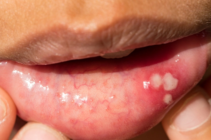
Frictional Keratosis
Definition
Frictional keratosis is a reactive lesion caused by chronic mechanical irritation of the oral mucosa.
Etiology
Common causes include:
- Sharp or fractured teeth
- Ill-fitting dentures
- Orthodontic appliances
- Chronic cheek or lip biting (morsicatio)
Clinical Features
Appears as a localized white plaque
Surface may be rough or shredded
Frequently seen on:
Buccal mucosa along the occlusal plane
Lateral tongue
Lesion corresponds anatomically to the source of trauma
Usually asymptomatic
Management
- Removal of the source of irritation
- Lesions typically resolve completely
- If the lesion persists after elimination of trauma, biopsy is mandatory
Special Considerations
May be associated with:
- Psychiatric disorders
- Anxiety-related habits
- Learning disabilities (self-mutilation behaviors)
Malignant Potential
None, provided the diagnosis is correct
Smokers’ Keratosis
Definition
Smokers’ keratosis refers to white lesions caused by chronic exposure to tobacco smoke, particularly heat and chemical irritants.
Etiology
- Cigarette smoking
- Pipe smoking (classically associated)
- Cigar smoking
Clinical Features
White patches affecting:
Buccal mucosa
Tongue
Palate
Lesions are often diffuse
Particularly common in pipe smokers
May be associated with mild epithelial thickening
Management
- Smoking cessation leads to complete resolution
- Lesions should be monitored during cessation period
Malignant Potential
- Generally low
- Important to differentiate from true leukoplakia
Stomatitis Nicotina (Smoker’s Palate)
Definition
Stomatitis nicotina is a distinctive lesion of the hard palate associated with smoking.
Pathogenesis
Chronic heat exposure leads to:
- Hyperkeratosis of palatal mucosa
- Inflammation and dilation of minor salivary gland ducts
Clinical Features
- Diffuse white or grey background
- Numerous small red papules
- Each papule has a central dark point representing the opening of a minor salivary gland duct
- Typically asymptomatic
Clinical Significance
Although the palatal lesion itself is generally benign, its presence may indicate:
Increased risk of dysplasia or carcinoma at high-risk sites, including:
- Floor of mouth
- Lateral tongue
- Retromolar trigone
Management
- Smoking cessation
- Careful oral examination and follow-up
Syphilitic Leucoplakia
Definition
Syphilitic leucoplakia is a classical manifestation of tertiary syphilis, presenting as a white plaque on the tongue.
Clinical Features
- White patch on the dorsum of the tongue
- May be well-defined
- Often asymptomatic
Diagnostic Considerations
Confirmed by:
- Serological tests
- Histopathology
- Dark-field microscopy (in earlier stages)
Management
- Active syphilis must be treated with appropriate antibiotic therapy
- The white lesion often persists despite treatment
Malignant Potential
- High
- The lesion has a tendency to undergo malignant transformation
- Long-term surveillance is essential
Chronic Hyperplastic Candidosis (Candidal Leucoplakia)
Definition
A persistent form of oral candidosis presenting as a non-scrapable white plaque.
Clinical Features
Common sites:
Commissures
Buccal mucosa
Tongue
Lesions are adherent and may be nodular or homogeneous
Significance
- Associated with epithelial dysplasia
- Considered a potentially malignant disorder
Management
- Antifungal therapy
- Elimination of predisposing factors
- Biopsy if lesion persists
Lichen Planus
Overview
Oral lichen planus is a chronic inflammatory mucocutaneous disorder with immunological pathogenesis.
Clinical Forms
- Reticular (most common)
- Erosive (painful)
- Atrophic
- Plaque-like
Reticular Lichen Planus
- Interlacing white lines known as Wickham’s striae
- Typically bilateral on buccal mucosa
- Asymptomatic
- No malignant potential
Erosive Lichen Planus
- Ulcerated, erythematous areas
- Painful
- Premalignant potential present
Management
- Topical corticosteroids
- Regular review
Lupus Erythematosus
Oral Manifestations
Seen in systemic lupus erythematosus (SLE) and discoid lupus.
Clinical Features
- White plaques with central erythema
- Ulceration may be present
- Often resemble lichen planus
Significance
- Part of a systemic autoimmune disease
- Requires multidisciplinary care
Leucoplakia
Definition
Leucoplakia is defined as a white patch or plaque that cannot be characterized clinically or pathologically as any other disease.
Epidemiology
- Strongly associated with tobacco use
- Alcohol is a synergistic risk factor
Clinical Types
- Homogeneous
- Non-homogeneous (speckled, nodular, verrucous)
Malignant Potential
Variable
Higher in:
Non-homogeneous lesions
Lesions on floor of mouth or lateral tongue
Management
- Biopsy
- Risk factor elimination
- Long-term follow-up
Hairy Leucoplakia
Etiology
Associated with Epstein–Barr virus (EBV) infection and immunosuppression.
Clinical Features
- Corrugated white plaques
- Typically on lateral borders of the tongue
- Seen in HIV/AIDS and transplant patients
Malignant Potential
None
Panoral Leucoplakia
Definition
Diffuse involvement of almost the entire oral mucosa.
Significance
- Represents a field change
- High risk of malignant transformation
Oral Carcinoma Presenting as a White Patch
Clinical Importance
- Oral squamous cell carcinoma may initially appear as a white lesion
- Differentiation from leukoplakia is critical
High-Risk Sites
- Floor of mouth
- Lateral tongue
- Retromolar area
Skin Grafts in the Oral Cavity
Clinical Relevance
- May appear as white patches
- Common diagnostic pitfall in exams
- History is key
White Patches Associated with Systemic Disease
Renal Failure
- Produces soft, oval white patches
- Resolve after treatment of renal disease
Darier’s Disease
- Genetic disorder
- White papules on gingivae and palate
- Associated with skin lesions
Pachyonychia Congenita
- Rare genetic condition
- White patches on tongue
- Nail and skin involvement present
Proliferative Verrucous Leucoplakia
Definition
An aggressive, progressive form of leukoplakia.
Clinical Features
- Multifocal
- Recurrent
- Resistant to treatment
Malignant Potential
Extremely high
Frequently progresses to:
Squamous cell carcinoma
Verrucous carcinoma
Management
- Aggressive surgical intervention
- Long-term surveillance
- Female predominance
Conclusion
White patches of the oral mucosa encompass a broad spectrum of conditions, ranging from harmless developmental anomalies to aggressive premalignant and malignant disorders. Accurate diagnosis requires a thorough history, careful clinical examination, awareness of risk factors, and judicious use of biopsy. For clinicians and students alike, understanding these lesions is essential for early detection of oral cancer and effective patient care.
Regular follow-up, patient education, and interdisciplinary collaboration remain the cornerstones of successful management in oral medicine.