
Tooth texture encompasses the intricate physical and microscopic characteristics of dental surfaces, influencing both their functional performance and aesthetic appeal. This comprehensive exploration delves into the multifaceted aspects of tooth texture, examining its structural components, developmental patterns, and implications for oral health and dental practices.
Table of Contents
ToggleIntroduction to Tooth Texture
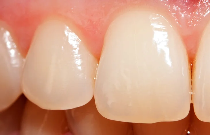
Tooth texture refers to the combination of visible and microscopic features that define the surface characteristics of a tooth. These include physical aspects like smoothness, roughness, ridges, grooves, and microscopic structures such as enamel rods, perikymata, and dentinal tubules. The texture is not merely a visual or tactile trait—it is deeply rooted in the functional biology of teeth and serves as a critical indicator of age, health status, and even evolutionary history.
Defining Texture in the Context of Dentistry
In dentistry, texture plays a critical role in multiple disciplines—restorative, cosmetic, preventive, and forensic. Texture encompasses both macrotexture (features visible to the naked eye) and microtexture (only visible under magnification). Macrotexture includes structures like cusps, fissures, and ridges, which aid in food processing. Microtexture includes enamel prisms, incremental growth lines, and wear facets that tell stories about an individual’s diet, oral hygiene practices, and age.
The scientific exploration of tooth texture is known as dental surface topography. Techniques such as scanning electron microscopy (SEM), laser profilometry, and dental microwear texture analysis (DMTA) allow researchers and clinicians to quantify these features with precision. Such studies reveal that even small changes in tooth texture can have major implications for function and aesthetics.
Importance in Evolution and Anthropology
Tooth texture is not just a contemporary concern, it has been a critical focus in paleoanthropology and evolutionary biology. The surface textures of fossilized teeth offer valuable insights into the diets and lifestyles of ancient species. For example, the presence of fine enamel striations or heavy pitting can reveal whether a hominid species was herbivorous, omnivorous, or carnivorous.
Researchers studying Neanderthal and early Homo sapiens teeth have used microwear texture analysis to determine shifts in diet due to environmental changes. These texture patterns offer direct, tangible evidence of how humans adapted to their environments—information not easily gleaned from bones or artifacts.
Functional Relevance of Texture
The mechanical functionality of teeth depends heavily on their textural characteristics. The ridges and cusps on molars help grind food, while the sharp edges of incisors and canines assist in biting and tearing. On a micro-level, the enamel’s organization into rods and interrod material distributes mechanical forces across the tooth structure, preventing fractures.
Additionally, texture contributes to the self-cleaning ability of teeth. The smoothness of enamel, combined with saliva flow, helps in reducing bacterial accumulation. However, when texture is altered—by erosion, abrasion, or developmental defects—the tooth becomes more susceptible to plaque retention and decay.
Texture and Aesthetic Perception
Texture also profoundly affects how we perceive teeth aesthetically. A natural, healthy tooth has a certain level of gloss, translucency, and surface irregularity that makes it appear “alive.” In contrast, overly smooth or unnaturally uniform textures (often seen in low-quality veneers or dental prosthetics) may look artificial.
In cosmetic dentistry, replicating the correct texture is vital for achieving natural-looking restorations. Modern dental laboratories and CAD/CAM systems are now programmed to simulate microtextures that mimic the surface patterns of enamel, enhancing both realism and patient satisfaction.
Texture in Diagnostics and Treatment Planning
Tooth texture provides diagnostic clues for clinicians. For example:
- Loss of enamel texture may indicate early stages of dental erosion or over-brushing.
- Excessively smooth or polished areas can suggest bruxism (tooth grinding).
- Rough or pitted surfaces might point to developmental disorders like amelogenesis imperfecta.
Understanding tooth texture enables more accurate treatment planning in fields such as:
- Prosthodontics (to match the surface of crowns or bridges to adjacent natural teeth)
- Orthodontics (to assess wear from brackets or appliances)
- Pediatric dentistry (to monitor enamel development)
Interdisciplinary Relevance
Tooth texture is a bridge between multiple scientific and clinical disciplines:
- In materials science, it informs the development of dental ceramics and composites.
- In biomechanics, it’s used to model stress distribution during chewing.
- In public health, texture analysis helps assess the impact of fluoride exposure or dietary patterns in populations.
From ancient fossil records to cutting-edge dental technologies, texture remains an essential aspect of our understanding of oral health and human biology.
Enamel: The Outer Shield
Tooth enamel is the hardest and most mineralized substance in the human body. It serves as the outermost layer of the crown of each tooth and functions as the first line of defense against mechanical, thermal, and chemical challenges. Enamel is also a central player in determining the surface texture of a tooth, both to the touch and under the microscope.
Composition and Structure of Enamel
Enamel is composed of approximately:
- 96% inorganic material, mainly in the form of hydroxyapatite crystals (Ca₁₀(PO₄)₆(OH)₂)
- 3% water
- 1% organic matrix
This extraordinarily high mineral content explains enamel’s hardness (Mohs hardness scale: 5–6) and brittleness. Despite this brittleness, the microstructural arrangement of enamel grants it impressive resistance to wear and fracture under physiological conditions.
Enamel Rods and Prisms
The most fundamental building block of enamel texture is the enamel rod (also known as an enamel prism). These tightly packed, elongated structures run from the dentinoenamel junction (DEJ) to the outer enamel surface. Each rod is about 4–8 microns in diameter and composed of thousands of hydroxyapatite crystallites.
Rods are organized in a keyhole shape, with the “head” facing the tooth surface and the “tail” nestled in between other rods. The way rods are arranged and oriented (often in a wavy or fish-scale pattern) contributes to the optical and mechanical properties of enamel texture.
Interrod Enamel
Between each enamel rod lies interrod enamel, a matrix of similar composition but with slightly different crystal orientation. The interplay between rods and interrod enamel introduces subtle variations in surface texture and reflects light differently, influencing tooth sheen and color.
Incremental Lines of Growth
As enamel is formed incrementally during tooth development, it retains markers of its formation:
- Striae of Retzius: These are internal growth lines that manifest as perikymata on the external surface.
- Neonatal lines: Special striae of Retzius found in the enamel of teeth that begin forming before and after birth. They provide a distinct texture in early-formed enamel.
These growth patterns are embedded into enamel and subtly influence surface texture, especially in younger individuals whose enamel has not undergone significant wear.
Surface Features: Perikymata and Retzius Lines
Among the most visible manifestations of enamel texture on the tooth surface are perikymata—fine, horizontal ridges seen on the labial (front) surfaces of incisors and canines, especially in younger individuals. These lines are the external expressions of the striae of Retzius, which are internally oriented, arc-shaped features within the enamel.
Perikymata: Surface Fingerprints of Tooth Growth
Perikymata are spaced approximately 30–40 micrometers apart and can be observed under good lighting or magnification. Their prominence varies by age, tooth type, and individual genetics. With age, these structures tend to wear away, either due to attrition (mechanical wear from chewing or grinding), abrasion (from brushing or external substances), or erosion (chemical dissolution).
Their clinical significance includes:
- Age estimation in forensic odontology.
- Identification of developmental enamel defects (e.g., hypoplasia, where perikymata are irregular or absent).
- Serving as a guide in cosmetic dentistry when replicating natural tooth appearance.
Striae of Retzius: Internal Growth Records
Internally, the striae of Retzius appear as dark lines under light microscopy or SEM. These lines reflect a rhythmic pattern of enamel secretion during amelogenesis (enamel formation by ameloblasts). When sectioned longitudinally, these lines can resemble concentric rings in tree trunks, each ring marking a developmental cycle.
In paleopathology, exaggerated striae may indicate systemic stress (e.g., malnutrition, illness) during tooth development, making enamel texture a reliable biological archive.
Enamel Surface Texture and Optical Properties
The arrangement of enamel rods and surface microfeatures directly influences how teeth reflect and refract light. This determines visual characteristics like:
- Translucency: Due to the way light scatters through varying rod orientation.
- Gloss: Highly polished enamel has a more reflective surface.
- Color: Enamel is semi-translucent; thus, its texture influences how the underlying dentin color appears.
This is why dentists carefully mimic enamel surface textures when restoring teeth with veneers, crowns, or composite fillings. High-end dental ceramics are engineered to reproduce rod-like microstructures and replicate the optical behavior of natural enamel.
Mechanical Implications of Enamel Texture
Enamel’s microtexture is not just aesthetically important—it plays a crucial mechanical role:
- Stress dissipation: The complex rod orientation and microcracks called enamel tufts and lamellae help manage and redistribute stress, preventing catastrophic failure.
- Wear resistance: Enamel texture affects how well the tooth resists surface wear. Smoother enamel tends to be more resistant to plaque but may wear faster in occlusal areas.
- Adhesive bonding: Surface texture influences how well dental adhesives bond to enamel. Acid-etching techniques, which intentionally roughen the enamel surface, rely on modifying microtexture to enhance adhesion.
Enamel Texture Through the Lifespan
Enamel is acellular and cannot regenerate. Once formed, its texture is modified only by external factors:
- In Children and Adolescents: Enamel appears more textured due to well-defined perikymata and less surface wear.
- In Adults: Normal wear from chewing, acidic foods, and brushing smooths the enamel surface. This alters the tooth’s gloss and may expose the underlying dentin in extreme cases.
- In Seniors: Enamel may become thinner, smoother, and more brittle. Texture loss can reduce bite efficiency and affect prosthodontic planning.
Developmental and Acquired Texture Disorders
Enamel Hypoplasia
A developmental condition where the enamel is deficient in quantity, often presenting as pits, grooves, or missing sections. This changes both the functional and aesthetic texture of teeth.
Dental Fluorosis
Dental fluorosis is caused by excessive fluoride intake during tooth development. It creates mottled enamel texture with white spots, lines, or in severe cases, brown discoloration and pitting.
Erosion and Abrasion
Both processes alter enamel texture significantly:
- Erosion smooths and flattens the enamel.
- Abrasion (from toothbrushes, toothpaste, or external particles) can create grooves and rough patches.
Clinical Implications for Enamel Texture
- Restorative Dentistry: Accurately replicating enamel’s surface texture improves aesthetic integration. Dentists use finishing and polishing instruments to mimic natural grooves, ridges, and gloss.
- Preventive Dentistry: Maintaining enamel texture involves fluoride treatments, dietary counseling, and proper oral hygiene to avoid erosion and wear.
- Pediatric Dentistry: Monitoring enamel development is essential to detect early signs of systemic issues.
- Cosmetic Dentistry: Texture manipulation via microabrasion or polishing is commonly used to enhance smile aesthetics.
Research Frontiers in Enamel Texture
Modern research is pushing the boundaries of our understanding of enamel:
- Nano-mechanical testing: Measures how variations in microtexture affect mechanical strength.
- 3D digital mapping: High-resolution scans can replicate enamel textures for personalized restorations.
- Bioengineered enamel: Scientists are developing synthetic versions that mimic natural texture at a molecular level.
Enamel’s complex texture isn’t just a passive trait—it’s a biologically engineered marvel optimized over millions of years of evolution.
Dentin: The Underlying Support
Dentin is the second major calcified tissue of the tooth, located beneath the enamel and cementum, and surrounding the central pulp chamber. While enamel is known for its hardness and brittle nature, dentin provides resilient support, acting as a shock absorber during mastication (chewing) and a critical mediator of sensory perception. Dentin is also central to the overall texture of a tooth—especially once enamel begins to wear down with age or damage.
Composition and Structure of Dentin
Dentin is a mineralized connective tissue with a different composition than enamel:
- 70% inorganic material (mainly hydroxyapatite crystals)
- 20% organic material (predominantly type I collagen)
- 10% water
This makes dentin softer and less brittle than enamel, allowing it to deform slightly under stress. Its unique composition also provides the foundation for the texture that emerges as teeth age or become worn.
Types of Dentin
There are three primary types of dentin, each influencing texture differently:
- Primary dentin: Formed during tooth development, it makes up the bulk of the tooth’s dentinal structure.
- Secondary dentin: Deposited after the tooth erupts, continuing throughout life. This slow deposition alters the internal architecture of the tooth and affects the pulp chamber shape.
- Tertiary dentin (also called reparative dentin): Formed in response to trauma, caries, or other stimuli. It has an irregular texture and is often less mineralized, contributing to clinical changes in surface hardness.
Each of these types can impact how dentin presents clinically—whether it’s polished and smooth, irregular and bumpy, or deeply grooved depending on age, health, and history of dental interventions.
Dentin Tubules and Microscopic Texture
One of the most distinctive features of dentin is the presence of dentinal tubules—microscopic channels that radiate from the pulp chamber outward to the enamel or cementum border. There are about 15,000 to 65,000 tubules per square millimeter, depending on their location.
Dentinal Tubules and Texture Formation
These tubules are critical in defining dentin’s microtexture:
- The density and diameter of the tubules change with proximity to the pulp.
- Closer to the pulp, tubules are wider and more numerous, making the texture more porous.
- Near the outer dentin, tubules are more compressed and the surface is smoother.
The orientation and size of these tubules influence how dentin behaves:
- In sensitivity: Exposed tubules allow stimuli (temperature, pressure, chemicals) to reach nerve endings, leading to dentin hypersensitivity.
- In restorative bonding: Adhesive dentistry depends on interacting with tubules; their presence and pattern determine the mechanical interlocking of dental materials.
The Dentinoenamel Junction (DEJ): A Crucial Interface
The dentinoenamel junction (DEJ) is the boundary between enamel and dentin. It is not a flat, simple interface but rather a scalloped, textured layer that significantly affects tooth durability and function.
Role of DEJ in Texture and Function
- The scalloped nature enhances mechanical interlocking between enamel and dentin.
- Acts as a stress distributor: It absorbs and spreads forces from biting or grinding.
- In microscopy, the DEJ has a unique wavy texture, which increases the surface area of contact and reinforces the tooth against fracture.
In teeth where enamel has been lost (due to erosion, abrasion, or decay), the texture and resilience of dentin and its DEJ become even more crucial.
Sensory and Biological Functions of Dentin
While enamel is insensate (has no nerve supply), dentin is biologically active. Odontoblasts, specialized cells located at the pulp end of dentinal tubules, play key roles in sensation, defense, and repair.
Sensory Role and Texture Perception
- Dentin is capable of transmitting pain, pressure, and thermal signals.
- Exposed dentin—often due to enamel wear or gum recession—feels rougher and more sensitive to touch, temperature, and acidic foods.
- Patients often describe exposed dentin as having a “chalky” or “grainy” texture compared to smooth enamel.
Clinically, assessing dentin texture can help diagnose the cause of hypersensitivity, tooth wear, or non-carious cervical lesions.
Aging and Dentin Texture
As people age, dentin undergoes several structural and compositional changes:
- Sclerosis of dentinal tubules: Mineral is deposited within the tubules, leading to sclerotic dentin that appears glassy and translucent. This type of dentin is smoother but harder and more brittle.
- Secondary dentin accumulation narrows the pulp chamber, changing the internal architecture and altering the surface texture beneath the enamel.
These changes influence:
- Bonding efficacy during restorative procedures (as aged dentin is less reactive).
- The clinical feel and resistance encountered during drilling and instrumentation.
Clinical Texture of Dentin in Dentistry
In Restorative Procedures
Dentists encounter various dentin textures when preparing cavities:
- Sound dentin: Feels slightly resilient under a dental explorer.
- Caries-affected dentin: Softer, moist, and may have a leathery texture.
- Caries-infected dentin: Even softer, discolored, and often flaky.
Identifying the texture helps clinicians decide how much material to remove and how best to restore the tooth.
In Bonding and Adhesion
Adhesive bonding to dentin is more complex than bonding to enamel due to:
- The presence of moisture in tubules.
- The organic collagen matrix.
- The need for etching or conditioning agents to modify the surface for better mechanical and chemical adhesion.
Different bonding systems (etch-and-rinse, self-etch, or universal adhesives) are designed to work with the specific textural characteristics of dentin to optimize outcomes.
Dentin Texture in Disease and Trauma
Erosion and Abrasion
Dentin exposed due to loss of enamel is more susceptible to erosion. This results in:
- Smoother surface initially due to demineralization.
- Later, irregular and pitted surface as organic components break down.
- Clinical signs: Yellowish appearance, dull surface luster, and increased roughness.
Attrition and Tooth Grinding
Chronic bruxism or tooth grinding leads to:
- Flattened dentin surfaces.
- Formation of wear facets with polished, shiny textures.
- Cracks and chipping at the DEJ, further compromising enamel integrity.
Root Surface Exposure
Gum recession reveals cementum-covered root dentin, which is naturally rougher and softer than crown dentin. This area is more prone to:
- Caries
- Sensitivity
- Abrasion from brushing
Texture here is critical for diagnosing and managing non-carious cervical lesions (NCCLs).
Dentin in Regenerative and Future Dentistry
The field of regenerative dentistry is working toward:
- Dentin-pulp complex regeneration using stem cells and growth factors.
- Bioactive materials that mimic natural dentin texture and promote integration with pulp tissues.
- Nanotechnology-enhanced composites that replicate the elastic modulus and surface granularity of dentin.
These developments hinge on a clear understanding of how texture contributes to function, and how mimicking natural microarchitecture leads to better clinical outcomes.
Microscopic Textures and Their Significance
While the macroscopic features of teeth—like cusps, ridges, and grooves—are crucial for mastication and aesthetics, the microscopic textures embedded within the tooth’s structure provide the real engineering behind its remarkable functionality. These ultra-fine features play a key role in how teeth manage forces, resist damage, and maintain their integrity over a lifetime. The importance of these microtextures extends from biological development to clinical dentistry and even into paleontological research.
Enamel Rods and Interrod Substance
Enamel, though highly mineralized, is not a homogeneous surface. At the microscopic level, it is composed of enamel rods (prisms)—bundle-like structures formed by tightly packed hydroxyapatite crystals. Each enamel rod is about 4–8 micrometers in diameter and spans from the dentinoenamel junction (DEJ) to the external surface.
Functional Significance of Enamel Rods
- Mechanical reinforcement: The rod-interrod orientation distributes forces during mastication and biting, preventing localized stress that could lead to cracking or fracture.
- Light manipulation: The microscopic texture of enamel rods affects the way light is refracted and reflected, contributing to the optical properties of teeth (gloss, translucency, brightness).
- Etching patterns: In clinical dentistry, acid etching alters the enamel rod texture to enhance bonding. Different etching patterns (Type I, II, III) are defined by how the acid affects rod and interrod enamel.
Rod Orientation Patterns
In certain areas of the tooth, particularly in deciduous teeth and the cervical region of permanent teeth, enamel rods tilt outward. In the occlusal regions, they run perpendicular to the surface. This spatial arrangement creates variable textural resistance and has clinical implications for:
- Cavity design (to preserve enamel strength)
- Bonding techniques (to avoid marginal leakage)
Enamel Tufts, Lamellae, and Spindles
These are structural “defects” or variations within enamel that play surprisingly important roles in tooth durability and texture.
Enamel Tufts
- Definition: Hypomineralized, ribbon-like structures that originate at the DEJ and extend into enamel.
- Microscopic appearance: Resemble “grass tufts” under scanning electron microscopy.
- Function: Act as stress absorbers that prevent crack propagation. They may appear as localized softer areas, subtly influencing tactile texture in worn enamel.
Enamel Lamellae
- Definition: Thin, linear faults that extend from the enamel surface toward the DEJ.
- Texture contribution: They serve as pathways for potential crack initiation under excessive forces or thermal changes.
- Clinical impact: Lamellae can be sites of bacterial ingress or restoration failure, especially in cosmetic veneers.
Enamel Spindles
- Definition: Short, linear extensions of dentinal tubules that cross into enamel.
- Significance: These may act as sites of increased permeability and sensitivity, and though invisible to the naked eye, they contribute to the biological texture of teeth.
The Dentinoenamel Junction (DEJ) Micromorphology
The DEJ is far from a smooth, flat boundary. It is a scalloped and highly textured interface, resembling a jigsaw connection between enamel and dentin. Its undulating morphology is critically important.
Stress Resistance
- Scalloped design increases surface area, improving the mechanical adhesion between enamel and dentin.
- Resists delamination: The interlocking nature of the DEJ texture prevents enamel from peeling off or fracturing easily during mastication.
Optical and Imaging Relevance
- The DEJ often appears as a distinct band on radiographs and optical coherence tomography (OCT) scans due to its unique structural orientation.
- High-resolution microscopy shows this junction as a transitional texture zone, where enamel rods bend and blend into dentinal tubules.
Dental Microwear Texture Analysis (DMTA)
DMTA is a relatively recent and highly valuable method used to study the microscopic textures on the occlusal (chewing) surfaces of teeth. It uses 3D surface topography analysis to quantify microscopic wear patterns caused by food, grit, or non-dietary behavior (e.g., tool use, pipe smoking).
Scientific Applications
- Anthropology: Used to infer the diets of early humans and extinct animals.
- Paleontology: Helps determine whether a species was herbivorous, carnivorous, or omnivorous based on enamel abrasion and pitting.
- Forensics: Aids in identifying lifestyle and age from teeth in human remains.
Quantifying Texture Parameters
DMTA uses statistical parameters to describe texture:
- Complexity (Asfc): Reflects how varied the surface is—higher in species that chew hard foods.
- Anisotropy (epLsar): Measures the orientation of wear—high values indicate shearing action.
- Textural fill volume (Tfv): Assesses how much “roughness” is present in surface pits and valleys.
Clinical Significance of Microscopic Texture
Understanding and preserving microscopic texture has multiple clinical implications:
Adhesion in Restorative Dentistry
- Etching and bonding techniques rely on modifying enamel’s microscopic texture to allow adhesives to penetrate and interlock with enamel rods and interrod spaces.
- Poor etch patterns lead to weaker bonds, increasing the risk of marginal leakage or debonding.
Diagnostic Imaging
Texture anomalies—like those caused by fluorosis or early caries—can be detected by modern techniques such as:
Scanning electron microscopy (SEM)
Atomic force microscopy (AFM)
Optical coherence tomography (OCT)
Confocal laser scanning microscopy (CLSM)
These tools highlight subtle surface changes long before they become visible or symptomatic.
Reproduction of Natural Texture in Cosmetic Dentistry
- For crowns, veneers, or composite fillings, mimicking microscopic texture is vital for a natural appearance.
- Master ceramists use specialized burs, brushes, and glazing techniques to create fine grooves, ridges, and light-scattering features.
Future Directions in Texture-Based Research
Emerging fields continue to investigate how to better replicate or restore natural tooth textures:
- Biomimetic materials: Engineered to replicate the microstructure of enamel and dentin for restorations that look and behave like natural teeth.
- Nano-textured dental surfaces: Dental implants and restorations are being developed with textures that encourage better tissue integration and plaque resistance.
- AI-based texture recognition: Algorithms are being trained to detect early texture changes indicative of disease or wear using intraoral scanners and photographic data.
Dental Microwear Texture Analysis (DMTA)
Dental Microwear Texture Analysis (DMTA) is a cutting-edge technique used to analyze the microscopic wear features on the surfaces of teeth. These features develop as a result of interactions between the tooth and various elements in the environment, primarily diet. By studying microwear textures—scratches, pits, ridges, and other microscopic features—scientists can reconstruct dietary habits, infer behavioral patterns, and assess health status.
Unlike traditional dental wear analysis, which relied heavily on qualitative assessments, DMTA is quantitative and three-dimensional, providing high-resolution data that are reproducible and statistically robust. As such, it plays a major role in anthropology, evolutionary biology, dentistry, forensics, and paleontology.
The Basics of DMTA
What Is Being Measured?
Microwear refers to microscopic abrasions on the surface of enamel or dentin, typically on the occlusal (chewing) surfaces of molars and premolars. These textures result from:
- Food particles (especially hard or abrasive items like seeds, bone, and grit)
- Exogenous materials (dust, sand, dietary inclusions)
- Non-dietary behaviors (tool use, habits like nail-biting or pipe smoking)
DMTA quantifies these features using precise metrics:
- Complexity (Asfc): Measures surface roughness; high complexity often indicates hard-object feeding.
- Anisotropy (epLsar): Measures the alignment of surface features; high anisotropy reflects directional movement like shearing.
- Textural Fill Volume (Tfv): Quantifies how much material would be needed to fill in pits and valleys on the tooth surface.
- Scale of maximum complexity (Smc): Indicates the scale at which surface textures are most variable.
These parameters are computed from 3D scans obtained via white-light confocal microscopy or laser scanning confocal microscopy.
Historical Context and Evolution
DMTA evolved from earlier studies that used scanning electron microscopy (SEM) to describe dental wear patterns. While SEM provided detailed images, it was limited by:
- Subjectivity in interpretation
- Two-dimensional imaging
- Limited ability to quantify depth or volume
The development of scale-sensitive fractal analysis in the late 20th century revolutionized the field. Researchers could now describe tooth surfaces using mathematical metrics, improving objectivity and reproducibility. Today, DMTA is a standard tool in evolutionary research, often used alongside isotopic or anatomical data to reconstruct ancient diets.
DMTA in Human Evolution and Anthropology
Reconstructing Diets of Early Hominins
Microwear texture patterns have been used to distinguish between:
- Graminivores (grass-eaters): Typically show low complexity, dominated by parallel scratches.
- Frugivores (fruit-eaters): Show higher complexity due to tougher, irregular foods.
- Hard-object feeders (e.g., nuts, seeds): Show high pit density and surface roughness.
Famous examples:
- Paranthropus boisei was once believed to be a hard-object feeder based on jaw anatomy, but DMTA revealed a diet of soft vegetation.
- Neanderthals showed variable wear patterns depending on region and climate, indicating dietary flexibility.
Cultural and Tool-Use Behaviors
DMTA has revealed non-dietary wear caused by:
- Use of teeth as tools (e.g., hide processing)
- Pipe smoking
- Occupational habits (e.g., holding items between teeth)
These behaviors leave distinct textural signatures, enabling researchers to infer aspects of lifestyle beyond diet.
Clinical Relevance of DMTA
Diet and Tooth Wear in Modern Populations
In contemporary humans, DMTA is used to:
- Monitor wear patterns in children and adults to evaluate occlusal function.
- Study how processed diets (which are softer) lead to reduced enamel wear and changes in occlusal morphology.
- Identify excessive wear or pathology from behaviors like bruxism (teeth grinding), where repeated microabrasions leave characteristic textures.
Monitoring Tooth Erosion and Restoration Failure
Dentists can use surface texture analysis to:
- Assess the progression of acid erosion, which smooths and dulls enamel texture.
- Evaluate the performance of dental materials: how well do crowns or fillings maintain or mimic the microtexture of natural enamel?
- Determine abrasivity of toothpastes or brushing habits, based on roughness scores post-cleaning.
Limitations and Considerations in DMTA
Like any scientific technique, DMTA has limitations:
- Preservation bias in fossil teeth may obscure original textures.
- Some foods leave no microwear trace, especially soft or non-abrasive items.
- The timescale represented by wear textures is often short (weeks to months), making DMTA a snapshot rather than a full dietary biography.
- The technique requires expensive equipment and expert interpretation, limiting its routine use in clinical settings.
Despite these challenges, ongoing improvements in imaging and software analysis are making DMTA faster, more affordable, and more accessible to clinicians and researchers alike.
Integration with Other Analytical Tools
Modern researchers often integrate DMTA with other methods to get a fuller picture of oral health and dietary behavior:
- Isotope analysis: Provides information about long-term dietary trends.
- Tooth shape and enamel thickness measurements: Give context to wear patterns.
- Genetic data: May explain susceptibility to wear or enamel structure variations.
Together, these methods form a multi-disciplinary approach to understanding human biology through teeth.
Factors Influencing Tooth Texture
Tooth texture is not a static trait; it is dynamic and evolves over the course of a lifetime due to a variety of influences, both natural and external. While the basic enamel and dentin textures are genetically and developmentally programmed, they are significantly altered by environmental exposure, personal habits, dietary choices, and even medical interventions.
Understanding these factors is crucial not only for clinicians who wish to preserve or restore natural tooth surfaces but also for researchers who use texture as a window into diet, behavior, and health.
Age and Natural Tooth Wear
Aging is one of the most predictable factors in the transformation of tooth texture.
In Children and Adolescents
- Enamel texture is typically well-defined, with visible perikymata (horizontal ridges) and a high degree of surface gloss.
- Microscopic features like striae of Retzius are more likely to influence enamel reflectivity.
- Primary (baby) teeth show less mineralized enamel and dentin, giving them a different tactile and optical texture.
In Adults
- Regular wear from mastication smooths the enamel surface over time.
- Texture becomes less defined, and the tooth may feel more polished but slightly less reflective.
- Enamel may become thinner, exposing the underlying dentin, which has a rougher, more porous texture.
In Older Adults
- Attrition, erosion, and abrasion accumulate, leading to a flatter occlusal surface and loss of detailed macro- and microtexture.
- In cases of extensive wear, the pulp may be exposed or covered with secondary/tertiary dentin, changing the tooth’s appearance and feel significantly.
Dietary Habits and Texture Modification
Diet is a powerful determinant of tooth texture, influencing both macroscopic wear and microscopic abrasions.
Hard and Abrasive Foods
- Foods like nuts, seeds, raw vegetables, and unprocessed grains introduce microwear in the form of pits and striations.
- These changes are often beneficial in evolutionary terms, as they indicate dietary adaptability.
Acidic Foods and Drinks
Citrus fruits, vinegar, soda, and wine cause enamel erosion, leading to:
Smoother, glossier surfaces in early stages
Dull, softened, and eventually pitted enamel in later stages
Erosion undermines enamel’s natural ridges and optical characteristics, reducing both function and beauty.
Processed and Soft Diets
- Modern diets high in processed, soft foods result in less dental wear and may lead to over-retained enamel textures that were naturally meant to wear down.
- This lack of natural abrasion can also contribute to dental crowding and changes in occlusion over time.
Oral Hygiene and Mechanical Abrasion
How we care for our teeth dramatically influences texture.
Brushing Technique
- Aggressive brushing with firm-bristled toothbrushes can wear away enamel, particularly at the cervical (neck) area.
- This can result in abrasion lesions, which feel rough and may be hypersensitive due to exposed dentin.
Toothpaste Abrasivity
Toothpastes vary in their Relative Dentin Abrasivity (RDA) score.
- High-RDA pastes (e.g., whitening products) remove surface stains but may roughen enamel.
- Low-RDA pastes are gentler, helping preserve natural gloss and texture.
Use of Dental Tools
- Over-polishing during professional cleaning may temporarily increase smoothness but remove fine enamel texture if not carefully controlled.
- Prophylaxis pastes and ultrasonic scalers, while effective for cleaning, can alter the microtopography of enamel if overused.
Parafunctional Habits and Occupational Factors
Bruxism (Teeth Grinding)
This involuntary habit can result in:
Flat, polished occlusal surfaces
Exposed dentin
Stress cracks along the enamel
Texture in bruxed teeth is often significantly altered and can feel unnaturally smooth or irregular, depending on severity.
Occupational Exposure
- Jobs involving airborne grit (e.g., mining, sandblasting) can lead to exogenous wear, resulting in rougher surfaces and more frequent enamel loss.
- Occupational habits, like holding objects in the mouth (e.g., tailors, carpenters), can cause localized wear facets.
Developmental and Genetic Factors
Some individuals are born with variations in enamel and dentin texture due to genetic or systemic conditions.
Amelogenesis Imperfecta
A group of hereditary conditions affecting enamel formation.
Teeth may be:
Pitted or grooved
Hypoplastic (too thin)
Discolored and rough
Texture is often markedly irregular, requiring restorative treatment to regain function and appearance.
Dentinogenesis Imperfecta
Affects dentin development, resulting in:
Opalescent appearance
Weak, soft dentin that easily chips
Altered tactile texture (often chalky or brittle)
Environmental Disruptions During Development
Fever, malnutrition, trauma, or medication during tooth formation can cause:
- Enamel hypoplasia (quantitative defects) leading to grooves or pits
- Enamel hypocalcification (qualitative defects), resulting in porous, chalky enamel
Restorative and Cosmetic Interventions
Restorative treatments inevitably alter natural texture—but ideally in ways that replicate it.
Dental Fillings and Crowns
- The success of a restoration is judged not just by color, but by how well it mimics enamel and dentin texture.
- Advances in CAD/CAM dentistry and 3D milling now allow restorations to include fine grooves, scallops, and luster.
- Poorly textured restorations can feel too smooth or too rough and may be functionally inadequate.
Veneers and Cosmetic Bonding
- Achieving a lifelike appearance depends on mimicking light diffusion through natural microtexture.
- Skilled ceramists use layering techniques and custom staining/glazing to create enamel-like texture.
Whitening Treatments
- Bleaching can dehydrate enamel, temporarily changing its surface texture and making it feel rougher or more porous.
- Overuse can result in microcracks, increasing the enamel’s susceptibility to staining and erosion.
Systemic Health and Medication
Medical Conditions
- GERD (acid reflux) introduces stomach acid into the mouth, causing enamel erosion and texture changes.
- Bulimia similarly leads to acid-induced texture loss, especially on lingual surfaces.
Medications
- Some medications reduce saliva flow, increasing the risk of erosion and plaque buildup.
- Others (like tetracycline during development) can influence both color and texture through internal staining and hypocalcification.
Environmental and Lifestyle Factors
- Water fluoridation contributes to stronger enamel and a smoother surface in optimal doses.
- Smoking and tobacco use increase surface staining and may lead to rougher texture from plaque accumulation.
- Mouth breathing can dry out enamel, reducing the protective effects of saliva and increasing vulnerability to surface changes.
Clinical Implications of Tooth Texture
Tooth texture is more than an academic curiosity or evolutionary clue—it plays a critical role in clinical dentistry. From diagnostics to restoration design, from patient satisfaction to the longevity of dental work, the texture of teeth influences nearly every aspect of oral healthcare.
Modern dentistry now incorporates a deeper understanding of macrotexture and microtexture into treatment planning, restoration design, cosmetic procedures, and even preventive strategies. Mastery of this subject equips clinicians to deliver care that is both functional and aesthetically superior.
Diagnostic Value of Texture
Identifying Pathology
Changes in tooth texture often serve as early indicators of oral health problems:
- Smooth, glassy surfaces on anterior teeth may signal enamel erosion due to dietary acids or acid reflux.
- Chalky, porous textures could indicate enamel hypocalcification or fluorosis.
- Pitted or grooved surfaces may reflect developmental defects like enamel hypoplasia.
- Rough, irregular texture around restorations may suggest marginal breakdown or secondary caries.
Assessing Wear Patterns
The tactile and visual examination of surface texture helps identify:
- Bruxism: Smooth, flat occlusal surfaces and polished wear facets.
- Abrasion: V-shaped notches at the cervical areas of teeth due to overbrushing.
- Erosion: Softening and smoothing, especially on lingual/palatal surfaces of upper teeth.
- Attrition: Loss of enamel and dentin due to tooth-to-tooth contact.
These findings influence decisions regarding occlusal adjustment, protective appliances (e.g., night guards), or dietary counseling.
Tooth Texture and Restorative Dentistry
Bonding and Adhesion
Successful adhesion to tooth structure depends heavily on surface texture:
- Etching enamel with phosphoric acid creates microporosities in the enamel rod network, enhancing bond strength.
- Conditioning dentin modifies the smear layer and exposes tubules, allowing resin infiltration and hybrid layer formation.
Understanding natural texture ensures optimal surface preparation and stronger, longer-lasting bonds.
Replicating Natural Texture in Restorations
Highly polished restorations may look artificial if they lack enamel-like surface complexity:
- Composite fillings must be finished and polished to match adjacent enamel in luster and topography.
- Crowns and veneers should include subtle ridges, depressions, and gloss variations to mimic light scattering and feel natural.
Digital technologies (e.g., CAD/CAM milling, 3D printing) now allow technicians to scan and replicate natural textures with remarkable precision.
Aesthetic Dentistry and Texture Perception
Tooth texture significantly influences a smile’s appearance—even more so than color or alignment in many cases.
Surface Gloss and Light Reflection
Natural enamel has variable gloss across the surface due to the orientation of enamel rods and the presence of perikymata. This gloss is a key factor in the perceived vitality of teeth. Over-polished restorations may appear flat or fake because they reflect light too uniformly.
Texture and Age Appropriateness
- Younger teeth exhibit well-defined perikymata, high surface luster, and microgrooves.
- Older teeth show smoother, flatter surfaces with duller gloss.
In aesthetic restorations, matching age-appropriate texture can help create lifelike outcomes. A young adult with unnaturally smooth veneers may appear “overdone,” while mature patients benefit from restorations that reflect their natural dental history.
Smile Design and Customization
Advanced smile makeovers use microtexture tools (fine burs, brushes, lasers) to customize texture:
- Grooves, stippling, or micro-scratches can be added manually.
- Artistic adjustments are often based on patient personality, habits, and natural features.
Prosthodontics and Implantology
In full-mouth rehabilitations or prosthetic design, texture matching is critical for both function and feel.
Dentures and Bridges
- Smooth, textureless denture teeth are easily spotted as artificial.
- High-quality dentures replicate the microtexture of enamel using layered acrylics and ceramic processing techniques.
Dental Implants
Although the implant itself is embedded in bone, the visible prosthetic crown must replicate natural enamel:
- A mismatched surface texture can feel foreign in the mouth.
- Poor textural integration may also affect how food moves across the teeth, impacting speech and chewing efficiency.
Periodontics and Texture in Gingival Health
Rough surfaces promote plaque retention:
- Smooth enamel helps resist bacterial colonization.
- Roughened surfaces, whether from acid erosion, improper restorations, or developmental defects, are more prone to plaque buildup and inflammation.
Clinical interventions aim to preserve or restore smoothness in exposed root surfaces and around dental margins to maintain periodontal health.
Preventive Strategies Based on Texture
Texture-related interventions are part of many preventive care programs:
Remineralization
- Use of fluoride varnishes or calcium phosphate products can restore surface smoothness and resist future demineralization.
- Regular application can reduce hypersensitivity caused by exposed dentin tubules.
Desensitizing Treatments
Gels and varnishes that occlude dentin tubules smooth out tactile texture, decreasing sensitivity.
Diet and Hygiene Counseling
- Patients are educated about acidic foods and habits that alter surface texture.
- Recommendations may include low-abrasivity toothpastes, gentle brushing, and frequent hydration to maintain natural enamel luster.
Texture-Guided Technology in Clinical Practice
Modern tools now allow precise assessment and replication of dental texture:
- Intraoral scanners provide 3D models that capture surface topography.
- Optical coherence tomography (OCT) enables non-invasive evaluation of microtexture.
- AI and texture-recognition software are being developed to assist with early diagnosis and treatment planning.
These technologies promise more personalized, predictive, and effective care.
Preservation and Enhancement of Tooth Texture
Preserving and enhancing tooth texture is a cornerstone of modern dental care. As texture plays a vital role in function, comfort, and aesthetics, protecting it is essential to long-term oral health. Whether the goal is to maintain natural enamel, reduce sensitivity, prevent erosion, or replicate natural texture in restorations, dentists use a range of strategies—from simple daily habits to advanced biomimetic treatments.
This section outlines the preventive measures, clinical interventions, and emerging technologies that help preserve or restore natural tooth texture.
Preventive Measures for Maintaining Natural Texture
The most effective strategy for preserving tooth texture is to prevent its loss in the first place. Prevention starts with lifestyle habits and daily oral hygiene.
Proper Brushing Technique
- Use soft-bristled toothbrushes to avoid mechanical abrasion.
- Avoid excessive horizontal brushing motions, especially near the gum line.
- Electric toothbrushes with pressure sensors can help reduce the risk of surface damage.
Low-Abrasivity Toothpastes
- Choose toothpaste with a low Relative Dentin Abrasivity (RDA), especially for people with enamel erosion, dentin exposure, or sensitive teeth.
- Whitening pastes, while effective for stain removal, often have higher abrasivity and may wear away enamel texture over time.
Protective Dietary Habits
- Limit acidic foods and beverages (e.g., soda, citrus, sports drinks). Rinse with water afterward if consumed.
- Avoid brushing immediately after acid exposure; enamel is temporarily softened and more prone to erosion.
- Chew sugar-free gum to stimulate saliva flow, which helps neutralize acid and protect enamel.
Fluoride Use
- Fluoride strengthens enamel by aiding in remineralization and forming fluorapatite, which is more resistant to acid attack.
- Use fluoridated toothpaste and consider topical fluoride treatments at dental visits for high-risk patients.
Remineralization Therapies and Texture Restoration
When texture loss has begun, remineralization therapies can help reverse early damage and restore smoothness and structure to enamel.
Topical Fluoride
- High-concentration fluoride varnishes or gels applied in-office can rebuild weakened enamel, smoothing surface roughness.
- Daily use of prescription-strength fluoride toothpaste is recommended for patients with moderate to high caries risk.
Calcium and Phosphate-Based Products
- Products containing calcium phosphate (CaP) compounds (e.g., CPP-ACP, nano-hydroxyapatite) mimic the natural building blocks of enamel.
- These help fill in microscopic surface defects, restoring texture and decreasing sensitivity.
MI Paste and Tooth Mousse
Derived from casein (milk protein), these products are rich in calcium and phosphate and are especially useful in remineralizing hypomineralized enamel and early erosion.
Cosmetic and Clinical Texture Enhancement
When texture has been permanently altered or lost, dental professionals can recreate or improve surface topography using cosmetic and restorative methods.
Enamel Microabrasion
A minimally invasive procedure that removes a superficial enamel layer (about 50–200 microns) using a combination of acidic slurry and abrasive materials.
Often used to treat:
Mild fluorosis
Superficial stains
Textural irregularities
Leaves a smoother, more uniform surface and improves gloss.
Composite Bonding and Finishing Techniques
- Texturing can be manually sculpted using specialized finishing burs, sandpaper discs, and polishing pastes.
- Skilled dentists create subtle grooves, perikymata, and gloss variations to mimic adjacent natural teeth.
Ceramic Veneers and Crowns
Dental ceramists apply multi-layered ceramics to simulate the translucency, microtexture, and reflectivity of enamel.
Surface texture is added by:
Fine bur detailing
Hand-sculpted anatomical features
Controlled glazing and staining
Managing Texture in Dentin and Root Surfaces
In cases where enamel is lost and dentin is exposed, preservation of surface quality is vital to reduce sensitivity and prevent further damage.
Desensitizing Agents
- Seal off dentinal tubules to reduce fluid movement, which causes pain.
- Create a smoother surface, which is more resistant to plaque and biofilm formation.
Periodontal Therapy
- Exposed root surfaces are more porous and irregular in texture.
- Scaling and root planing remove calculus but must be done carefully to avoid creating excessive roughness.
- Adjunct therapies (e.g., laser treatment, smoothing agents) may be used to optimize surface finish.
Professional Maintenance and Polishing
Routine dental cleanings play a critical role in preserving and restoring healthy texture:
Selective Polishing
- Not all teeth need polishing at every cleaning.
- Over-polishing can remove microtexture and reduce enamel thickness.
- Selective polishing targets only stained or rough areas, using fine-grit pastes to preserve natural luster.
Air Polishing and Prophylaxis
- Air polishing units use water, air, and fine powder (e.g., glycine or erythritol) to clean without aggressive abrasion.
- Ideal for maintaining biofilm control without compromising enamel texture.
Emerging Technologies for Texture Enhancement
Advances in dental materials and imaging are opening new frontiers for texture preservation and restoration.
Biomimetic Restorative Materials
- Newer composite resins and ceramics are designed to simulate the mechanical and optical properties of enamel and dentin.
- Some incorporate nanoparticles that replicate natural texture and mineral content.
Intraoral Scanners and Digital Design
- 3D intraoral scanners capture high-resolution surface textures.
- Digital smile design systems allow clinicians to preview and customize restorations to match or enhance texture.
AI and Texture Mapping
- Machine learning tools are being developed to detect subtle texture changes indicating early decay or erosion.
- Can aid in preventive diagnostics and restoration planning.
Patient Education and Texture Awareness
Educating patients about the importance of preserving texture can lead to better outcomes:
- Explain why teeth may feel “rough” or “chalky” after acidic exposure or whitening.
- Emphasize the value of protective habits to keep enamel intact.
- Encourage regular checkups where texture can be evaluated as part of a full oral health assessment.
Conclusion
Tooth texture is a complex interplay of structural and functional elements that are vital to dental health and aesthetics. A comprehensive appreciation of the factors influencing tooth texture informs better clinical practices and promotes strategies for preserving the natural integrity of teeth.