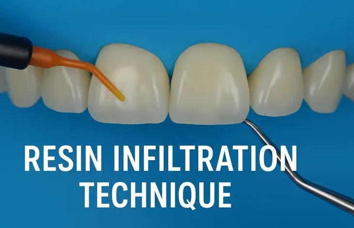
The pursuit of minimally invasive dentistry has reshaped modern clinical practice. As the profession shifts from a “drill and fill” approach toward preventive and micro-invasive strategies, new materials and techniques have emerged to treat early carious lesions while preserving sound tooth structure. One such innovation is the resin infiltration technique (RIT), a procedure that fills the gap between preventive non-operative care and conventional restorative treatment.
Resin infiltration is particularly effective in managing non-cavitated proximal caries and white spot lesions (WSLs), which traditionally present diagnostic and therapeutic challenges. It provides a method of sealing the porous enamel structure with a low-viscosity resin that penetrates the lesion body, halting demineralization and improving esthetics without mechanical removal of tissue.
Table of Contents
ToggleHistorical Background
Historically, the management of early carious lesions involved either remineralization strategies (fluoride, casein phosphopeptide-amorphous calcium phosphate, etc.) or surgical removal followed by restoration. Proximal early lesions, often detected radiographically, presented a dilemma: non-surgical therapies could slow but not always arrest progression, while restorative treatment often required significant loss of tooth structure.
In 2009, the commercial introduction of Icon® (DMG, Hamburg, Germany) marked the first standardized resin infiltration system. It was designed to treat incipient proximal and smooth-surface lesions, especially those confined to the enamel or reaching the outer third of dentin. This technique represented a paradigm shift: instead of waiting for progression or surgically intervening, clinicians could now arrest lesions micro-invasively.
Theoretical Basis
Caries Progression in Enamel
Enamel caries begins with subsurface demineralization beneath an intact surface layer. This results in increased porosity within the lesion body. Over time, without intervention, the lesion may progress into dentin, necessitating restorative care.
Principle of Resin Infiltration
The resin infiltration technique works on three principles:
- Capillary Action: A low-viscosity resin penetrates the porous lesion body via capillary forces.
- Diffusion Barrier: Once polymerized, the infiltrant occludes micro-porosities, reducing diffusion pathways for acids and dissolved minerals.
- Refractive Index Matching: By filling the porosities, the resin alters light scattering within enamel, improving esthetics (e.g., masking white spot lesions).
Materials Used
The primary commercially available product is Icon®, which includes:
- Icon-Etch (15% HCl): Removes the pseudo-intact surface layer to expose lesion porosities.
- Icon-Dry (99% ethanol): Desiccates the lesion, enabling better resin penetration.
- Icon-Infiltrant: A low-viscosity, unfilled, light-curable resin (triethylene glycol dimethacrylate, TEGDMA) that infiltrates the lesion.
Indications
1. Proximal Non-Cavitated Carious Lesions
- Lesions extending into the enamel or outer third of dentin.
- Detected radiographically or clinically.
2. Smooth-Surface White Spot Lesions (WSLs)
- Post-orthodontic decalcifications.
- Developmental defects (mild fluorosis, hypomineralization).
3. Esthetic Applications
- Masking enamel opacities.
- Improving surface translucency.
Contraindications
- Cavitated lesions.
- Lesions extending into the inner third of dentin.
- Poor moisture control (saliva contamination).
- Patients with very high caries risk requiring broader preventive management.
Step-by-Step Clinical Procedure
1. Proximal Lesions
- Isolate with rubber dam or retraction aids.
- Place wedge or separator to improve access.
- Apply Icon-Etch (15% HCl) for 2 minutes to erode the surface layer.
- Rinse thoroughly and dry.
- Apply Icon-Dry (ethanol) for 30 seconds; assess lesion penetration visually.
- Apply Icon-Infiltrant resin and allow 3 minutes for penetration.
- Remove excess resin and light cure.
- Repeat resin application for 1 minute, then cure again.
2. Smooth-Surface Lesions (e.g., White Spots)
- Clean and isolate the tooth.
- Apply Icon-Etch for 2 minutes; repeat up to 3 times if necessary.
- Rinse and dry with Icon-Dry.
- Apply Icon-Infiltrant, allow penetration, then light cure.
- Repeat resin application and cure.
- Polish to enhance smoothness and gloss.
Advantages
- Minimally Invasive: Preserves healthy tooth structure.
- Arrests Progression: Creates a barrier to acid diffusion.
- Immediate Esthetic Improvement: Masks white spot lesions effectively.
- Painless: No drilling, anesthesia, or postoperative sensitivity.
- Single-Visit Procedure: Quick and efficient.
- Cost-Effective: Less expensive than restorative treatment in the long run.
Limitations
- Technique-sensitive (requires excellent isolation).
- Limited to non-cavitated lesions.
- Long-term color stability is still under investigation.
- Does not restore lost tooth structure, only arrests progression.
- May require repeated etching for deeper or more resistant lesions.
Clinical Evidence
Numerous studies have evaluated resin infiltration’s effectiveness:
- Caries Progression Arrest: Randomized controlled trials show significant reduction in lesion progression compared to fluoride varnish or sealants.
- Esthetic Outcomes: Multiple studies confirm improved esthetics in WSLs after orthodontic treatment, with results often visible immediately.
- Longevity: Follow-up studies (up to 7 years) suggest stable arrest of caries and sustained esthetic improvement, though slight color regression may occur.
- Comparison with Fluoride Therapy: Infiltration shows superior results in halting lesion progression when compared with fluoride varnish alone.
Application in Orthodontics
White spot lesions are a common complication of fixed orthodontic appliances, with prevalence rates between 25–45%. Resin infiltration offers:
- Immediate esthetic improvement.
- Better patient satisfaction compared to bleaching or remineralization agents.
- Potential to reduce long-term restorative needs.
Esthetic Dentistry Applications
Resin infiltration is gaining popularity for cosmetic enhancement:
- Mild Fluorosis: By blending opacities with surrounding enamel.
- Hypomineralization Lesions: Particularly in anterior teeth, improving confidence and smile esthetics.
- Alternative to Microabrasion: Less aggressive, avoids unnecessary enamel removal.
Future Directions
- Improved Materials: Research into infiltrants with enhanced penetration, antibacterial activity, and color stability.
- Digital Integration: Incorporating AI-based radiographic analysis to detect candidate lesions early.
- Combination Therapies: Using infiltration with remineralization agents or bioactive materials for synergistic effects.
- Broader Esthetic Applications: Expanding use in cosmetic dentistry for generalized opacities and developmental defects.
Conclusion
The resin infiltration technique is a revolutionary tool in modern minimally invasive dentistry. It bridges the gap between preventive and restorative care by halting lesion progression and improving esthetics without sacrificing sound enamel. Clinical evidence supports its efficacy in both caries management and cosmetic dentistry, particularly for orthodontic-induced white spot lesions.
While not without limitations, resin infiltration represents a significant advancement, aligning with dentistry’s overarching goal of preserving natural tooth structure and enhancing patient quality of life. With ongoing innovation and broader adoption, it is poised to become a standard of care in managing early enamel lesions.
References
- Kielbassa, A. M., Müller, J., & Gernhardt, C. R. (2009). Closing the gap between oral hygiene and minimally invasive dentistry: A review on the resin infiltration technique of incipient (proximal) enamel lesions. Quintessence International, 40(8), 663–681.
- Paris, S., & Meyer-Lueckel, H. (2009). Inhibition of caries progression by resin infiltration in situ. Caries Research, 43(5), 383–388.
- Meyer-Lueckel, H., & Paris, S. (2010). Improvement of resin infiltration technique with ethanol drying. Journal of Dental Research, 89(10), 1068–1072.
- Paris, S., Hopfenmuller, W., & Meyer-Lueckel, H. (2010). Resin infiltration of caries lesions: An efficacy randomized trial. Journal of Dental Research, 89(8), 823–826.
- Ekstrand, K. R., & Martignon, S. (2013). Resin infiltration for proximal caries lesions: Clinical and radiographic evaluation after 1 year. Caries Research, 47(4), 364–372.
- Kim, S., Kim, E. Y., Jeong, T. S., & Kim, J. W. (2011). The evaluation of resin infiltration for masking labial enamel white spot lesions. International Journal of Pediatric Dentistry, 21(4), 241–248.
- Knösel, M., Eckstein, A., & Helms, H. J. (2013). Durability of esthetic improvement following Icon® resin infiltration of multibracket-induced white spot lesions compared with no therapy over 6 months: A randomized clinical trial. American Journal of Orthodontics and Dentofacial Orthopedics, 144(1), 86–96.
- Yetkiner, E., Wegehaupt, F. J., & Attin, T. (2015). The effect of different pretreatment methods on resin infiltration of white spot lesions. Clinical Oral Investigations, 19(9), 2203–2209.
- Meyer-Lueckel, H., Paris, S., & Kielbassa, A. M. (2007). Surface layer erosion of natural carious lesions with hydrochloric acid gels in preparation for resin infiltration. Caries Research, 41(3), 223–230.
- Borges, A. B., Caneppele, T. M. F., Masterson, D., & Maia, L. C. (2017). Effectiveness of resin infiltration on caries progression in proximal lesions: A systematic review and meta-analysis. International Journal of Paediatric Dentistry, 27(4), 277–285.
- Cazzolla, A. P., et al. (2021). Resin infiltration in enamel white spot lesions: A systematic review and meta-analysis. Journal of Esthetic and Restorative Dentistry, 33(5), 712–722.
- Son, J., Kim, J., Lee, S., & Kang, M. (2022). Comparison of resin infiltration and remineralization treatments for post-orthodontic white spot lesions: A randomized clinical trial. Scientific Reports, 12, 8139.
- Paris, S., Bitter, K., & Meyer-Lueckel, H. (2020). Resin infiltration of caries lesions: An updated systematic review and meta-analysis. Journal of Dentistry, 96, 103326.
- Albelasy, N. M., & Zidan, A. Z. (2022). Resin infiltration as a micro-invasive technique for enamel caries: Current concepts and clinical relevance. Journal of Conservative Dentistry, 25(3), 227–235.
- Langer, M., Paris, S., & Meyer-Lueckel, H. (2018). Resin infiltration of naturally stained enamel lesions: Clinical evaluation after 1 year. Clinical Oral Investigations, 22(2), 701–708.

