
The removal of third molars, commonly referred to as wisdom teeth, is one of the most frequently performed procedures in oral and maxillofacial surgery and general dental practice. Despite its routine nature, third molar surgery remains a complex and sometimes controversial topic due to variations in indications, surgical difficulty, patient morbidity, and evolving clinical guidelines. Not all third molars require removal, and inappropriate extraction can expose patients to unnecessary risks, while delayed or missed intervention can lead to significant pathology.
Table of Contents
ToggleBackground and Clinical Guidelines
Historically, third molars were frequently removed prophylactically, often during adolescence or early adulthood. However, modern guidelines—most notably those from the National Institute for Health and Care Excellence (NICE)—recommend a more conservative approach. Current guidance advises against the routine prophylactic removal of asymptomatic, pathology-free impacted third molars.
The rationale for this shift includes:
- Recognition that many third molars remain asymptomatic for life
- Awareness of surgical risks such as nerve injury and infection
- Improved understanding of long-term outcomes
Despite guidelines, significant variation in clinical practice persists. In the United Kingdom, the absolute number of third molar extractions has not markedly declined, but the average age at which patients undergo surgery has increased. This trend has important implications for surgical difficulty and complication rates, as bone quality and healing potential change with age.
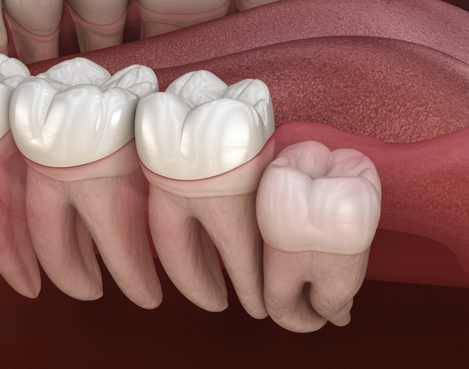
Aetiology of Third Molar Impaction
Third molars are the last teeth to erupt, usually between the ages of 17 and 25. Their delayed eruption places them at high risk of impaction, particularly in modern human populations.
Key Aetiological Factors
- Jaw–Tooth Size Discrepancy
Inherited mismatch between tooth size and jaw length is one of the most significant contributors. When the mandibular or maxillary arch lacks sufficient space, the third molars fail to erupt normally. - Dietary Influences
A soft Western diet reduces occlusal and interproximal wear. Historically, tougher diets produced natural abrasion at contact points, gradually creating space in the dental arch. The absence of this mechanism contributes to crowding. - Evolutionary Changes
Anthropological evidence suggests a gradual reduction in jaw size over evolutionary time, while tooth size has remained relatively constant. This evolutionary lag predisposes modern humans to third molar impaction. - Local Mechanical Factors
Abnormal angulation, early loss of adjacent teeth, or dense overlying bone can physically obstruct eruption.
Clinical Symptoms Associated with Third Molars
Not all impacted third molars produce symptoms. However, when pathology develops, it is often recurrent and progressive.
Common Symptoms
- Pain, often intermittent initially, becoming persistent with infection
- Swelling of the gingiva or face
- Pericoronitis, the most common presentation
- Trismus, due to inflammation of masticatory muscles
- Foul taste or halitosis, caused by purulent discharge
Complications and Associated Pathology
- Dento-facial infections that may spread to fascial spaces
- Caries of the third molar or distal surface of the second molar
- External or internal resorption of the adjacent second molar
- Cystic change, particularly dentigerous cysts
- Periodontal disease distal to the second molar
Importantly, third molars completely covered by bone are unlikely to become infected. In contrast, partially erupted teeth with communication to the oral cavity are highly susceptible to recurrent infection. In older patients, reduced vascularity of bone impairs immune response and healing, making infections more difficult to manage.
Indications for Removal
The decision to remove a third molar should be based on clear clinical or radiographic pathology rather than prophylaxis alone.
Accepted Indications
- Recurrent or severe pericoronitis
- Unrestorable caries involving the third molar or second molar
- Resorption of adjacent teeth
- Cystic or neoplastic change
- Periodontal disease affecting the distal aspect of the second molar
- Fracture management or orthognathic considerations (less common)
Non-Indications
- Prevention of dental crowding alone
- Vague or non-specific temporomandibular joint pain
- Asymptomatic, pathology-free impacted third molars
Understanding these distinctions is essential to avoid overtreatment and medico-legal risk.
Timing of Third Molar Removal
The timing of extraction plays a crucial role in surgical difficulty and patient outcomes.
Optimal Age Range
Symptoms most commonly occur in the late teens and early twenties. At this stage:
- Bone is softer, more elastic, and less mineralized
- Roots are often incompletely formed
- Healing potential is high
- Complication rates are lower
For these reasons, when extraction is indicated, early adulthood is often the most favourable time.
Prophylactic Removal
Routine prophylactic removal of asymptomatic third molars is currently difficult to justify based on available evidence. Long-term studies and systematic reviews, including Cochrane analyses, have not demonstrated sufficient benefit to outweigh surgical risks in symptom-free patients.
Choice of Anaesthesia
The choice of anaesthetic modality should be individualized based on surgical complexity, patient factors, and operator experience.
Local Anaesthesia (LA)
- Suitable for most uncomplicated unilateral impactions
- Often combined with vasoconstrictors to improve haemostasis
- May be supplemented with oral or intravenous sedation
Sedation
- Appropriate for anxious patients or moderately complex procedures
- Allows cooperation while reducing psychological stress
General Anaesthesia (GA)
Indicated for:
Bilateral impacted third molars
Technically difficult extractions (e.g. disto-angular impactions)
Patients with special needs
Requires careful patient selection and hospital facilities
A thorough medical history is essential to identify contraindications to sedation or GA.
Pre-Operative Assessment
Effective third molar surgery begins with meticulous assessment.
Clinical Examination
- Assess eruption status: unerupted, partially erupted, or fully erupted
- Evaluate soft tissue condition and signs of infection
- Note mouth opening, gag reflex, and patient cooperation
Radiographic Assessment
A dental panoramic tomograph (DPT) is the imaging modality of choice.
Key features to assess include:
Angulation
Vertical
Mesio-angular
Disto-angular
Horizontal
Transverse
Depth of Impaction
Relationship of crown to alveolar crest
Degree of Impaction
Against bone, second molar, or ramus
Root Morphology
Number, curvature, divergence, fusion
Bone Density
Inferior Dental (ID) Canal Relationship
Proximity, overlap, diversion, or narrowing
High-risk signs may necessitate further imaging (e.g. CBCT)
Associated Pathology
Cysts, caries, resorption, or infection
Surgical Planning and Path of Withdrawal
Successful third molar removal depends on understanding biomechanics and minimizing trauma.
Path of Withdrawal
The natural path of withdrawal is often obstructed by:
- Overlying bone
- Adjacent second molar
- Tooth angulation
Attempting to remove the tooth without addressing these obstacles increases the risk of:
- Excessive force
- Fracture
- Nerve injury
Bone Removal and Tooth Sectioning
Key planning considerations include:
- How much bone to remove to allow elevator placement
- Whether sectioning the crown or roots will reduce trauma
- Direction of sectioning to protect the inferior dental and lingual nerves
Rotational movements may assist disimpaction, but compensatory movements can place the nerve at risk if the tooth is not appropriately sectioned.
Patient Information and Consent
Informed consent is both an ethical and legal requirement.
Patients should be warned about:
- Post-operative pain and swelling
- Trismus
- Infection
- Dry socket
- Temporary or permanent damage to the inferior dental or lingual nerves
- Bleeding and delayed healing
Clear communication improves patient cooperation, satisfaction, and medico-legal protection.
Conclusion
Third molar removal is a cornerstone of dento-alveolar surgery that requires careful case selection, thorough assessment, and meticulous planning. Modern practice emphasizes conservative management, evidence-based indications, and individualized care. While many third molars never require removal, timely intervention for pathological teeth can prevent significant morbidity.
For dental students and clinicians alike, understanding the biological, anatomical, and surgical principles underlying third molar management is essential for safe and effective practice. As guidelines continue to evolve, ongoing education and critical appraisal of evidence remain vital.
References
- National Institute for Health and Care Excellence (NICE).
Guidance on the Extraction of Wisdom Teeth (TA1).
London: NICE; 2000. - Hay DM.
Third molar surgery: variation in clinical decision-making and practice.
Australian Dental Journal. 2018;63(4):519–525.
doi:10.1111/adj.12620 - McArdle M, Renton T.
The effects of NICE guidelines on the management of third molar teeth.
British Dental Journal. 2012;213:E9.
doi:10.1038/sj.bdj.2012.806 - Mettes TG, Nienhuijs ME, van der Sanden WJ, Verdonschot EH, Plasschaert AJ.
Interventions for treating asymptomatic impacted wisdom teeth in adolescents and adults.
Cochrane Database of Systematic Reviews. 2012;Issue 6:CD003879.
doi:10.1002/14651858.CD003879.pub3 - Peterson LJ, Ellis E, Hupp JR, Tucker MR.
Contemporary Oral and Maxillofacial Surgery.
6th ed. St. Louis: Mosby Elsevier; 2014. - Hupp JR, Ellis E, Tucker MR.
Contemporary Oral and Maxillofacial Surgery.
7th ed. Philadelphia: Elsevier; 2019. - Renton T.
Prevention of iatrogenic inferior alveolar nerve injuries in relation to third molar surgery.
British Dental Journal. 2010;209(10):481–486.
doi:10.1038/sj.bdj.2010.978 - Bataineh AB.
Sensory nerve impairment following mandibular third molar surgery.
Journal of Oral and Maxillofacial Surgery. 2001;59(9):1012–1017. - Chiapasco M, De Cicco L, Marrone G.
Side effects and complications associated with third molar surgery.
Oral Surgery, Oral Medicine, Oral Pathology. 1993;76(4):412–420. - Fielding AF, Whitley D.
Pathological sequelae of impacted third molars.
Oral Surgery, Oral Medicine, Oral Pathology. 1988;65(3):265–271.