
Successful complete denture fabrication begins with an accurate and logical approach to recording the patient’s maxillomandibular relationship. For edentulous patients, whose natural tooth guidance is absent, the dentist must rely on anatomical landmarks, clinical judgement, and systematic procedures to establish proper occlusion, aesthetics, comfort, and function. Poorly recorded occlusion affects every stage downstream—from tooth arrangement to the final prosthesis—and contributes significantly to patient dissatisfaction.
Table of Contents
ToggleWhy Occlusal Records Matter in Complete Dentures
In natural dentition, teeth provide proprioception, occlusal stops, and cues that help the mandible find a stable position. Once the teeth are lost, this sensory input disappears. The dentist must compensate by creating occlusion based on repeatable jaw relationships and functional anatomy.
Accurate occlusal records affect:
- Retention and stability of the dentures
- Chewing efficiency
- Speech and phonetics
- Facial esthetics and lip support
- Temporomandibular joint (TMJ) comfort
- Patient confidence and satisfaction
Errors in vertical dimension, occlusal plane, or mandibular position often become amplified during tooth setup or fabrication of the final prosthesis. Thus, achieving correct occlusal records during the record block stage is arguably one of the most critical steps in complete denture construction.
Preparing the Patient and Record Bases
Before occlusal records can be taken, the record bases (also known as trial plates) must be stable, retentive, and accurately adapted to the patient’s mucosa. Poor record bases lead to errors at every stage, because unstable foundations cause shifting of the wax rims during jaw registration.
Positioning the Patient
The patient must be seated correctly before beginning measurements:
- The Frankfort plane (tragus → infraorbital rim) should be parallel to the floor.
- The head must not be tilted upward or downward, as this affects perceived lip support and vertical dimension.
- The patient should be relaxed and upright, not reclined unless absolutely necessary.
Correct posture ensures that the soft tissues, muscles, and lips assume their natural resting position—crucial for accurate measurement of facial height and tooth visibility.
Assessing the Fit of Record Bases
Ideally, the record bases should be rigid acrylic or shellac with well-adapted borders. If the bases are unstable:
- A new impression may be taken using zinc oxide eugenol (ZOE) or light-bodied elastomer while the base is in the mouth.
- This reline allows a stable platform for recording occlusion.
A clinician should never proceed with occlusal registration if the bases rock, lack retention, or distort upon removal.
Establishing the Upper Wax Rim
The upper wax rim forms the foundation for determining lip support, tooth positioning, and the orientation of the occlusal plane.
1. Adjusting the Upper Rim for Lip Support
Lip support is essential for natural facial appearance:
- Insufficient support makes the upper lip appear sunken or aged.
- Excessive support makes the lip appear tense or protrusive.
To check support:
- Ask the patient to pronounce words like “Emma,” “Pea,” or “Mee.”
- Observe the nasolabial angle and fullness of the philtrum.
- The rim should lightly contour the inner surface of the upper lip.
2. Trimming the Occlusal Plane of the Upper Rim
The occlusal plane is a crucial aesthetic and functional determinant.
Aesthetics (Anterior Height)
About 1–2 mm of wax (simulating future tooth display) should be visible below the relaxed upper lip.
This gives a youthful smile; with age, lip length increases and tooth display decreases, so individual adjustment may be necessary.
Orientation of the Occlusal Plane
The upper occlusal plane must be parallel to:
- The interpupillary line (anterior reference)
- The ala–tragus line (ala of nose → tragus of ear) (posterior reference)
These serve as external facial landmarks ensuring the teeth appear level when the patient smiles or talks.
Lingual–Palatal Position
At rest, the patient’s tongue should:
- Just touch behind the lower occlusal plane
- Not rest excessively on the rim or push it outward
This ensures the future denture has appropriate phonetic and functional positioning.
Marking Lines on the Wax Rim
Once the occlusal plane is set, mark:
- Centre line (aligned with facial midline)
- Canine lines (for width of anterior teeth)
- Smile line (for incisal edge curvature)
These guides assist the technician during tooth arrangement.
Adjusting the Lower Wax Rim
Once the upper rim is established, the lower rim must be shaped to meet it harmoniously.
1. Lip Support and Cheek Contour
The lower rim should support the lower lip without displacing it.
Incorrect shaping leads to:
- Drooping chin
- Inward collapsing lips
- Poor esthetics
2. Bucco-Lingual Positioning
Posterior segments must be positioned:
- Over the crest of the ridge for stability
- In harmony with tongue space and cheek contours
A lower rim placed too far lingually restricts the tongue; too far buccally causes cheek biting and instability.
3. Meeting the Upper Rim Evenly
The lower rim should contact the upper rim evenly in the posterior and anterior regions when the mandible is guided into retruded contact position (RCP).
Uneven contact leads to errors in vertical dimension and mandibular relationship.
Vertical Dimension: Understanding FWS and OVD
Establishing a proper vertical dimension of occlusion (OVD) is critical for function, speech, comfort, and esthetics.
Definitions
- Resting Face Height (RV or RVD): Distance between two facial points when the mandible is at rest (usually nose → chin).
- Occlusal Vertical Dimension (OVD): Distance between the same points when the teeth (or rims) are in contact.
- Freeway Space (FWS):
FWS = RVD – OVD
Typically 2–4 mm.
If OVD is too large → clicking, muscle fatigue, pain, difficulty closing lips.
If OVD is too small → aged appearance, reduced chewing efficiency, TMJ compression.
Methods of Measuring RVD
1. Willis Gauge
- Measures chin → base of nose distance.
- Quick but not highly accurate (±1 mm).
2. Spring Dividers
- Measures between facial dots on nose tip and chin.
- Very simple but unsuitable for patients with facial hair.
3. Phonetic Method
Observe speech patterns during:
- “F” and “V” sounds (evaluate incisal edge placement)
- “S” sounds (assess closest speaking space)
4. Esthetic and Facial Profile Assessment
Consider:
- Lower facial height
- Nasolabial angle
- Lip competence
- Mandibular posture
Often a combination of methods yields the most accurate result.
Recording the Horizontal Jaw Relationship (RCP)
The most reproducible mandibular position without tooth guidance is Retruded Contact Position (RCP).
RCP is a musculoskeletal position, independent of occlusal contacts.
Why RCP?
- It is repeatable.
- Less influenced by muscle tension.
- Provides a reliable reference for denture construction.
Procedure
- Stabilize record bases.
- Place occlusal registration paste or wax.
- Guide the mandible gently into RCP—do not push or force.
- Hold rims lightly until material sets.
Relationship Between RCP and ICP
- In natural dentition, Intercuspal Position (ICP) lies about 1 mm anterior to RCP.
- Some prosthodontists set dentures to allow small anterior freedom for comfort (“freedom in centric”).
Verifying Accuracy
Repeat closure several times; if contacts shift, adjust rims or remake the registration.
Selecting Artificial Teeth
Choosing the correct mould, size, and shade of artificial teeth is an important part of denture esthetics.
1. Shade Selection
Consider:
- Age
- Skin tone
- Eye colour
- Patient preference
- Lighting conditions (preferably natural light)
2. Mould and Size
Anterior teeth selection depends on:
- Facial shape (square, ovoid, tapering)
- Width between canine marks
- Vertical display needed for phonetics and smile
- Ridge resorption pattern
Posterior teeth selection focuses on:
- Function
- Masticatory efficiency
- Occlusal scheme
Low-cusped or cuspless teeth are used when:
- Ridge height is poor
- Patient has difficulty finding ICP
- Balanced occlusion is challenging
Document any disagreement between practitioner and patient.
Positioning Anterior and Posterior Artificial Teeth
The aim is to restore natural appearance and functional efficiency while respecting anatomical changes from resorption.
1. Anterior Teeth Positioning
In natural dentition:
Upper incisors are approximately 10 mm anterior to the incisive papilla.
With ridge resorption:
- The papilla appears more posterior.
- Artificial incisors must be placed labial to the ridge for lip support and esthetics.
Target esthetic features:
- Nasolabial angle ~90°
- Natural smile curvature
- Symmetry with facial midline
2. Posterior Teeth Positioning
Posterior teeth should be:
- Narrow (to reduce lateral forces and improve stability)
- Positioned centrally over the ridge
- Set within the neutral zone (balance between tongue and cheeks)
Broader teeth increase lateral forces, destabilizing dentures during function.
3. Cusped vs. Cuspless Teeth
- Cusped teeth provide better chewing efficiency but may introduce lateral forces.
- Cuspless teeth increase stability and are ideal for patients with poor ridge retention or neuromuscular control issues.
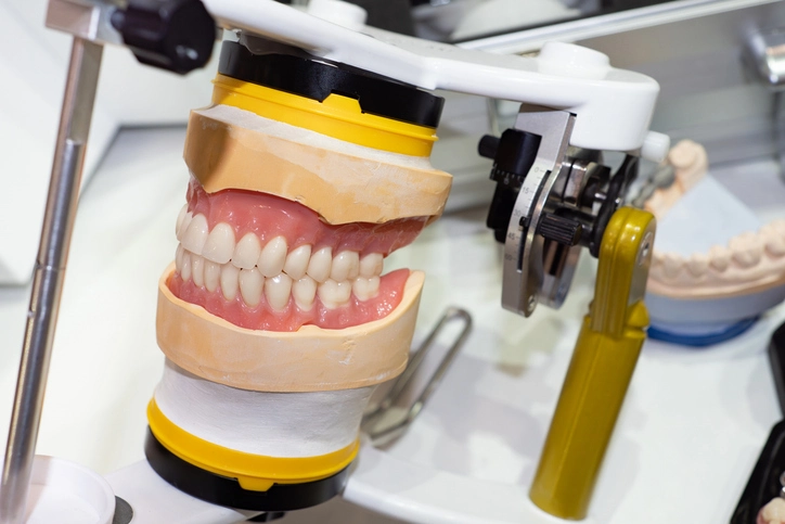
The Role of the Articulator
An articulator allows the technician to mount casts accurately and set teeth in a functional occlusal scheme.
Types of Articulators
1. Simple Hinge Articulator
- Easiest and most commonly used
- Does not simulate condylar guidance
- Often adequate because most patients adapt well
2. Average-Value Articulator
- Uses standardized averages for condylar angles
- Provides some degree of balanced articulation
- Ideal compromise between simplicity and function
3. Semi-Adjustable Articulator
- Allows individualized condylar settings
- Recommended by textbooks
- Useful for complex cases or to reduce post-delivery adjustments
Facebow Registration
A facebow record helps mount the maxillary cast in its correct spatial relationship to the hinge axis.
Advantages include:
- Better esthetic outcome
- Reduced occlusal errors
- More accurate tooth setup
However, many satisfactory dentures are made without a facebow due to patient adaptability.
Common Clinical Pitfalls
Understanding common errors helps practitioners avoid them.
1. Poorly Fitting Bases
Unstable record bases create inaccurate jaw relations.
2. Premature Contacts
Rims touching:
- Posteriorly prematurely → rim tips up anteriorly
- Anteriorly prematurely → posterior tilt up
Both distort the vertical dimension and mandibular position.
3. Incorrect Freeway Space
Too little → clicking, fatigue, discomfort
Too much → aged appearance, reduced function
4. Over-correction in Patients with Old Dentures
Patients adapt to worn dentures; drastic changes in OVD or tooth position may overwhelm their neuromuscular system.
Conclusion
Recording occlusion for complete dentures is both an art and a science. It requires understanding facial aesthetics, anatomy, mandibular dynamics, and the patient’s functional needs. Success depends on attention to detail in each stage—from trimming wax rims to capturing accurate jaw positions. With careful technique, effective communication with the technician, and consideration of patient preferences, clinicians can provide dentures that offer stability, comfort, efficient mastication, and natural appearance.
Mastering these principles ensures greater success in complete denture treatment and improves patient satisfaction significantly.
References
- Zarb, G. A., Hobkirk, J., Eckert, S., & Jacob, R. (2013). Prosthodontic Treatment for Edentulous Patients: Complete Dentures and Implant-Supported Prostheses (13th ed.). Elsevier Mosby.
- Basker, R. M., Davenport, J. C., & Thomason, J. M. (2011). Prosthetic Treatment of the Edentulous Patient (5th ed.). Wiley-Blackwell.
- McCord, J. F., & Grant, A. A. (2000). Complete Dentures: A Clinical Manual. Quintessence Publishing.
- Watt, D. M., & MacGregor, A. R. (1976). Designing Complete Dentures. Saunders.
- Boucher, C. O. (1990). Boucher’s Prosthodontic Treatment for Edentulous Patients (11th ed.). Mosby.
- McCord, J. F., & Tyson, J. (2011). “A review of techniques for recording the maxillomandibular relationship in complete denture construction.” Journal of Prosthetic Dentistry, 106(3), 153–160.
- Atwood, D. A. (1971). “Reduction of residual ridges: a major oral disease entity.” Journal of Prosthetic Dentistry, 26(3), 266–279.
- Silverman, M. M. (1951). “The speaking method in measuring vertical dimension.” Journal of Prosthetic Dentistry, 1(4), 464–471.
- Tallgren, A. (1957). “Changes in adult facial structure as related to aging, wear and loss of teeth.” American Journal of Physical Anthropology, 15(1), 265–275.
- Hickey, J. C., & Zarb, G. A. (1969). “Current concepts in occlusal rehabilitation.” Journal of Prosthetic Dentistry, 22(4), 324–336.
- The British Society for the Study of Prosthetic Dentistry (BSSPD). Guidelines for the Use of Complete Dentures.
- Academy of Prosthodontics. The Glossary of Prosthodontic Terms (GPT-9), 2017.
- National Institute for Health and Care Excellence (NICE). Guidance on Oral Health and Prosthodontic Rehabilitation.
- Jacob, R. F. (2016). “The functional denture patient: A holistic approach.” Dentistry Today, 35(4), 112–118.
- Rahn, A. O., Ivanhoe, J. R., & Plummer, K. D. (2009). Textbook of Complete Dentures. PMPH USA.