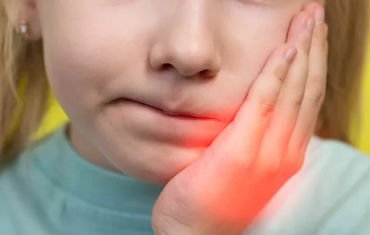
Pulpitis, an inflammation of the dental pulp tissue, is a common and complex clinical entity that presents significant diagnostic and therapeutic challenges in dentistry. The dental pulp is a vascular, nerve-rich soft tissue located within the center of the tooth, providing sensation and nourishment to the surrounding hard tissues. Pulpitis can lead to pain, infection, and tooth loss if untreated. A deep understanding of the pathophysiology, clinical presentation, diagnostic modalities, and treatment options is essential for dental professionals managing patients with pulpitis.
This article delves into every aspect of pulpitis, from its etiology to clinical management, aiming to equip professionals with comprehensive insights to optimize patient outcomes.
Table of Contents
ToggleAnatomy of the Dental Pulp
The dental pulp is the innermost part of the tooth, residing in the pulp chamber and root canals, and is enveloped by dentin. This soft tissue comprises cells, extracellular matrix, nerves, and blood vessels. Major cell types include odontoblasts, fibroblasts, macrophages, and stem cells, each playing distinct roles in tooth vitality. Odontoblasts, for instance, line the dentin-pulp interface and are essential for dentin formation, responding to damage with the production of reparative dentin.
The vascular supply of the pulp is rich, receiving blood from arterioles that enter through the apical foramen. The nerve supply, largely derived from the trigeminal nerve, provides sensation, including pain, and serves as a conduit for neural responses in inflammation.
Etiology and Pathophysiology of Pulpitis
Pulpitis primarily arises due to infection, trauma, or a combination of both. The primary etiological factors include:
- Microbial Infection
- Mechanical Trauma
- Chemical Irritants
- Thermal Insults
Microbial Infection
Dental caries, the most common cause, provides a pathway for bacterial invasion into the pulp. As bacteria penetrate dentinal tubules, they release toxins that trigger an inflammatory response.
Mechanical Trauma
Trauma from dental procedures (e.g., cavity preparation, orthodontic forces) can lead to pulp inflammation, especially when excessive force or inadequate cooling is applied.
Chemical Irritants
Restorative materials and dental bleaching agents can lead to chemical insult if not adequately isolated.
Thermal Insults
Heat generated from procedures, such as high-speed drilling without water cooling, may damage the pulp tissue.
When these factors compromise the integrity of the dentin-pulp complex, bacteria and toxins reach the pulp, leading to an inflammatory response. This response initiates an immune cascade, releasing mediators like histamines, prostaglandins, and cytokines, which result in vasodilation and increased vascular permeability. The confined pulp cavity does not permit tissue expansion, leading to increased intrapulpal pressure, compression of blood vessels, and ischemia, further exacerbating tissue injury.
Classification of Pulpitis
Pulpitis is broadly classified into two main categories based on its clinical presentation and histopathological features:
- Reversible Pulpitis
- Irreversible Pulpitis
Reversible Pulpitis
Characterized by mild inflammation, reversible pulpitis is a transient condition where the pulp retains its vitality. Symptoms include mild pain or sensitivity to stimuli (e.g., cold, sweets), which subsides upon removal of the irritant.
Irreversible Pulpitis
Marked by significant, often irreversible damage to the pulp, this condition results in severe pain that may be spontaneous, persistent, and exacerbated by thermal stimuli. Irreversible pulpitis indicates a compromised pulp with a high likelihood of necrosis if untreated.
Irreversible pulpitis can further be sub-classified into:
- Acute Pulpitis: Intense, throbbing pain that can be challenging to localize, often without visible external swelling.
- Chronic Pulpitis: A lower-grade, longstanding inflammation that may present with intermittent pain, particularly upon biting or consuming hot or cold foods.
Clinical Presentation and Diagnosis
Diagnosing pulpitis involves evaluating the patient’s symptoms, performing clinical tests, and using diagnostic tools. A thorough examination includes:
- Patient History
- Clinical Examination
- Advanced Imaging
Patient History
Understanding the patient’s pain characteristics (e.g., duration, intensity, triggers, and relief factors) is crucial. Reversible pulpitis typically presents with short-lasting pain, while irreversible pulpitis often involves prolonged, spontaneous pain.
Clinical Examination
Checking for visible signs of caries, restorations, or fractures, as well as assessing the periodontal status, helps identify potential causative factors.
Diagnostic Tests:
- Thermal Testing: Cold testing (e.g., with endo-ice) can elicit a sharp pain in reversible pulpitis, whereas irreversible pulpitis may cause prolonged or intense pain.
- Electric Pulp Testing (EPT): Used to assess pulp vitality, EPT may help confirm pulpal health in borderline cases, though it has limitations in very young or heavily restored teeth.
- Percussion and Palpation: Tenderness on percussion or palpation often indicates periapical involvement and is typically associated with irreversible pulpitis or apical periodontitis.
- Radiographic Examination: While pulpitis itself does not produce radiographic changes, radiographs are vital in detecting carious lesions, periapical pathologies, or fractures.
Advanced Imaging
Cone-beam computed tomography (CBCT) offers three-dimensional views, assisting in the evaluation of complex cases, especially in the presence of periapical pathology or in assessing tooth fractures.
Differential Diagnosis
Pulpitis must be differentiated from other causes of dental pain, such as:
- Periapical Abscess: A periapical abscess typically presents with localized pain, swelling, and is often associated with a necrotic pulp.
- Cracked Tooth Syndrome: Characterized by sharp pain upon biting, cracked tooth syndrome may mimic pulpitis but usually lacks the thermal sensitivity.
- Trigeminal Neuralgia: Distinguished by episodic, electric-shock-like pain, trigeminal neuralgia affects multiple teeth and is triggered by even light touch, differentiating it from pulpitis.
Accurate diagnosis is essential, as treatment varies significantly among these conditions.
Treatment of Pulpitis
Management of pulpitis depends on whether it is reversible or irreversible:
Reversible Pulpitis
Treatment aims to remove the irritant, allowing the pulp to heal. Steps include:
- Removal of Caries: In cases where decay is present, conservative removal followed by restoration with materials that provide a proper seal and adequate protection is essential.
- Desensitizing Agents: Application of desensitizing agents may provide relief for patients with mild symptoms.
- Indirect Pulp Capping: In cases where caries removal is close to the pulp but does not expose it, pulp capping by applying a protective liner (e.g., calcium hydroxide or mineral trioxide aggregate) can facilitate dentin repair and protect the pulp.
Irreversible Pulpitis
Irreversible pulpitis necessitates the removal of inflamed pulp tissue to alleviate symptoms and prevent further complications. Treatment options include:
- Pulpectomy: Removing the entire pulp tissue, commonly used in emergency settings to relieve pain, is followed by either root canal therapy or extraction.
- Root Canal Therapy (RCT): Root canal treatment is the standard treatment for irreversible pulpitis in patients wishing to retain the tooth. The process involves cleaning and shaping the canals, disinfecting, and then filling them with an inert material.
- Extraction: In cases where the tooth is non-restorable or the patient opts for tooth removal, extraction is the only option. This is typically followed by a discussion on replacement options.
Medications in Pulpitis Management
While antibiotics are often overprescribed, they are not indicated for pulpitis unless there is systemic involvement or significant infection. Pain management includes non-steroidal anti-inflammatory drugs (NSAIDs) like ibuprofen, which reduce inflammation and alleviate pain.
Complications of Pulpitis
Untreated pulpitis can lead to various complications:
- Pulp Necrosis
- Apical Periodontitis
- Systemic Complications
Pulp Necrosis
Continued inflammation and ischemia can cause pulp death, leading to necrosis and infection spread beyond the tooth.
Apical Periodontitis
Bacterial invasion into periapical tissues can result in periapical granuloma, cyst formation, or abscess.
Systemic Complications
Though rare, untreated pulpitis can cause cellulitis, Ludwig’s angina, or even systemic spread, requiring prompt intervention.
Prevention of Pulpitis
Preventative strategies include:
- Oral Hygiene Education: Promoting effective oral hygiene practices reduces the risk of caries and pulpitis.
- Regular Dental Check-ups: Early detection of caries and restoration failures enables timely intervention.
- Protective Measures during Dental Procedures: Using adequate water cooling, protective liners, and ensuring minimal invasive techniques reduce pulpal damage risks.
Emerging Technologies and Future Directions
Advances in dental technology and materials hold promise for improving pulpitis management:
- Biomaterials
- Minimally Invasive Techniques
- Regenerative Endodontics
Biomaterials
Novel pulp-capping materials like bioactive glass and stem cell therapies are showing potential in enhancing pulp tissue regeneration.
Minimally Invasive Techniques
Techniques like laser therapy and photobiomodulation have emerged as potential adjuncts to traditional treatments.
Regenerative Endodontics
Regenerative dentistry focuses on revitalizing the pulp tissue through the use of stem cells, growth factors, and scaffolds, potentially offering a biological alternative to conventional root canal therapy.
Frequently Asked Questions (FAQs)
What is pulpitis?
Pulpitis is the inflammation of the dental pulp, a soft tissue inside the tooth that contains nerves, blood vessels, and connective tissues. It can be reversible or irreversible, depending on the extent of the damage.
What causes pulpitis?
Pulpitis is primarily caused by bacterial infection due to deep dental caries, but it can also result from trauma, chemical irritation from restorative materials, excessive heat from dental procedures, and repeated dental interventions.
What is the difference between reversible and irreversible pulpitis?
- Reversible pulpitis: The pulp is inflamed but can heal if the irritant (e.g., caries) is removed. Pain is usually short-lived and triggered by stimuli like cold or sweets.
- Irreversible pulpitis: The inflammation is severe and leads to pulp necrosis. Pain may be spontaneous, prolonged, and exacerbated by hot stimuli.
How is pulpitis diagnosed?
Diagnosis involves patient history, clinical examination, and special tests such as:
- Thermal tests (cold/hot sensitivity tests)
- Electric Pulp Testing (EPT)
- Percussion and palpation tests
- Radiographic imaging (X-rays or CBCT for advanced cases)
Can pulpitis heal on its own?
Reversible pulpitis can heal if the underlying cause (e.g., caries, trauma) is treated early. However, irreversible pulpitis cannot heal on its own and requires root canal treatment or extraction.
What are the treatment options for pulpitis?
- For reversible pulpitis: Removing the cause (e.g., caries removal, protective restorations, desensitizing agents) can help the pulp recover.
- For irreversible pulpitis: Root canal therapy (RCT) is required to remove the inflamed pulp and disinfect the root canals. In some cases, tooth extraction may be necessary.
Does pulpitis always require a root canal?
No. Reversible pulpitis can be managed with conservative treatments, but irreversible pulpitis requires a root canal to save the tooth or extraction if the tooth is non-restorable.
Can antibiotics treat pulpitis?
Antibiotics are not effective for pulpitis unless there is a spreading infection (e.g., cellulitis, systemic symptoms). Pain management usually involves NSAIDs like ibuprofen.
How can pulpitis be prevented?
- Maintain good oral hygiene (brushing, flossing, fluoride use)
- Regular dental check-ups to detect early caries
- Using protective barriers during dental procedures to minimize pulp irritation
- Avoiding excessive force and temperature changes in dental treatments
What happens if pulpitis is left untreated?
Untreated pulpitis can progress to pulp necrosis, leading to periapical abscess, apical periodontitis, or systemic complications such as cellulitis or Ludwig’s angina.
Can pulpitis cause facial swelling?
Yes, if pulpitis progresses to pulp necrosis and leads to a dental abscess, it can cause swelling in the face and jaw, requiring urgent treatment.
Can a cracked tooth lead to pulpitis?
Yes. A cracked tooth can expose the dentin and pulp to bacteria, leading to pulp inflammation and potential infection.
Is pain from pulpitis always severe?
No. Early-stage reversible pulpitis may cause mild discomfort, while irreversible pulpitis often presents with severe, throbbing pain.
Can stress cause pulpitis?
Stress itself does not cause pulpitis, but habits like bruxism (teeth grinding) can lead to microfractures and irritation, making the pulp susceptible to inflammation.
What are the latest advancements in pulpitis treatment?
- Regenerative endodontics: Using stem cells and biomaterials to restore pulp vitality.
- Bioceramic sealers: More biocompatible and effective root canal filling materials.
- Laser therapy: Emerging as an adjunct for pulp capping and disinfection.
Conclusion
Pulpitis is a multi-faceted dental condition with significant implications for oral and systemic health. For dental professionals, understanding the pathophysiology, clinical presentation, diagnostic tools, and appropriate management is crucial. Advances in diagnostic imaging, restorative materials, and regenerative endodontics are transforming pulpitis management, potentially offering patients enhanced outcomes. By embracing both traditional and emerging techniques, dental professionals can better address the challenges of pulpitis, preserving tooth vitality whenever possible and enhancing patient quality of life.

