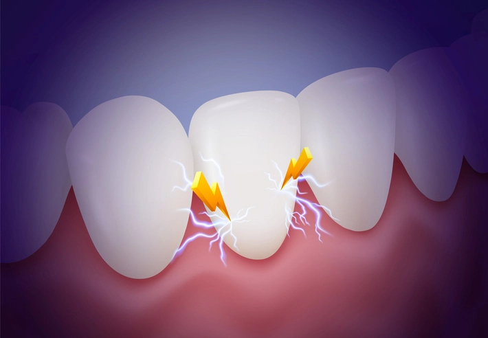
Traumatic injuries to the teeth are a major cause of pulpal and periapical pathosis, particularly in the pediatric population. Epidemiological studies estimate that up to 30% of children experience some form of dental trauma before adulthood (Andreasen & Andreasen, 2007). Such injuries may lead to various pulpal sequelae, ranging from reversible inflammation to irreversible necrosis and resorption. The long-term viability of the pulp and periodontal ligament (PDL) depends largely on the extent of injury to the apical neurovascular supply and the body’s ability to initiate repair.
The pulp–dentine complex, though enclosed within rigid dentinal walls, possesses a remarkable capacity for healing. However, the closed environment also makes it highly susceptible to ischemic and inflammatory damage. In the immature permanent tooth, the presence of an open apex allows for greater healing potential and revascularization compared to the mature tooth, in which the apical constriction is narrow and the collateral circulation limited.
Table of Contents
TogglePathophysiology of Pulpal Injury
Disruption of Apical Vessels
One of the most critical determinants of pulpal prognosis after trauma is the extent of apical vascular disruption. Luxation and avulsion injuries often result in tearing or compression of the apical vessels, leading to ischemia of the pulp. If revascularization fails to occur within days, necrosis becomes inevitable. Immature teeth, however, demonstrate a greater capacity for revascularization due to their wide apical foramina, which facilitate re-entry of blood vessels and undifferentiated mesenchymal cells (Cvek, 1992).
Pulp Exposure and Inflammatory Strangulation
Crown fractures involving pulp exposure introduce bacterial contamination, eliciting inflammatory responses within the pulp tissue. Even in the absence of direct exposure, pulpal inflammation may arise from microcracks or dentinal tubule communication. Hemorrhage and edema within the rigid pulp chamber elevate intrapulpal pressure, compressing the apical microcirculation—a phenomenon known as inflammatory strangulation. If unrelieved, this leads to ischemic necrosis.
Diagnosis of Pulpal Status Following Trauma
Clinical and Radiographic Evaluation
Accurate diagnosis of pulpal status is fundamental to treatment planning. Immediately after trauma, vitality testing may yield false-negative results due to transient neural shock. Therefore, multiple follow-up assessments are recommended at 4–6 week intervals.
Clinical parameters include:
- Colour change: grey discoloration indicates necrosis; yellow suggests calcific metamorphosis.
- Mobility: often reflects PDL injury rather than pulpal status.
- Percussion sensitivity: may indicate periapical inflammation.
- Sinus tract formation or swelling: signs of chronic infection.
- Radiographic findings: widening of the PDL space, periapical radiolucency, or root resorption.
Pulp Sensibility and Vitality Testing
Traditional electric and thermal tests assess sensory response rather than true vitality. Laser Doppler flowmetry and pulse oximetry offer more accurate measures of pulpal blood flow but are not widely available in clinical settings. Consequently, vitality testing should be interpreted in conjunction with clinical and radiographic findings rather than in isolation.
Pulpal Necrosis and Management
Pulp Death
Pulp necrosis following trauma may result from vascular compromise or secondary bacterial infection. Once confirmed, endodontic therapy is indicated to prevent periapical pathology. In mature teeth, conventional root canal therapy (RCT) with chemo-mechanical debridement and obturation using gutta-percha is sufficient. In immature teeth, however, the absence of a natural apical constriction complicates obturation, necessitating alternative procedures such as apexification or regenerative endodontics.
Apexification and Alternative Endodontic Approaches
Traditional Calcium Hydroxide Apexification
Historically, calcium hydroxide apexification has been the standard treatment for immature non-vital teeth. The procedure involves the placement of a non-setting calcium hydroxide paste within the canal to induce the formation of a calcific apical barrier. The paste is replaced at 3-month intervals, and barrier formation typically occurs within 6–12 months (Sheehy & Roberts, 1997).
Clinical protocol:
- Access under rubber dam isolation.
- Extirpation of necrotic pulp and irrigation with sodium hypochlorite.
- Working length determination 1–2 mm short of the radiographic apex.
- Filling of the canal with calcium hydroxide paste (e.g., Hypocal™, UltraCal™).
- Radiographic monitoring every 3 months until barrier formation.
- Final obturation with gutta-percha following confirmation of closure.
Although calcium hydroxide remains effective, prolonged use may weaken dentinal walls, predisposing teeth to cervical root fractures (Andreasen et al., 2002). Therefore, shorter-term alternatives are now preferred.
Mineral Trioxide Aggregate (MTA) Apexification
Mineral Trioxide Aggregate (MTA), introduced by Torabinejad et al. (1995), has become a superior alternative for apical barrier formation. Its biocompatibility, high pH, and excellent sealing properties allow for the formation of an artificial apical stop within one or two visits.
Advantages:
- Rapid treatment (completion within 1–2 appointments).
- Reduced risk of root fracture.
- Excellent long-term success rates (>85%).
Limitations:
- Cost and handling difficulties.
- Potential for tooth discoloration with certain MTA formulations.
MTA apexification represents a paradigm shift from traditional multi-visit therapy to a more efficient and predictable single-visit approach.
Regenerative Endodontics and Revascularization
Regenerative endodontic procedures (REPs) or revascularization aim to restore pulp vitality and promote continued root development. These biologically based treatments rely on the recruitment of stem cells from the apical papilla and formation of new vital tissue within the canal.
Procedure steps:
- Disinfection using low-concentration sodium hypochlorite and antibiotic paste.
- Induction of apical bleeding to provide a scaffold of blood clot.
- Sealing the canal with MTA or bioceramic material.
- Coronal restoration with a tight seal to prevent reinfection.
Clinical studies have demonstrated significant increases in root length and wall thickness following REPs (Banchs & Trope, 2004). This approach is particularly valuable for immature teeth where preservation of structural integrity is paramount.
Root Resorption Following Trauma
Resorption refers to the pathological loss of dental hard tissue mediated by osteoclastic or odontoclastic activity. It can be internal or external, depending on the site of initiation.
Internal Root Resorption
Etiology and Pathogenesis:
Internal resorption is initiated by chronic pulpal inflammation that stimulates odontoclastic activity within the pulp chamber or canal. It typically occurs after trauma, caries, or restorative procedures that expose dentinal tubules.
Clinical and Radiographic Features:
- Pink spot discoloration due to granulation tissue.
- Radiographically, a well-defined circular radiolucency continuous with the canal walls.
- Usually asymptomatic unless perforation occurs.
Management:
Immediate root canal therapy is required to remove inflamed pulp tissue and arrest the resorptive process. Calcium hydroxide dressing assists in inactivating clastic cells, followed by obturation with thermoplasticized gutta-percha or MTA if perforation is present (Patel et al., 2018).
External Root Resorption
External resorption originates from the root surface and may be surface, inflammatory, or replacement type.
1. Surface Resorption
A self-limiting process resulting from minor PDL damage. It usually repairs spontaneously with new cementum deposition and requires no intervention.
2. Inflammatory External Resorption
Commonly associated with infected necrotic pulp and bacterial by-products penetrating through dentinal tubules. Radiographically, irregular radiolucent areas are visible along the root surface. Treatment involves endodontic therapy to eliminate the source of infection, typically with calcium hydroxide dressing to arrest clastic activity.
3. Replacement Resorption (Ankylosis)
Replacement resorption occurs when extensive PDL damage leads to direct fusion between alveolar bone and root dentine. The root is gradually replaced by bone, leading to infraocclusion, particularly in growing children. Radiographically, the PDL space disappears, and the root outline merges with the surrounding bone.
No effective curative treatment exists once ankylosis occurs. The management strategy focuses on maintaining aesthetics and alveolar bone volume until prosthetic replacement becomes feasible.
Root Canal Obliteration (Calcific Metamorphosis)
Root canal obliteration, also known as calcific metamorphosis, represents a reparative response of the pulp following trauma. The odontoblasts or odontoblast-like cells deposit tertiary dentine within the canal space, resulting in progressive narrowing or complete obliteration of the pulp chamber.
Epidemiology:
Occurs in approximately 4–35% of luxation injuries (Andreasen et al., 1987). Despite the radiographic loss of canal space, only 13–16% of affected teeth develop pulp necrosis.
Clinical Presentation:
- Yellow discoloration due to increased dentine deposition.
- Usually asymptomatic.
- Radiographs reveal partial or complete canal obliteration.
Management:
Prophylactic endodontic treatment is not recommended unless signs of periapical pathology arise. If endodontic intervention becomes necessary, magnification and CBCT imaging are invaluable for locating the obliterated canal. Success rates for subsequent RCT remain high (approximately 80%), provided that the canal can be negotiated (Malmgren et al., 2012).
Long-Term Outcomes and Prognosis
Colour Changes
Crown discoloration serves as an important indicator of pulpal response. Grey discoloration typically denotes necrosis, whereas yellow suggests calcific metamorphosis. Bleaching or veneer restoration may be considered after completion of endodontic therapy.
Root Fracture and Structural Weakness
Long-term calcium hydroxide therapy can lead to alteration of dentinal properties, predisposing to cervical root fracture. MTA-based techniques significantly reduce this risk by minimizing treatment duration.
Ankylosis and Growth Disturbance
In the mixed dentition, ankylosed teeth may interfere with alveolar growth, resulting in infraocclusion. Early recognition through regular follow-up allows timely intervention, such as decoronation to preserve alveolar ridge contour.
Preventive and Supportive Strategies
Immediate and appropriate emergency management of dental trauma is essential to reduce pulpal sequelae.
- Replantation of avulsed teeth should occur within 15–30 minutes to maximize PDL survival.
- Storage media such as milk, saliva, or Hank’s Balanced Salt Solution maintain cell viability during transport.
- Splinting: Flexible splints for 1–2 weeks promote physiologic healing of the PDL.
- Follow-up schedule: Clinical and radiographic review at 3, 6, and 12 months, and annually thereafter.
Advances in Biomaterials and Regenerative Science
The evolution of endodontic biomaterials has greatly improved outcomes in traumatized immature teeth.
- Bioceramic materials (e.g., Biodentine™, EndoSequence®) offer excellent biocompatibility and sealing with easier handling than MTA.
- Stem cell–based regenerative endodontics shows promise in restoring pulp-dentine complex functionality.
- 3D imaging (CBCT) enhances detection of early resorptive lesions and guides minimally invasive treatment.
Continued research into biomimetic materials and tissue engineering is likely to redefine the management of pulpal sequelae in the coming decade.
Conclusion
Pulpal sequelae following trauma encompass a diverse range of pathologies arising from disruption of the pulp’s vascular and neural integrity. Successful management requires a sound understanding of the biological mechanisms of injury, appropriate diagnostic tools, and tailored therapeutic interventions.
In pediatric dentistry, the preservation of pulpal vitality and continued root development are paramount goals. Calcium hydroxide and MTA apexification remain foundational techniques, while regenerative endodontics offers a biologically advanced alternative. Vigilant monitoring and timely intervention can prevent complications such as resorption and ankylosis, thereby preserving both function and aesthetics of the developing dentition.
References
- Andreasen, J. O., & Andreasen, F. M. (2007). Textbook and Color Atlas of Traumatic Injuries to the Teeth (4th ed.). Wiley-Blackwell.
- Andreasen, F. M., Farik, B., & Munksgaard, E. C. (2002). Long-term calcium hydroxide as a root canal dressing may increase risk of root fracture. Dental Traumatology, 18(3), 134–137.
- Banchs, F., & Trope, M. (2004). Revascularization of immature permanent teeth with apical periodontitis: new treatment protocol? Journal of Endodontics, 30(4), 196–200.
- Cvek, M. (1992). Prognosis of luxated non-vital maxillary incisors treated with calcium hydroxide and filled with gutta-percha. Endodontics & Dental Traumatology, 8(2), 45–55.
- Malmgren, B., Andreasen, J. O., Flores, M. T., Robertson, A., DiAngelis, A. J., Andersson, L., & Bourguignon, C. (2012). International Association of Dental Traumatology guidelines for the management of traumatic dental injuries: 3. Dental Traumatology, 28(3), 174–182.
- Patel, S., Saberi, N., & Mannocci, F. (2018). Internal root resorption: a review. International Endodontic Journal, 51(12), 1206–1228.
- Sheehy, E. C., & Roberts, G. J. (1997). Use of calcium hydroxide for apical barrier formation and healing in non-vital immature permanent teeth: a review. British Dental Journal, 183(7), 241–246.
- Torabinejad, M., Watson, T. F., & Pitt Ford, T. R. (1995). Sealing ability of a mineral trioxide aggregate when used as a root-end filling material. Journal of Endodontics, 21(3), 109–112.*

