
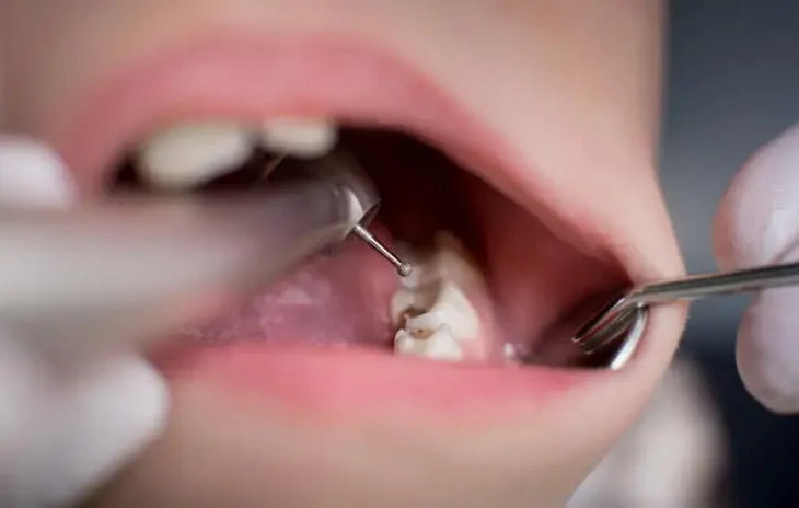
Pulp capping is a widely practiced dental procedure aimed at preserving the vitality of the tooth’s pulp. The process is employed when the dental pulp, which contains the nerve and blood supply of the tooth, is exposed or nearly exposed due to trauma, caries, or cavity preparations. This procedure is critical in preventing the need for more invasive treatments like root canal therapy (RCT), which might otherwise be required if the pulp becomes irreversibly inflamed or necrotic.
This article provides a comprehensive understanding of pulp capping, examining the procedure’s rationale, types, materials used, success rates, and potential complications. The goal is to explore the procedure’s clinical relevance and to provide insight into how advancements in dental materials and techniques have shaped the modern approach to pulp capping.
Anatomy and Physiology of the Dental Pulp
To appreciate the importance of pulp capping, it’s essential to first understand the structure and function of the dental pulp. The pulp resides within the tooth’s central chamber, commonly referred to as the pulp chamber, and extends through the root canals. It consists of nerves, blood vessels, lymphatics, and connective tissue that are responsible for:
- Nutritional supply: The pulp provides essential nutrients to the dentin, which forms the bulk of the tooth structure.
- Sensory function: The pulp contains sensory nerves that respond to temperature changes, mechanical stimuli, and chemical irritants.
- Defensive response: The pulp can initiate reparative processes, including the production of tertiary dentin, when exposed to injury or decay.
Any damage to the pulp can compromise these functions, leading to inflammation, infection, or necrosis if left untreated. Pulp capping attempts to address this by protecting and preserving the pulp’s health.
Indications for Pulp Capping
The goal is to encourage the pulp to heal itself and avoid the need for more invasive treatments like root canal therapy. There are two types of pulp capping: direct and indirect.
Here are the indications for pulp capping:
Indications for Direct Pulp Capping
Direct pulp capping is used when the pulp is exposed due to injury or decay. The goal is to cover the exposed pulp with a protective material and encourage the formation of reparative dentin. Indications include:
- Small, mechanical exposure
- Traumatic exposure
- Carious exposure in a young tooth
- Vital pulp without signs of irreversible damage
Small, mechanical exposure
This occurs when the pulp is accidentally exposed during dental procedures such as drilling. The exposure is usually clean and minimal.
Traumatic exposure
When the pulp is exposed because of a fracture or injury to the tooth (like a broken tooth from an accident).
Carious exposure in a young tooth
In young permanent teeth, where the pulp can heal more efficiently, pulp capping is an option to prevent further pulp damage.
Vital pulp without signs of irreversible damage
The pulp should show no signs of irreversible pulpitis (which is characterized by intense, spontaneous pain or an inability to respond properly to thermal testing). There should be no infection, abscess, or other indicators that the pulp is severely damaged.
Indications for Indirect Pulp Capping
Indirect pulp capping is used when the pulp is nearly exposed (but not fully) due to deep caries. Here, decayed dentin is left near the pulp, and a medicament is applied to allow healing. Indications include:
- Deep carious lesions
- Vital pulp
- No spontaneous pain
- Absence of periapical pathology
Deep carious lesions
When the caries are close to the pulp but the pulp has not been exposed. Removing all of the decay may risk exposing the pulp, so some decayed dentin is left in place.
Vital pulp
The pulp should still be vital and able to heal. Signs of irreversible pulpitis or necrosis indicate that the pulp is not capable of healing, and pulp capping would not be successful.
No spontaneous pain
The tooth should not present with spontaneous, prolonged, or severe pain. The presence of mild discomfort to stimuli like cold is acceptable, but spontaneous pain may indicate that pulp capping is not the best option.
Absence of periapical pathology
There should be no radiographic evidence of periapical (around the tooth’s root) pathology, which would indicate advanced pulp damage or infection.
Contraindications of Pulp Capping
Pulp capping is not recommended in the following scenarios:
- Irreversible pulpitis
- Necrotic pulp
- Persistent infection
Irreversible pulpitis
Once the pulp becomes inflamed beyond repair, the only solution is root canal therapy or extraction.
Necrotic pulp
If the pulp has died, pulp capping would be ineffective.
Persistent infection
Teeth with active infections should not undergo pulp capping, as this could lead to failure of the treatment and further complications.
Types of Pulp Capping
There are two primary types of pulp capping: direct and indirect. The choice between these depends on whether the pulp is exposed or only at risk of exposure.
Indirect Pulp Capping
In indirect pulp capping, the dental pulp is not yet exposed, but decay has progressed close enough to the pulp chamber that further removal of caries could lead to an exposure. The goal here is to stop the progression of decay, allowing the pulp to remain vital and stimulate the formation of tertiary (reparative) dentin. This procedure is typically performed as follows:
- Decay removal
- Material placement
- Final restoration
Decay removal
The dentist removes as much carious dentin as possible without exposing the pulp.
Material placement
A protective dressing is placed over the remaining thin layer of dentin to act as a barrier and promote healing.
Final restoration
The tooth is restored with a filling material, such as composite resin, amalgam, or a crown, depending on the extent of decay.
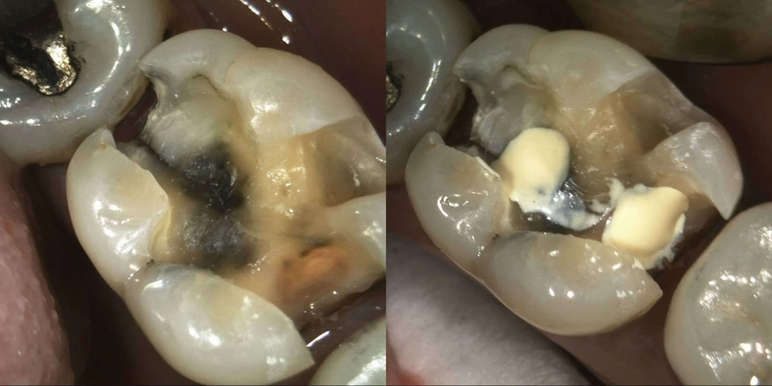
Direct Pulp Capping
Direct pulp capping is employed when the dental pulp has been directly exposed, either due to trauma, deep caries removal, or cavity preparation. In this case, the exposed pulp is protected with a biocompatible material to encourage healing and prevent infection or necrosis.
This procedure is typically performed as follows:
- Hemostasis
- Material application
- Final restoration
Hemostasis
Any bleeding from the exposed pulp is controlled to create a clean, dry environment.
Material application
A biocompatible material (usually MTA, calcium hydroxide, or newer bioceramics) is placed directly over the exposed pulp to stimulate reparative dentin formation and seal the pulp from bacterial contamination.
Final restoration
The tooth is then restored with an appropriate restorative material, such as composite resin or amalgam.
Success Rates of Pulp Capping
The success of pulp capping depends on several factors, including the material used, the condition of the pulp, and the clinician’s skill. Generally, indirect pulp capping has a higher success rate than direct pulp capping because the pulp is not directly exposed.
Indirect pulp capping
Studies have shown success rates ranging from 70% to 90%, with better outcomes in younger patients and when modern biocompatible materials are used.
Direct pulp capping
Success rates for direct pulp capping can be more variable, with reported rates ranging from 50% to 80%. MTA and other bioceramic materials tend to offer better outcomes than traditional calcium hydroxide.
Factors Affecting Success
- Pulp vitality
- Sterility
- Restorative seal
Pulp vitality
The health of the pulp at the time of the procedure is crucial. Teeth with vital, non-inflamed pulps are more likely to have successful outcomes.
Sterility
Keeping the treatment site free from contamination by saliva and bacteria is essential for preventing post-operative infections.
Restorative seal
The final restoration must provide a tight seal to prevent bacterial leakage, which could compromise the pulp and lead to failure.
Materials for Pulp Capping
The choice of material plays a significant role in the long-term success of pulp capping procedures. Over the years, several materials have been developed, each with its unique set of properties.
- Calcium Hydroxide
- Mineral Trioxide Aggregate (MTA)
- Biodentine
- Other Bioceramics
Calcium Hydroxide
Calcium hydroxide has been the traditional material for pulp capping for decades, primarily due to its ability to stimulate the formation of a dentin bridge and its antibacterial properties. However, it has several limitations, including poor sealing ability, dissolution over time, and a higher likelihood of creating a brittle dentin bridge that is prone to fractures.
Pros
- Stimulates dentin formation.
- Antibacterial properties.
- Inexpensive and easy to use.
Cons
- Can dissolve over time, leading to treatment failure.
- Provides a less reliable seal, increasing the risk of bacterial infiltration.
Mineral Trioxide Aggregate (MTA)
MTA is considered the gold standard for pulp capping in modern dentistry. It has been shown to create a superior seal, promote the formation of a more robust dentin bridge, and exhibit excellent biocompatibility.
Pros
- Promotes the formation of a high-quality dentin bridge.
- Excellent sealing properties.
- Biocompatible with the pulp and surrounding tissues.
- Stimulates a more predictable healing response.
Cons
- More expensive than calcium hydroxide.
- Longer setting time, which can be inconvenient in some clinical situations.
- Handling properties can be challenging for some practitioners.
Biodentine
Biodentine is a relatively newer material that has gained attention for its bioactivity and ease of use. It has properties similar to MTA but sets faster, making it more convenient for clinical applications.
Pros
- Similar bioactive properties to MTA.
- Faster setting time.
- Good sealing ability.
Cons
- Still relatively new, with fewer long-term studies compared to MTA and calcium hydroxide.
- More expensive than traditional materials like calcium hydroxide.
Other Bioceramics
Newer bioceramic materials are being developed with the goal of offering better sealing, biocompatibility, and handling than traditional pulp capping materials. These materials show great promise for the future of pulp therapy.
Pros
- Biocompatible and promote healing.
- Better handling properties than MTA.
- Potential for faster and more predictable results.
Cons
- High cost.
- Limited long-term clinical studies to evaluate their success compared to more established materials.
Advancements in Pulp Capping Techniques
In recent years, the field of pulp capping has evolved significantly with advancements in materials, techniques, and clinical approaches, driven by ongoing research and innovation. These developments have contributed to improved outcomes, especially in cases where the pulp was previously considered at high risk of irreversible damage. Below are some of the key advancements that have reshaped pulp capping:
- Bioactive Materials
- Guided Tissue Engineering
- Laser-Assisted Pulp Capping
- Improved Diagnostic Tools
- Enhanced Clinical Techniques
Bioactive Materials
The introduction of bioactive materials has revolutionized pulp capping. Materials like MTA, Biodentine, and other bioceramics not only protect the pulp but also encourage biological healing. These materials interact with the body’s natural processes to form a dentin bridge, providing a durable barrier between the pulp and external factors like bacteria or temperature changes.
Compared to traditional materials like calcium hydroxide, bioactive materials are more likely to create a high-quality, durable dentin bridge that is less susceptible to breakdown or fracture. They also exhibit excellent sealing capabilities, reducing the chances of microleakage and subsequent infection.
Guided Tissue Engineering
Recent advances in tissue engineering and biomaterials science offer exciting possibilities for pulp capping. Growth factors, such as bone morphogenetic proteins (BMPs) and vascular endothelial growth factor (VEGF), can be combined with pulp capping materials to stimulate more precise and controlled reparative dentin formation. These growth factors promote the regeneration of pulp tissue, potentially allowing clinicians to repair larger defects while maintaining pulp vitality.
Furthermore, stem cell therapies are being explored as a way to regenerate dental tissues, particularly in younger patients. When combined with appropriate scaffolding materials and growth factors, stem cells could provide a way to completely regenerate the damaged pulp, offering an alternative to root canal therapy.
Laser-Assisted Pulp Capping
Laser technology has also found its way into pulp capping. Low-level laser therapy (LLLT), also known as photobiomodulation, has been studied for its ability to reduce inflammation, enhance tissue healing, and promote pulpal regeneration. This technology has been used as an adjunct to traditional pulp capping materials to accelerate the healing process, particularly when pulp exposure is small and the tissue is vital.
By controlling bleeding more effectively and disinfecting the area, lasers offer a more sterile environment, reducing the risk of contamination during pulp capping procedures.
Improved Diagnostic Tools
Accurately diagnosing the condition of the pulp is crucial for the success of pulp capping. Advancements in diagnostic technologies, such as cone-beam computed tomography (CBCT) and thermal pulp vitality testing, allow clinicians to make more informed decisions about whether pulp capping is a viable treatment option. These tools offer high-resolution imaging that can help detect periapical pathology, pulp chamber integrity, and the exact depth of carious lesions.
In some cases, pulpal blood flow measurement with laser Doppler flowmetry or pulse oximetry may be used to assess the vitality of the pulp more accurately, further improving the success rates of pulp capping procedures.
Enhanced Clinical Techniques
The success of pulp capping is not only dependent on materials but also on clinical technique. Advances in minimally invasive dentistry have led to the development of more conservative methods for caries removal, such as air abrasion or ozone therapy. These approaches aim to preserve as much healthy tooth structure as possible, reducing the likelihood of pulp exposure in the first place.
Moreover, modern adhesive systems allow for stronger bonds between the restorative material and the tooth, improving the overall seal and longevity of the pulp cap. The use of high-quality bonding agents helps ensure that the restoration remains intact, reducing the risk of bacterial infiltration that could compromise the treatment.
Success Rates and Prognosis
The overall success rates of pulp capping are highly variable, depending on several factors, such as:
- Age of the patient
- Size and extent of pulp exposure
- Sterility
- Material choice
Age of the patient
Younger patients, especially those with open apices, tend to have higher success rates due to their pulp’s greater healing capacity and ability to form dentin.
Size and extent of pulp exposure
Smaller exposures with less tissue damage have higher success rates. Large exposures or those that involve significant bleeding may have a poorer prognosis.
Sterility
Ensuring that the pulp is not contaminated during the procedure is critical. Any exposure to bacteria can lead to pulp inflammation, infection, and eventual necrosis.
Material choice
Materials like MTA and Biodentine have consistently shown better outcomes than calcium hydroxide, particularly in direct pulp capping procedures.
Long-Term Success and Follow-Up
While the initial success of pulp capping is important, the long-term success and monitoring of the treated tooth are equally crucial. Studies have shown that teeth treated with bioactive materials have a higher chance of remaining vital over time, compared to those treated with calcium hydroxide, which may degrade or fail after several years.
It is recommended that teeth treated with pulp capping be monitored through regular dental check-ups, including radiographic examinations to assess the formation of the dentin bridge, as well as clinical evaluation for symptoms of pulpitis or periapical inflammation. Early detection of any problems can allow for intervention before the tooth requires more invasive treatment.
Clinical Success Rates
While success rates for indirect pulp capping often reach as high as 90% when performed under ideal conditions, direct pulp capping tends to be less predictable, with success rates ranging from 50% to 80%. The variability is largely due to the degree of pulp exposure, pulp health at the time of the procedure, and the specific technique and materials used. MTA and Biodentine have shown success rates at the higher end of this spectrum, largely due to their superior sealing abilities and bioactive properties.
Potential Complications and How to Mitigate Them
Despite the benefits of pulp capping, complications can occur, some of which may lead to treatment failure. The most common complications include:
- Pulpal Inflammation and Infection
- Lack of Dentin Bridge Formation
- Restorative Failure
- Resorption or Calcification
Pulpal Inflammation and Infection
If the pulp becomes inflamed or infected post-treatment, pulp capping can fail. This can occur if bacteria were not adequately removed or if the sealing material was insufficient to block microbial infiltration.
Mitigation: Proper isolation of the tooth using a rubber dam, thorough cleaning of the cavity, and the use of high-quality sealing materials can help reduce the risk of post-treatment infection.
Lack of Dentin Bridge Formation
In some cases, the pulp may not respond as expected, leading to an insufficient or absent dentin bridge. This can result in continued sensitivity or the eventual death of the pulp.
Mitigation: Using materials with a proven track record of inducing dentinogenesis, such as MTA or Biodentine, can improve the likelihood of successful bridge formation.
Restorative Failure
A poorly placed or compromised final restoration can allow bacterial leakage, which in turn can compromise the pulp capping treatment.
Mitigation: Ensuring a tight seal with a well-placed final restoration, coupled with the use of strong bonding systems, is essential to prevent leakage.
Resorption or Calcification
In rare cases, the pulp may undergo internal resorption or excessive calcification, leading to complications that may require root canal treatment.
Mitigation: Early detection through regular radiographs and clinical assessments can help manage these rare but serious complications before they lead to the loss of the tooth.
Future Directions in Pulp Capping
The field of pulp capping is rapidly advancing, with ongoing research focusing on improving success rates, developing regenerative treatments, and refining materials. Some future directions include:
Regenerative Pulp Therapies
The goal of regenerative dentistry is to restore the natural structure and function of the tooth. Advances in stem cell biology and tissue engineering hold the potential to move beyond pulp capping toward pulp regeneration, where damaged or necrotic pulp could be replaced with living, functional tissue.
Research into dental pulp stem cells (DPSCs) and their potential applications in regenerative endodontics is expanding, offering hope that one day, pulp capping may evolve into a full regenerative process that can heal even severely compromised pulps.
Nano-Materials
Nanotechnology is increasingly being explored in dental materials, including those used in pulp capping. Nanoparticles can be incorporated into existing materials to improve their biocompatibility, antibacterial properties, and mechanical strength. These advancements could lead to more effective and long-lasting pulp capping solutions.
Digital Dentistry and Robotics
The use of digital tools, such as 3D printing and robotic-assisted procedures, could enhance the precision of pulp capping treatments. AI-powered diagnostic tools may also improve clinicians’ ability to assess pulp vitality and predict the best course of treatment.
Frequently Asked Questions (FAQs)
Is pulp capping better than a root canal?
Pulp capping is a conservative treatment used when only a small area of the pulp is exposed and the tissue is still healthy enough to heal. It aims to preserve the tooth’s vitality. In contrast, a root canal is performed when the pulp is irreversibly inflamed or infected. Therefore, pulp capping isn’t “better” overall—it’s simply appropriate for specific, less severe cases, while a root canal is needed when the pulp can no longer be saved.
Is pulp capping the same as a filling?
No. A filling restores a tooth after decay or structural damage, whereas pulp capping involves applying a protective material directly over an exposed pulp to encourage healing and maintain vitality. They are different procedures with distinct goals.
How long does it take for a pulp cap to heal?
Healing times vary based on the extent of the exposure and individual healing response. Initial healing might be noticed within several weeks, but complete recovery is often monitored over a few months. Follow-up exams and radiographs are used to confirm that the pulp is healing properly.
Is pulp capping painful?
Most patients experience minimal discomfort during the procedure because local anesthesia is used. Some sensitivity or mild discomfort afterward can occur, but it is generally temporary and manageable with over-the-counter pain relievers.
What are the disadvantages of pulp capping?
Potential drawbacks include:
- Limited case selection: Success depends on the health of the pulp and the size of the exposure.
- Risk of failure: If the pulp is contaminated or the exposure is too large, the treatment might not prevent future infection, possibly necessitating more extensive procedures later.
- Uncertainty: In some cases, it can be less predictable than a root canal treatment, especially if proper protocols aren’t followed.
How much does a pulp cap cost?
Costs can vary widely depending on geographic location, the dental practice, and insurance coverage. Generally, the cost for a pulp capping procedure may range from a few hundred dollars to several hundred dollars. For an exact estimate, it’s best to consult with your dental provider.
What not to do after pulp capping?
After the procedure, you should:
- Avoid chewing on hard or crunchy foods with the treated tooth.
- Steer clear of very hot or very cold foods and beverages that could trigger sensitivity.
- Follow your dentist’s advice on oral hygiene and avoid habits (like chewing gum) that might stress the area.
- Attend all follow-up appointments to ensure proper healing.
What is the success rate of pulp capping?
When used in appropriately selected cases—typically with a small, controlled exposure—the success rate can range from about 70% to 90%. Outcomes depend on factors such as the extent of pulp damage, infection control, and adherence to post-procedure care.
How do you know if an infection has reached the pulp?
Signs that an infection has affected the pulp include:
- Spontaneous or lingering pain, especially in response to hot or cold stimuli
- Sensitivity when biting or chewing
- Throbbing pain or discomfort that doesn’t subside
- In some cases, swelling or discoloration of the tooth may be evident
A dentist can confirm the extent of the infection using clinical examinations, pulp vitality tests, and radiographs.
Conclusion
Pulp capping remains a critical tool in modern dentistry for preserving the vitality of the tooth and avoiding more invasive treatments like root canal therapy. With advancements in bioactive materials, clinical techniques, and regenerative therapies, the success and reliability of pulp capping continue to improve. The shift toward materials such as MTA and Biodentine has shown significant promise in enhancing outcomes, and future innovations in the field could further revolutionize this procedure, making it an even more effective and durable solution for managing pulp exposure.
By staying at the forefront of emerging technologies and materials, clinicians can ensure they are offering the best possible care for their patients, helping to preserve the natural dentition and maintain long-term oral health.