
Complete denture therapy remains one of the core clinical skills in prosthodontics, and despite advancements in implant dentistry, millions of patients worldwide rely on well-made complete dentures for functional and aesthetic rehabilitation. The success of a complete denture depends on a deep understanding of oral anatomy, biomechanics, impression techniques, and the dynamic relationship between the denture and the surrounding musculature. Poorly designed or ill-fitting dentures can compromise mastication, phonetics, aesthetics, and patient comfort, ultimately reducing quality of life.
Table of Contents
TogglePrinciples of Complete Denture Retention and Stability
Retention
Retention refers to the ability of a denture to resist vertical displacement away from its supporting tissues, particularly during functional activities such as speaking, swallowing, and mastication. Several factors contribute to denture retention:
1. Peripheral Seal
A well-developed peripheral seal is essential for creating a partial vacuum between the denture base and the underlying mucosa. This vacuum forms due to close adaptation of the denture borders, preventing ingress of air. The width, depth, and contour of the sulcus influence how well this seal is achieved.
2. Contact Area Between Denture and Mucosa
The greater the surface area of intimate contact between the denture and the underlying tissues, the more effective the retention. Smooth, well-adapted denture bases help retain the thin saliva film necessary for adhesion and cohesion.
3. Accuracy of Fit
A denture that conforms precisely to the shape of the supporting tissues retains more effectively. Tissue movement, ridge resorption, and impression inaccuracies can all compromise this fit.
4. Saliva Quality and Quantity
Saliva plays a critical role through:
- Adhesion: attraction between saliva and denture base.
- Cohesion: attraction of saliva molecules to one another.
- Surface tension: helps maintain contact between denture and tissues.
Very thick saliva may prevent adequate seating, while very dry mouths (xerostomia) significantly impair retention.
5. Neuromuscular Control
Although more relevant to stability than retention, neuromuscular coordination is essential. Patients with compromised muscle tone, neurological disorders, or reduced proprioception may struggle to maintain denture position during function.
Stability
Stability refers to the denture’s resistance to horizontal or rotational movement under functional forces. Without stability, even a highly retentive denture will feel uncomfortable and insecure.
Important factors influencing stability include:
1. Form of Supporting Tissues
Broad, firm, well-keratinized ridges enhance stability, while flabby or severely resorbed ridges compromise it.
2. Occlusal Scheme
Balanced occlusion ensures simultaneous bilateral contacts in excursive movements, reducing tipping forces.
3. Polished Surface Contours
The external denture contours affect how the lips, cheeks, and tongue guide and stabilize the denture. Proper contouring helps the musculature “seat” rather than displace the denture.
The Neutral Zone
The neutral zone is the dynamic area in the oral cavity where the forces of the tongue acting outward and the lips and cheeks acting inward are in equilibrium. Positioning teeth and denture contours within this zone is essential for:
- Enhanced stability
- Improved phonetics
- Natural appearance
- Reduction of dislodging muscular forces
Patients with severely resorbed ridges particularly benefit from neutral zone recording, as soft tissue forces become the primary determinants of stability.
Optimizing Retention and Stability
Clinicians must carefully design and adjust dentures to maximize retention and stability. Key strategies include the following:
1. Maximum Extension of the Denture Base
Extending the denture base to cover the permissible anatomical areas increases support, retention, and stability.
Upper Denture Extension
Should include:
- The tuberosities
- The vibrating line region (posterior palatal seal)
- Compressible tissues slightly beyond the vibrating line for post-damming
Proper extension also improves suction and reduces posterior dislodgement.
Lower Denture Extension
Should extend to:
- Full depth and width of the lingual pouch (retromylohyoid area)
- Halfway across the retromolar pad
- Buccal shelves for support
The retromylohyoid space is particularly important as it offers exceptional retention for lower dentures when fully utilized.
Caution: Overextending the flange results in discomfort, sore spots, and muscle-induced dislodgement.
2. Close Adaptation of the Denture Base
Close adaptation ensures effective surface tension and prevents rocking or lifting during function. Any gaps between the denture base and mucosa allow air entry and reduce retention.
3. Tooth Position Within the Neutral Zone
Teeth placed outside the neutral zone are prone to being displaced by the tongue, cheeks, or lips. Proper positioning:
- Enhances stability
- Improves speech
- Restores natural appearance
- Prevents cheek or tongue biting
4. Adequate Polished Surface Contours
Polished surfaces should:
- Guide muscles to support the denture
- Avoid deflective surfaces that cause displacement
- Encourage neuromuscular harmony
5. Good Border Seal
A good border seal prevents air from breaking suction. Achieved by:
- Properly contoured flanges that fill the sulcus
- A functional post-dam on compressible palatal tissues
6. Balanced Occlusion
Balanced occlusion minimizes tipping during lateral or protrusive movements. It is especially important in complete dentures where no natural teeth remain to provide proprioception.
Common Denture Faults
Complete dentures commonly fail due to technical errors. Common faults include:
- Insufficient freeway space (FWS): leads to discomfort, clicking teeth, or muscular fatigue.
- Inadequate replication of previously successful dentures in long-term wearers.
- Occlusal discrepancies, such as premature contacts or uneven guidance.
- Incorrect denture extension: under-extension reduces support, while over-extension causes displacement and pain.
Understanding these faults helps improve clinical troubleshooting and enhances patient outcomes.
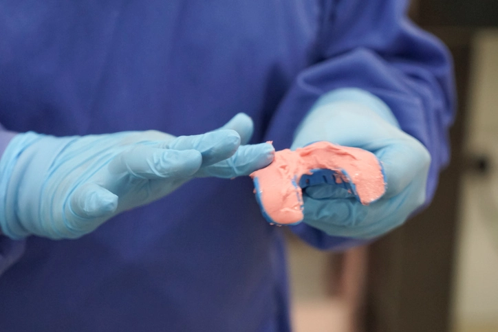
Impression Procedures in Complete Dentures
Impression making is a foundational step that determines the denture’s fit, comfort, and overall success.
Tissue Health Before Impressions
Inflamed tissues distort during impression taking. A tissue conditioner may be required to return tissues to a healthy state, particularly in patients with prolonged denture wear or ill-fitting dentures.
Two-Stage Impression Approach
Traditionally, complete denture impressions involve:
1. Preliminary Impressions
Recorded with:
- Alginate
- Elastomer
- Impression compound
Using an edentulous stock tray, the aim is to capture general anatomy to allow fabrication of a special tray.
Marking the desired extension on the preliminary impression guides the technician.
Special Trays
Fabricated using:
- Self-cure acrylic
- Light-cure acrylic
Spacing for impression materials:
- ZOE: 0.5 mm
- Elastomer: 0.5–1.5 mm
- Alginate: 3 mm
Greenstick stops assist with accurate positioning.
2. Master Impressions
The purpose of the master impression is to record:
- Maximum denture-bearing area
- Functional sulcus width and depth
- Accurate peripheral seal
Adjusting the Special Tray
Before recording the impression:
- Over-extensions must be trimmed
- Edges refined through selective border molding
Perforated trays result in a more mucostatic impression by reducing tissue compression.
Manipulating Soft Tissues
Patients may be asked to:
- Say “oooh” and “eee”
- Protrude or lateralize the tongue
- Move lips and cheeks gently
This simulates functional movements and shapes the impression borders.
Muco-compressive vs Muco-static Impressions
Muco-compressive
- Records tissues under functional load
- Potentially enhances stability under chewing
- Risk of over-compression leading to denture movement during rest
Muco-static
- Records mucosa at rest
- Generally associated with better retention
- May lead to discomfort if load distribution is uneven
Alginate is considered more muco-static, whereas ZOE tends to record tissues with minimal distortion over time.
Clinical reality often blends these approaches depending on patient anatomy and tissue quality.
Special Impression Techniques
Neutral Zone Impression Technique
Indicated in:
- Severely resorbed ridges
- Highly unstable mandibular dentures
- Patients with strong or uncoordinated musculature
Procedure
- Obtain secondary impressions and record occlusion.
- Construct a lower baseplate with wire loops over the occlusal plane.
- Place tissue conditioner around loops to act as moldable material.
- Insert into mouth and ask patient to perform functional movements: Swallow, Purse lips, Say “Oooh” and “Eee”
- Remove and trim impression.
- Use the impression to guide tooth and flange placement.
- Convert impression into a wax mold to position denture teeth accurately.
This technique ensures that the denture occupies a zone compatible with neuromuscular forces, enhancing stability.
Flabby Ridge Management
A flabby ridge is highly compressible tissue, often occurring in the maxillary anterior region opposing natural lower teeth. Traditional impressions distort such tissues, leading to unstable dentures.
Management Options
- Mild flabbiness → elastomer in perforated tray
- Moderate/severe → two-stage impression with window technique
Two-Stage Window Technique
- Record first impression with tray incorporating a window over flabby area.
- Trim away impression material in window.
- Reseat tray.
- Record flabby tissue using low-viscosity elastomer or plaster.
Combination cases may require semi-adjustable articulator mounting to control occlusal forces during function.
Functional Impression
Functional impressions use tissue conditioner placed inside a patient’s existing denture. After several days of wear:
- The lining adapts to functional tissue form
- Provides a more physiologic foundation
- Useful for compromised ridges or trial dentures
This technique often improves comfort and stability without complete remake.
Common Impression Problems and How to Address Them
1. Feather Edge (Under-extension)
Indicates inadequate tray extension.
Solution: Extend tray borders using greenstick and retake impression.
2. Tray Border Visible Through Impression Material
Indicates over-extension or inadequate space.
Solution: Reduce tray borders and repeat impression.
3. Air Blows
Small → fill with wax.
Large → retake impression or adjust mix.
4. Tray Not Centered
Often due to too much material obscuring landmarks.
Solution: Align handle with patient’s nose; use appropriate amount of material.
5. Retching
Common during upper impressions.
Solutions:
- Build rapport and maintain calm demeanor
- Take lower impression first
- Use fast-setting materials
- Apply distraction techniques (e.g., wiggle toes or fingers)
6. Xerostomia (Dry Mouth)
ZOE contraindicated; use elastomers.
7. Tray Showing Through in Otherwise Good Areas
Solve by prescribing tin-foil relief during processing.
Conclusion
Mastery of denture retention, stability, impression techniques, and special procedures such as neutral zone recording and flabby ridge management is crucial for providing functional, comfortable, and retentive complete dentures. Successful outcomes depend on understanding anatomy, patient-specific factors, biomechanics, and the interplay between tissues and impressions.
By applying these principles meticulously, clinicians can dramatically enhance denture performance and patient satisfaction. Continuous refinement of these skills remains essential for every practitioner involved in prosthodontic rehabilitation.
References
- Zarb GA, Hobkirk J, Eckert S, Jacob R. Prosthodontic Treatment for Edentulous Patients: Complete Dentures and Implant-Supported Prostheses. 13th ed. St. Louis: Mosby; 2012.
- McCord JF, Grant AA. Impression making. Br Dent J. 2000;188(9):484–492.
- Chaffee NR, Felton DA, Cooper LF. Protocols for impression procedures for complete dentures. J Am Dent Assoc. 1999;130(12):1759–1764.
- Boucher CO. Complete denture impressions based upon the anatomy of the mouth. J Am Dent Assoc. 1944;31(16):1174-1181.
- The Academy of Prosthodontics. The Glossary of Prosthodontic Terms (GPT-9). J Prosthet Dent. 2017;117(5S):e1–e105.
- Johnson A, Wildgoose D. Complete denture retention and stability. In: Nairn RI, ed. Restorative Dentistry: An Integrated Approach. Oxford: Wright; 2008. p. 301–320.
- Basker RM, Davenport JC, Thomason JM. Prosthetic Treatment of the Edentulous Patient. 5th ed. Oxford: Wiley-Blackwell; 2011.
- Felton D, Cooper L, Duqum I, McGarry T, Ochoa L, et al. Evidence-based guidelines for the care and maintenance of complete dentures: A publication of the American College of Prosthodontists. J Prosthodont. 2011;20(S1):S1-S12.
- Jacobson TE, Krol AJ. A contemporary review of the factors involved in complete dentures. Part III: stability. J Prosthet Dent. 1983;49(3):306-313.
- Jacobson TE, Krol AJ. A contemporary review of the factors involved in complete dentures. Part II: retention. J Prosthet Dent. 1983;49(2):165-172.
- Wilson J, MacEntee M. Neutral zone in complete dentures: a review. J Prosthet Dent. 1982;48(3):356-360.
- Lynch CD, Allen PF. Management of the flabby ridge: using a two-material impression technique. Br Dent J. 2006;200(5):258-261.
- Kumar M, Somani R, Jaimini A, et al. Complete denture impression techniques—An overview. Int J Contemp Dent. 2011;2(2):34-42.
- Boucher’s impression philosophy revisited. Jpn Dent Sci Rev. 2014;50(4):95-106.
- Phoenix RD, Cagna DR, DeFreest CF. Stewart’s Clinical Removable Partial Prosthodontics. 4th ed. Chicago: Quintessence; 2008.
- Singh K, Gupta N. Impression making in complete dentures: A literature review. J Dent Med Sci. 2014;13(2):30–36.