
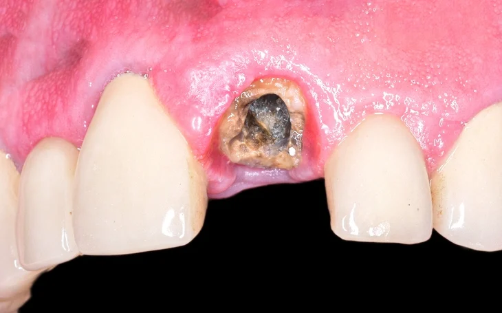
Dento-alveolar surgery forms a core component of oral and maxillofacial surgical practice and is an essential skill for dental practitioners. Among its most common procedures is the removal of retained tooth roots. Root removal may appear deceptively simple; however, it often presents significant clinical challenges due to anatomical limitations, pathological changes, patient-related factors, and proximity to vital structures. Poor planning or technique can lead to complications such as root displacement, nerve injury, infection, excessive bone loss, or delayed healing.
Table of Contents
ToggleIndications for Root Removal
The decision to remove a retained root should never be taken lightly. In certain circumstances, it may be preferable to leave a root fragment in situ, particularly when removal poses a higher risk than benefit. However, several clear indications necessitate removal.
Pathological Indications
Roots associated with pulpal or apical pathology should generally be removed. These include:
- Chronic apical periodontitis
- Acute periapical abscess
- Periapical granulomas or cysts
- Persistent infection following incomplete extraction
Retained infected roots act as a nidus for bacterial colonization and can lead to pain, swelling, sinus formation, or spread of infection.
Symptomatic Roots
Roots causing pain, tenderness, swelling, or recurrent infection require removal. Symptomatology indicates ongoing inflammation or infection, and conservative retention is inappropriate in these cases.
Prosthodontic Considerations
Roots that interfere with denture construction or stability should be removed. Sharp or prominent root fragments may lead to mucosal ulceration, instability of prostheses, and patient discomfort.
Medically Compromised Patients
In patients where even minor local infection cannot be tolerated, such as those who are:
- Immunocompromised
- Undergoing chemotherapy
- Poorly controlled diabetics
- At risk of infective endocarditis
the threshold for removing potentially problematic roots is lower.
Size and Position of the Root
Large roots, particularly those close to the alveolar crest, are more likely to cause future problems and are often best removed proactively.
Initial Assessment and Treatment Planning
Before attempting root removal, thorough assessment is critical.
Clinical Examination
The clinician should evaluate:
- Visibility of the root
- Mobility
- Surrounding soft tissue condition
- Presence of infection or sinus tract
- Proximity to adjacent teeth
Palpation and gentle probing can provide valuable information regarding accessibility.
Radiographic Assessment
A preoperative radiograph is mandatory unless the root is fully visible and straightforward. Radiographs help determine:
- Root length and curvature
- Degree of bone coverage
- Proximity to vital structures (mental nerve, inferior alveolar nerve, maxillary sinus)
- Presence of pathology
- Root morphology and number
Failure to adequately interpret radiographs is a common cause of surgical difficulty and complications.
Non-Surgical Methods of Root Removal
Non-surgical methods should always be attempted first when appropriate, as they minimize trauma and preserve alveolar bone.
Use of Root Forceps
Root forceps may be effective when:
- The root is close to the alveolar margin
- Adequate crown or root structure is exposed
- The forceps blades can securely engage the root
Proper visualization is essential. Blind forceps application increases the risk of slipping, fracturing the root further, or damaging adjacent tissues.
Use of Elevators
Elevators are commonly used to apply controlled leverage to deliver roots along their natural path of withdrawal.
For elevator use to be effective:
- A path of withdrawal must exist
- The elevator must be placed between bone and root
Elevators should be used gently and deliberately. Excessive force can result in:
- Root displacement into adjacent spaces
- Fracture of alveolar bone
- Damage to adjacent teeth
When to Abandon Non-Surgical Attempts
Persistence with unsuccessful non-surgical attempts is discouraged. Prolonged manipulation:
- Increases patient discomfort
- Causes unnecessary trauma
- May complicate subsequent surgical removal
If initial non-surgical methods fail, a surgical approach should be adopted promptly.
Surgical Methods of Root Removal
Surgical removal of roots requires meticulous planning and sound knowledge of anatomy and surgical principles.
Preoperative Planning
Before commencing surgery, the clinician should be able to answer the following questions:
- Why cannot the root be delivered?
- Where exactly is the root located?
- What anatomical structures are nearby?
If uncertainty exists, further radiographic assessment should be obtained.
Flap Design and Soft Tissue Management
Principles of Flap Design
An ideal surgical flap:
- Provides adequate access
- Preserves blood supply
- Avoids damage to vital structures
- Allows tension-free closure
Contrary to common misconception, larger flaps heal just as well as smaller ones, provided they are well designed and handled atraumatically.
Types of Flaps
Envelope Flap
Incision along the gingival margin or alveolar crest
No vertical releasing incisions
Suitable for simple access
Two-Sided Flap
One vertical releasing incision
Improved access and visibility
Three-Sided Flap
Two vertical releasing incisions
Maximum access
Used for difficult or deeply embedded roots
Anatomical Considerations
- Mental nerve: Releasing incisions should be avoided in this region to prevent sensory disturbance.
- Buccal branch of the facial artery: Vertical incisions near the mesial root of the lower second molar should be avoided where possible.
Vertical incisions should start two-thirds of the way distal to the included papilla, and interdental papillae should be preserved to ensure optimal healing and gingival contour.
Bone Removal (Osteotomy)
Principles of Bone Removal
The goal of bone removal is not to remove excessive bone, but to:
- Expose the maximum diameter of the root
- Create a point of application for elevators
Only the minimum amount of buccal bone necessary should be removed.
Technique
A round surgical bur (e.g., No. 8 round T-C bur) is commonly used under copious irrigation to prevent thermal damage. Bone removal should be:
- Controlled
- Conservative
- Focused on improving access
Preservation of alveolar bone is essential for future prosthetic or implant rehabilitation.
Root Elevation and Delivery
Elevator Application
The elevator is placed:
- Between bone and root
- Against the convex surface of curved roots
The root should be guided along its natural path of withdrawal, rather than forced in an unnatural direction.
Root Sectioning
When obstructions exist:
- The obstruction may be removed, or
- The root may be sectioned
Sectioning is often less traumatic and allows for controlled removal of individual root segments.
Wound Management and Closure
Following root removal:
- The socket should be thoroughly debrided
- Any granulation tissue should be removed
- The area should be irrigated with saline
- The flap should be repositioned and closed with minimal tension
Proper closure promotes hemostasis, reduces infection risk, and improves wound healing.
Special Clinical Situations
Small Apical Fragments
Small apical root fragments may be difficult to remove through conventional approaches. An apicectomy approach may be more appropriate, particularly when the fragment is deeply seated or close to vital structures.
Multirooted Teeth
For multirooted teeth:
- Roots should always be divided
- Attempting removal without division significantly increases difficulty and trauma
Division simplifies removal and reduces the risk of root fracture or bone damage.
Missing or Displaced Roots
If a root cannot be located:
- Obtain a repeat radiograph
- Examine the soft tissues carefully
Roots may be displaced into:
- The maxillary sinus
- Submandibular or sublingual spaces
- The aspirator
While careful searching is necessary, discretion is the better part of valour. Prolonged exploration can cause more harm than benefit. Transparency with the patient is essential.
Patient Refusal
If a patient refuses surgical removal:
- Respect their autonomy
- Clearly document the advice given
- Record the patient’s decision
Informed refusal is a valid outcome when appropriately documented.
Complications and Risk Management
Potential complications include:
- Infection
- Dry socket
- Root displacement
- Nerve injury
- Excessive bleeding
- Delayed healing
Most complications can be avoided through proper planning, gentle technique, and adherence to surgical principles.
Conclusion
The removal of retained roots is a common but technically demanding aspect of dento-alveolar surgery. Successful outcomes depend on accurate assessment, sound decision-making, and meticulous surgical technique. Non-surgical methods should be attempted where appropriate, but clinicians must recognize when surgical intervention is necessary. Adequate access, conservative bone removal, and respect for anatomical structures are paramount.
By adhering to these principles, clinicians can minimize complications, ensure patient comfort, and achieve predictable, safe results in the management of retained roots.
References
- Peterson, L. J., Ellis, E., Hupp, J. R., & Tucker, M. R.
Contemporary Oral and Maxillofacial Surgery.
7th ed. St. Louis: Elsevier; 2019. - Hupp, J. R., Ellis, E., & Tucker, M. R.
Contemporary Oral and Maxillofacial Surgery.
6th ed. Elsevier; 2014. - Howe, G. L., & Poyton, H. G.
Minor Oral Surgery.
3rd ed. Wright; 1990. - McGregor, A. D.
Fundamentals of Oral and Maxillofacial Surgery.
Churchill Livingstone; 1990. - Malamed, S. F.
Handbook of Local Anesthesia.
6th ed. Elsevier; 2013. - Kruger, G. O.
Textbook of Oral and Maxillofacial Surgery.
6th ed. CV Mosby; 1984. - British Association of Oral and Maxillofacial Surgeons (BAOMS).
Dento-alveolar Surgery Guidelines.
London: BAOMS; latest edition. - National Institute for Health and Care Excellence (NICE).
Surgical Management of Dental Conditions.
NICE Clinical Guidelines, UK.
Oral and Maxillofacial Surgery.
Mosby; 1985. - Field, E. A., & Longman, L. P.
Tyldesley’s Oral Medicine.
6th ed. Oxford University Press; 2013.