
Soft tissue swellings of the oral cavity are common findings in dental and medical practice. While the discovery of a lump in the mouth often raises concern about malignancy, the majority of oral soft tissue lumps are non-tumorous. These lesions arise from a variety of causes, including inflammatory reactions, developmental anomalies, reactive hyperplasias, vascular malformations, and systemic disease manifestations.
Understanding non-tumour soft tissue lumps is essential for accurate diagnosis, appropriate management, and reassurance of patients. Many of these lesions share similar clinical appearances, making careful history taking, examination, and sometimes histological investigation vital.
Table of Contents
ToggleGeneral Principles of Assessment
Before discussing individual lesions, it is important to outline general principles for evaluating oral soft tissue swellings.
History
Key points include:
- Duration and rate of growth
- Presence of pain or bleeding
- History of trauma or irritation
- Changes during pregnancy
- Medical history (e.g. endocrine disorders, Crohn’s disease, sarcoidosis)
- Habits such as denture use, smoking, or cheek biting
Clinical Examination
Important observations include:
- Site (gingiva, lip, floor of mouth, tongue, palate)
- Colour (red, pink, blue, white)
- Surface texture (smooth, ulcerated, papillated)
- Consistency (soft, firm, fluctuant)
- Mobility and attachment (sessile vs pedunculated)
- Blanching on pressure (suggestive of vascular lesions)
Investigations
Not all lesions require investigation. Options include:
- Radiographs (when bone involvement is suspected)
- Blood tests (e.g. calcium, phosphate, PTH)
- Ultrasound or MRI (for vascular or deep lesions)
- Biopsy (except where contraindicated, e.g. haemangioma)
Inflammatory and Infective Lesions
Abscess
An abscess is a localized collection of pus resulting from bacterial infection. In the oral cavity, abscesses commonly originate from dental infections, particularly apical abscesses secondary to pulp necrosis.
Clinical Features
- Diffuse or localized gingival swelling
- Pain and tenderness
- Erythema and warmth
- Possible discharge of pus
- Associated gingivitis or periodontal disease
Management
Management focuses on eliminating the source of infection:
- Drainage of pus
- Root canal treatment or extraction of the offending tooth
- Antibiotics only when systemic signs are present
- Improvement of oral hygiene
Metabolic and Endocrine-Related Lesions
Brown Tumour
Despite its name, a brown tumour is not a true neoplasm. It is a reactive giant cell lesion associated with hyperparathyroidism, either primary or secondary.
Pathophysiology
Elevated parathyroid hormone (PTH) levels lead to increased osteoclastic activity and bone resorption. The resulting lesions contain:
- Multinucleated giant cells
- Fibrous stroma
- Areas of haemorrhage (responsible for the brown colour)
Although more common in bone, brown tumours may occasionally present in soft tissues.
Clinical Features
- Swelling of jaw or oral tissues
- May be discovered incidentally
- Often painless
Diagnosis
Diagnosis is often suggested histologically and confirmed biochemically:
- Elevated calcium
- Reduced phosphate
- Raised alkaline phosphatase
- Elevated PTH
Management
- Treat the underlying hyperparathyroidism
- Surgical removal is often unnecessary as lesions regress once metabolic control is achieved
Developmental Cysts and Lesions
Dermoid Cyst
A dermoid cyst is a developmental lesion resulting from entrapment of ectodermal tissue during embryonic development.
Common Sites
- Lateral canthus of the eye
- Midline of the neck
- Floor of the mouth above the mylohyoid muscle
Clinical Features
- Slow-growing, painless swelling
- Doughy or fluctuant consistency
- In the floor of the mouth, may elevate the tongue and interfere with speech or swallowing
Management
- Complete but conservative surgical excision
- Careful removal to prevent recurrence
Congenital Epulis
Congenital epulis is a rare lesion present at birth, most commonly affecting the alveolar ridge of newborns.
Clinical Features
- Pedunculated or sessile mass
- Pink or red in colour
- May interfere with feeding or respiration
Histology
- Large granular cells
- Similar in appearance to granular cell tumours but with distinct clinical behaviour
Management
- Conservative surgical excision
- Excellent prognosis with no recurrence
Reactive Gingival Lesions
Peripheral Giant Cell Granuloma (Giant Cell Epulis)
This lesion is a reactive hyperplasia, not a true neoplasm, arising from the gingiva or alveolar mucosa.
Aetiology
- Chronic irritation
- Plaque, calculus
- Trauma from restorations or dentures
Clinical Features
- Deep red or purple gingival swelling
- May ulcerate or bleed
- Often found on the interdental papilla
Histology
- Multinucleated giant cells
- Vascular stroma
- Hemosiderin deposits
Management
- Surgical excision
- Removal of periosteum
- Curettage of underlying bone
- Elimination of local irritants
Pregnancy Epulis
Also known as a pregnancy tumour, this lesion represents a hormone-mediated inflammatory response to plaque during pregnancy.
Clinical Features
- Indistinguishable from pyogenic granuloma
- Commonly arises during the third month
- Bleeds easily
Management
- Emphasis on oral hygiene instruction
- Avoid surgery if possible, as lesions often regress postpartum
- Surgical excision only if function or comfort is compromised
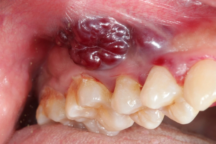
Pyogenic Granuloma
Despite the name, pyogenic granuloma is neither pyogenic nor a true granuloma. It is a highly vascular reactive lesion.
Aetiology
- Trauma
- Chronic irritation
- Hormonal influences
Clinical Features
- Red, fleshy, nodular mass
- Rapid growth
- Profuse bleeding on minor trauma
Histology
- Proliferation of capillaries
- Loose connective tissue stroma
Management
- Complete excision
- Removal of causative irritants
- Good oral hygiene
Fibroepithelial Polyp
This lesion represents an exaggerated fibrous tissue response to low-grade chronic trauma.
Clinical Features
- Sessile or pedunculated
- Firm, smooth surface
- Colour similar to adjacent mucosa
Common Causes
- Cheek or lip biting
- Ill-fitting dentures
- Sharp restorations
Management
- Surgical excision including base
- Correction of traumatic factors
Irritation (Denture) Hyperplasia
This is a common condition in denture wearers caused by repeated mechanical trauma.
Clinical Features
- Folds or rolls of fibrous tissue
- Typically in the sulcus
- Associated with over-extended denture flanges
Management
- Surgical excision
- Temporary removal or adjustment of dentures
- Pre-prosthetic measures and replacement of faulty dentures
Salivary-Related Lesions
Mucoceles
Mucoceles are mucous extravasation cysts caused by trauma to minor salivary gland ducts.
Clinical Features
- Soft, fluctuant swelling
- Bluish or translucent appearance
- Most common on the lower lip
Important Consideration
Upper lip swellings are less likely to be mucoceles and should raise suspicion of salivary gland tumours.
Management
- Surgical excision
- Removal of affected glands and ducts
Ranula
A ranula is a mucocele occurring in the floor of the mouth, arising from the sublingual gland.
Types
- Simple ranula: confined to the floor of the mouth
- Plunging ranula: extends beyond the mylohyoid into the neck
Management
- Excision of cyst and sublingual gland
- Submandibular gland removal may be required if duct damage occurs
Granulomatous Conditions
Granulomata
Granulomatous inflammation in the mouth may reflect systemic disease.
Causes
- Crohn’s disease
- Orofacial granulomatosis
- Sarcoidosis
- Foreign bodies (e.g. amalgam)
Clinical Importance
Oral manifestations may precede systemic symptoms, making dental professionals crucial in early diagnosis.
Management
- Treat underlying systemic condition
- Referral to medical specialists
Vascular and Lymphatic Lesions
Haemangioma
Haemangiomas are developmental vascular lesions present at or shortly after birth.
Clinical Features
- Red or blue swelling
- Blanch on pressure
- May grow, remain static, or regress
Important Rule
Do not biopsy due to risk of severe bleeding.
Management
- Observation (80% regress spontaneously)
- Laser therapy or cryotherapy
- Surgical excision only if very small
Lymphangioma
A rarer developmental lesion involving lymphatic vessels.
Types
- Microcystic: diffuse, infiltrative
- Macrocystic: well-defined cystic spaces
Clinical Features
- Enlarged tongue (macroglossia)
- Cheek or lip swelling
- Neck masses
Management
- Surgical excision when possible
- Sclerosing agents such as picibanil
- Treatment can be challenging due to infiltration
Vascular Malformations
Unlike haemangiomas, vascular malformations:
- Are present at birth
- Do not regress
- Grow with the patient
Management
- Interventional radiology
- Surgical management
- Multidisciplinary approach
Viral-Related Lesions
Warts and Squamous Papillomata
These lesions are associated with human papillomavirus (HPV).
Clinical Features
- Papillated, pink or white lesions
- Usually asymptomatic
- Can be solitary or multiple
Epidemiology
- True warts are rare in the mouth
- Oral papillomas are common
- Not always associated with STDs
Management
- Excision biopsy
- Ligation or diathermy if pedunculated
Conclusion
Non-tumour soft tissue lumps of the mouth encompass a wide spectrum of conditions, ranging from benign reactive lesions to manifestations of systemic disease. Although most are harmless, accurate diagnosis is essential to guide management, prevent recurrence, and identify potentially serious underlying conditions.
A systematic approach—combining history, examination, and appropriate investigation—allows clinicians to distinguish between lesions that require simple reassurance and those needing surgical or medical intervention. For dental professionals, familiarity with these conditions is a cornerstone of safe and effective oral healthcare.
References
- Cawson RA, Odell EW.
Cawson’s Essentials of Oral Pathology and Oral Medicine. 9th ed. London: Churchill Livingstone Elsevier; 2017. - Neville BW, Damm DD, Allen CM, Chi AC.
Oral and Maxillofacial Pathology. 4th ed. St. Louis: Elsevier; 2016. - Regezi JA, Sciubba JJ, Jordan RCK.
Oral Pathology: Clinical Pathologic Correlations. 7th ed. St. Louis: Elsevier; 2017. - Hupp JR, Ellis E, Tucker MR.
Contemporary Oral and Maxillofacial Surgery. 7th ed. St. Louis: Elsevier; 2019. - Scully C.
Oral and Maxillofacial Medicine: The Basis of Diagnosis and Treatment. 4th ed. Edinburgh: Churchill Livingstone Elsevier; 2013. - Langlais RP, Miller CS, Nield-Gehrig JS.
Color Atlas of Common Oral Diseases. 5th ed. Philadelphia: Wolters Kluwer; 2017. - Shafer WG, Hine MK, Levy BM.
Shafer’s Textbook of Oral Pathology. 8th ed. New Delhi: Elsevier India; 2016. - Greenberg MS, Glick M, Ship JA.
Burket’s Oral Medicine. 12th ed. Shelton, CT: PMPH USA; 2015. - Marx RE, Stern D.
Oral and Maxillofacial Pathology: A Rationale for Diagnosis and Treatment. 2nd ed. Hanover Park: Quintessence Publishing; 2012. - Eveson JW, Reichart PA, Sidransky D.
World Health Organization Classification of Tumours: Pathology and Genetics of Head and Neck Tumours. Lyon: IARC Press; 2005. - Batsakis JG.
Tumors of the Head and Neck: Clinical and Pathological Considerations. 2nd ed. Baltimore: Williams & Wilkins; 1979. - Woo SB.
Oral mucosal lesions: reactive, inflammatory and infectious conditions.
Dental Clinics of North America. 2014;58(2):403–425.