
Intraepithelial vesiculo-bullous disorders represent a clinically significant group of diseases in oral medicine due to their frequent oral involvement, painful presentation, and potential for serious systemic consequences. These disorders are characterized by the formation of vesicles and bullae within the epithelial layer as a result of disruption in intercellular adhesion. Because the oral mucosa is thin, moist, and constantly subjected to mechanical trauma, intraepithelial blisters often rupture rapidly, leaving erosions or ulcers as the predominant clinical findings.
The oral cavity often serves as the initial site of manifestation for many vesiculo-bullous disorders, particularly autoimmune conditions such as pemphigus vulgaris. As a result, dental professionals play a critical role in early detection, diagnosis, and referral, which can significantly influence patient outcomes. Understanding the pathogenesis, clinical features, diagnostic approaches, and management of intraepithelial vesiculo-bullous diseases is therefore essential for practitioners and students of oral medicine.
Table of Contents
ToggleBasic Terminology and Definitions
Vesicle
A vesicle is a small, circumscribed, fluid-filled lesion usually measuring a few millimetres in diameter. Vesicles contain clear serous fluid and are typically superficial. In the oral cavity, vesicles are rarely seen intact due to rapid rupture caused by mastication, speech, and friction against teeth and restorations.
Bulla
A bulla is a larger blister measuring 0.5 cm or more in diameter. Bullae may vary in consistency depending on the level at which they form. In intraepithelial disorders, bullae are thin-roofed and fragile, resulting in early breakdown and ulceration.
Erosion
An erosion is a superficial loss of epithelium that does not extend beyond the basement membrane. Erosions heal without scarring and are commonly observed after rupture of vesicles or bullae. In oral vesiculo-bullous disorders, erosions are among the most frequent clinical findings.
Ulcer
An ulcer is a breach of the epithelium extending into the underlying connective tissue. Ulcers are deeper than erosions, often painful, and may bleed or become secondarily infected. Persistent ulcers resulting from vesiculo-bullous diseases may significantly impair oral function.
Classification of Vesiculo-Bullous Disorders
Vesiculo-bullous disorders are commonly classified based on the level of epithelial separation:
- Intraepithelial vesiculo-bullous disorders
- Subepithelial vesiculo-bullous disorders
This classification has diagnostic and prognostic value. Intraepithelial disorders tend to produce fragile bullae that rupture easily, while subepithelial disorders form tense bullae that are more likely to remain intact.
Pathogenesis of Intraepithelial Bullae
The hallmark of intraepithelial vesiculo-bullous disorders is acantholysis, which refers to the loss of cohesion between epithelial cells. This process occurs due to damage or dysfunction of desmosomes, the specialized intercellular junctions responsible for maintaining epithelial integrity.
Desmosomes contain structural proteins such as desmogleins and desmocollins. When these proteins are targeted by autoimmune mechanisms or disrupted by genetic mutations, keratinocytes lose adhesion, leading to the formation of intraepithelial clefts and blisters. Because only a thin layer of epithelium covers these blisters, they are highly susceptible to rupture.
Immunopathology and Diagnostic Techniques
Immunofluorescence
Immunopathological testing is central to the diagnosis of many intraepithelial vesiculo-bullous disorders, particularly those of autoimmune origin.
- Direct immunofluorescence (DIF) is performed on fresh perilesional biopsy tissue and detects in-situ deposition of immunoglobulins and complement components.
- Indirect immunofluorescence (IIF) is performed on patient serum to identify circulating autoantibodies.
These techniques help differentiate between various vesiculo-bullous diseases and confirm the underlying pathogenic mechanism.
Histopathology
Routine histological examination reveals characteristic features such as suprabasal clefting, acantholytic cells, and preserved basal cell attachment in certain conditions. Correlation between clinical, histological, and immunological findings is essential for accurate diagnosis.
Pemphigus: The Prototypical Intraepithelial Disorder
Overview
Pemphigus is a group of chronic autoimmune vesiculo-bullous diseases characterized by intraepithelial blister formation. Without appropriate treatment, pemphigus can be life-threatening due to complications such as infection, dehydration, and adverse effects of immunosuppressive therapy.
Among the pemphigus variants, pemphigus vulgaris is the most common and clinically significant in oral medicine.
Pemphigus Vulgaris
Etiology and Immunopathogenesis
Pemphigus vulgaris is caused by IgG autoantibodies directed against desmosomal proteins, primarily desmoglein-3 and, in some cases, desmoglein-1. These autoantibodies disrupt intercellular adhesion between keratinocytes, leading to acantholysis and blister formation above the basal cell layer.
Epidemiology
Pemphigus vulgaris most commonly affects middle-aged adults and shows a female predominance. Increased prevalence has been reported in certain ethnic groups, including individuals of Jewish and Middle Eastern descent. Rarely, the condition may be drug-induced or associated with underlying malignancy.
Oral Manifestations
The oral cavity is involved in the vast majority of patients with pemphigus vulgaris and may represent the first site of disease in many cases. Commonly affected sites include the palate, buccal mucosa, labial mucosa, and gingiva.
Because oral bullae rupture rapidly, lesions usually present as painful erosions or ulcers rather than intact blisters. Gingival involvement may appear as desquamative gingivitis, characterized by erythema, epithelial sloughing, and bleeding.
Clinical Features
- Fragile intraepithelial bullae
- Painful erosions and ulcers
- Positive Nikolsky sign
- Difficulty eating, speaking, and maintaining oral hygiene
- Increased risk of secondary infection
Nikolsky Sign
The Nikolsky sign is positive when gentle lateral pressure causes epithelial separation. Although diagnostically helpful, intentional elicitation is discouraged due to patient discomfort and risk of tissue damage.
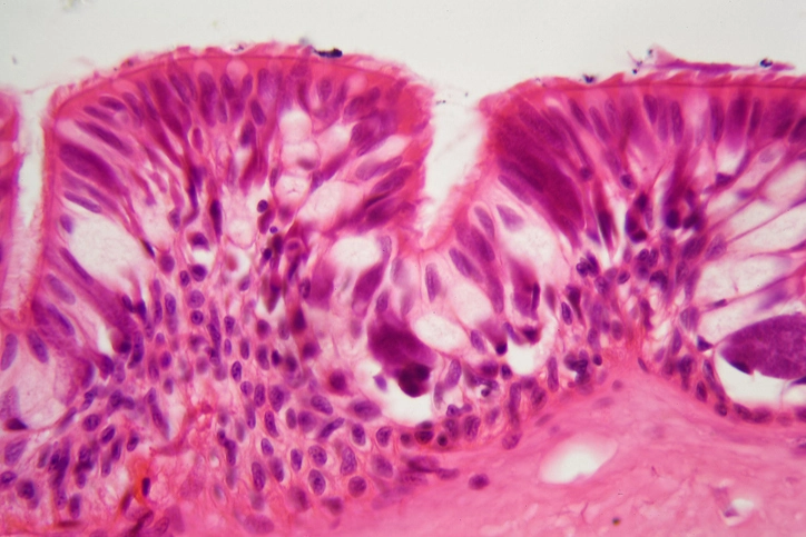
Histopathological and Immunofluorescence Findings
Histological examination typically shows suprabasal clefting with acantholytic cells and a “tombstone” appearance of basal cells. Direct immunofluorescence demonstrates intercellular deposition of IgG and complement in a characteristic net-like pattern.
Differential Diagnosis
Pemphigus vulgaris must be differentiated from other oral conditions presenting with erosions and ulcers, including aphthous stomatitis, erosive lichen planus, candidiasis, and subepithelial bullous disorders. Accurate diagnosis relies on biopsy and immunopathological testing.
Management of Pemphigus Vulgaris
Medical Treatment
Management focuses on suppressing the autoimmune response and preventing complications.
- Systemic corticosteroids remain the mainstay of treatment.
- Steroid-sparing agents such as azathioprine, mycophenolate mofetil, and cyclophosphamide are commonly used.
- Biologic therapies, particularly B-cell–targeting agents, have significantly improved disease control in refractory cases.
Monitoring
Disease activity is monitored through clinical assessment and measurement of circulating autoantibody levels.
Dental Considerations
Dental care should be conservative, with emphasis on minimizing trauma, maintaining oral hygiene, and coordinating treatment with medical specialists.
Benign Familial Chronic Pemphigus (Hailey–Hailey Disease)
Benign familial chronic pemphigus is a hereditary condition with autosomal dominant inheritance. Unlike pemphigus vulgaris, it is not autoimmune in nature and typically presents in young adults. Oral involvement is less common and milder. The disease follows a chronic, relapsing course but is not life-threatening.
Viral Infections with Intraepithelial Vesicles
Certain viral infections of the oral cavity produce intraepithelial vesicles that rupture to form ulcers. These conditions are usually acute, self-limiting, and associated with systemic symptoms. Accurate clinical history and presentation help distinguish viral lesions from autoimmune disorders.
Epidermolysis Bullosa Simplex
Epidermolysis bullosa simplex is an inherited intraepithelial blistering disorder caused by mutations affecting basal keratinocytes. Blisters form in response to minimal trauma. There is no definitive cure, and management is supportive, focusing on prevention of trauma and infection.
Dental Management Considerations
Patients with intraepithelial vesiculo-bullous disorders require special attention during dental treatment. Preventive care, atraumatic techniques, and interdisciplinary collaboration are essential to maintain oral health and quality of life.
Conclusion
Intraepithelial vesiculo-bullous disorders are a vital topic in oral medicine due to their frequent oral presentation, diagnostic complexity, and potential severity. Early recognition, accurate diagnosis, and appropriate management can significantly reduce morbidity and improve outcomes. Dental professionals are often the first to encounter these lesions and play a critical role in patient care.
A comprehensive understanding of the pathogenesis, clinical features, and management strategies of these disorders is essential for effective practice in oral medicine.