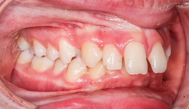
Overbite refers to the vertical overlap of the upper incisors over the lower incisors when the teeth are in centric occlusion. A normal overbite is generally defined as one-third to one-half overlap of the lower incisor crowns by the upper incisors. Variations beyond this normal range constitute either increased overbite (deep bite) or reduced overbite (open bite).
Measurement is often expressed qualitatively—such as one-quarter, one-half, or three-quarters—rather than numerically, as precise measurement with a ruler is seldom required for diagnostic or clinical purposes.
Increased overbite represents one of the most frequent vertical discrepancies encountered in orthodontic practice. While mild increases may be asymptomatic, excessive deep bites can cause esthetic, functional, and periodontal problems, making their identification and appropriate management essential to comprehensive orthodontic treatment.
Table of Contents
ToggleClassification
1. Skeletal vs. Dental Overbite
- Skeletal deep bite results from a vertical growth deficiency of the lower face, typically associated with a short lower anterior facial height (LAFH) and a horizontal growth pattern.
- Dental deep bite arises from abnormal tooth eruption patterns, particularly of the incisors or molars, rather than underlying skeletal discrepancies.
2. Associated Malocclusions
- Class II Division 2 malocclusion commonly presents with a markedly increased overbite, often accompanied by retroclined maxillary central incisors and upright or proclined lateral incisors.
- Class II Division 1 cases may exhibit an increased overbite secondary to protrusive maxillary incisors and a deep curve of Spee.
- Class III cases occasionally display an increased overbite as a compensatory mechanism that masks the underlying anteroposterior skeletal discrepancy. In such cases, the deep bite may actually be beneficial, contributing to occlusal stability.
Aetiology of Increased Overbite
The etiology of a deep overbite is multifactorial, involving a combination of skeletal, dental, muscular, and functional factors.
1. Dental Factors
- Eruption of Lower Incisors:
The lower incisors typically erupt until they make contact with either the palatal surfaces of the upper incisors or the palatal mucosa. If this eruption continues beyond the point of balanced occlusal contact, an excessive overbite develops. - Retroclined Upper Incisors:
Retroclination reduces the inter-incisal angle and encourages excessive overlap of the lower incisors, as seen in Class II Division 2 cases. - Curve of Spee:
A pronounced curve of Spee, particularly in the lower arch, can result from supraeruption of anterior teeth and infraeruption of posterior teeth, thereby deepening the overbite.
2. Skeletal Factors
- Decreased Lower Anterior Facial Height (LAFH):
A reduced LAFH and a low mandibular plane angle produce a short lower face and a horizontal growth pattern, which tend to deepen the bite. - Vertical Maxillary Deficiency:
Insufficient vertical development of the maxilla leads to decreased eruption of posterior teeth, contributing to overclosure of the mandible.
3. Muscular and Soft Tissue Factors
- Lip Morphology and Function:
A high lower lip line, firm lower lip musculature, and hyperactive mentalis muscle promote lingual tipping of the upper incisors and reduce the inter-incisal angle. - Strong Masticatory Muscles:
High muscle tonicity, often seen in brachyfacial (short-faced) individuals, can resist molar eruption, maintaining a deep bite relationship.
4. Inter-Incisal Angle
The normal inter-incisal angle is approximately 135°, while the highest acceptable value is about 145°. Angles above this threshold indicate excessive retroclination of the incisors and are strongly correlated with deep overbites.
5. Functional Influences
- Premature Incisal Contacts:
Anterior premature contacts can lead to functional closure of the mandible in a more posterior position, exacerbating the overbite. - Tongue and Habits:
Habits such as finger sucking or lip biting may cause dental positional changes that either deepen or maintain an excessive overbite.
Clinical Features
Patients with increased overbite may present with:
- Excessive vertical overlap of upper incisors on the lowers (>4 mm).
- Minimal display of lower incisors on smiling.
- Palatal trauma from lower incisor impingement.
- Mandibular posturing or restriction of movement.
- Reduced lower anterior facial height and an acute nasolabial angle.
- Associated dental wear and gingival trauma.
Clinically, a distinction must be made between traumatic and non-traumatic deep bites.
- Traumatic deep bite: The lower incisors contact the palatal mucosa, often causing ulceration or recession.
- Non-traumatic deep bite: The overlap is increased but does not cause soft tissue injury.
Diagnostic Considerations
A comprehensive diagnosis of increased overbite involves:
1. Clinical Examination:
- Measure vertical overlap in centric occlusion.
- Assess soft tissue impingement or traumatic contact.
- Evaluate facial proportions and lower anterior facial height.
2. Radiographic Evaluation:
- Lateral cephalogram for assessing skeletal vertical pattern, mandibular plane angle, and inter-incisal angle.
- Panoramic radiographs to evaluate eruption status and root morphology.
3. Model Analysis:
- Evaluate curve of Spee and overbite depth on study casts.
The goal of diagnosis is to determine whether the overbite is dentoalveolar or skeletal in origin, which directly dictates the treatment plan.
Approaches to Reducing Overbite
The choice of treatment depends on the underlying etiology, growth potential, and skeletal pattern of the patient. The primary approaches include extrusion of molars, intrusion of incisors, proclination of lower incisors, and surgical correction in severe adult cases.
1. Extrusion or Eruption of Molars
- Passive Eruption (with Bite Plane):
In growing patients, the use of an upper removable appliance (URA) with an anterior bite plane allows passive eruption of posterior teeth. This increases the vertical dimension, effectively reducing the overbite. - Active Extrusion (with Fixed Appliances):
Fixed appliances (FAs) may be used to extrude molars intentionally through controlled mechanics. However, in adults or nongrowing patients, molar extrusion may produce instability and relapse once the appliances are removed.
Advantages:
- Non-invasive and well-tolerated in young patients.
- Enhances facial vertical proportions.
Disadvantages:
- May produce clockwise rotation of the mandible.
- Risk of relapse in adults due to unstable vertical changes.
2. Intrusion of Incisors
Incisor intrusion is indicated when the deep bite results primarily from overeruption of the anterior teeth rather than deficiency of posterior eruption.
Methods:
- Utility Arches or Base Archwires: Used for true incisor intrusion.
- Segmental Arch Mechanics: Offers precise control with minimal side effects.
- Skeletal Anchorage (Mini-implants): Allows absolute intrusion without molar extrusion, ideal in adults.
Considerations:
- Intrusion should be limited to 2–3 mm to avoid root resorption.
- Forces must be light (10–20 g per tooth) and continuous.
Advantages:
- Effective and stable in adults.
- Maintains mandibular position and facial proportions.
Disadvantages:
- Technical complexity.
- Potential for root resorption or loss of vitality.
3. Proclination of Lower Incisors
Proclination of the lower incisors can reduce overbite by decreasing the inter-incisal angle and increasing incisor display. This is often achieved by using:
- Upper removable appliances with anterior bite planes.
- Lower utility arch or fixed appliances with light anterior forces.
This approach is indicated primarily in Class II Division 2 cases, where lower incisors are excessively retroclined and constrained by the upper incisors or lower lip.
However, this movement must be carefully controlled, as excessive proclination compromises long-term stability and periodontal health.
4. Surgical Intervention
In adults with severe skeletal deep bites and minimal growth potential, orthognathic surgery may be indicated.
Common Procedures:
- Anterior Maxillary Impaction: Elevation of the anterior maxilla, permitting autorotation of the mandible downward and backward to open the bite.
- Mandibular Advancement or Down-Graft Surgery: Adjusts vertical dimensions and corrects associated skeletal discrepancies.
Indications:
- Severe deep bites (>7 mm) with skeletal vertical deficiency.
- Cases associated with significant anteroposterior (AP) discrepancy.
- Adult patients where growth modification is no longer possible.
Management According to Malocclusion
Class II Division 2
This group frequently presents with deep bites due to retroclined upper incisors and increased lower lip pressure.
Treatment Goals:
- Relieve inter-incisal angle by proclining upper incisors.
- Encourage eruption or extrusion of molars to open the bite.
- Maintain or enhance facial esthetics by improving lip support.
Treatment Approach:
- Avoid extractions where possible to prevent further lingual tipping of lower incisors.
- Use upper removable appliances (URAs) with anterior bite planes or fixed appliances (FAs) for molar extrusion and incisor proclination.
- In growing patients, functional appliances can be employed to improve both vertical and AP dimensions.
Class II Division 1
In these cases, increased overbite often accompanies excessive overjet and reduced lower facial height.
Management:
- Functional appliances (e.g., Twin Block, Herbst) may be used to advance the mandible and stimulate molar eruption, indirectly reducing the overbite.
- Use of bite planes or vertical elastics to assist molar extrusion.
- Following AP correction, FAs can be employed for final bite opening and alignment.
Care must be taken to control incisor inclination and avoid relapse through retention of the corrected vertical dimension.
Class III
In Class III malocclusions, an increased overbite may serve a compensatory purpose, improving incisal contact and facial appearance.
In such cases, reducing the overbite is generally contraindicated, as it may worsen the occlusal and aesthetic relationships.
However, if the deep bite is traumatic or esthetically undesirable, careful adjustment may be undertaken in conjunction with skeletal correction.
Biomechanics of Overbite Correction
Orthodontic correction of a deep bite involves a delicate balance between:
- Incisor intrusion and molar extrusion,
- Facial esthetics and occlusal function, and
- Short-term success and long-term stability.
The treatment should aim to:
- Achieve a harmonious inter-incisal angle (120°–135°).
- Maintain a functional occlusal plane.
- Prevent undesirable mandibular rotations.
Biomechanical control can be achieved using modern appliances such as:
- Segmental arch technique (for controlled incisor intrusion).
- Mini-implants (for skeletal anchorage and absolute intrusion).
- Functional appliances (for vertical and sagittal modification in growing patients).
Stability of Overbite Reduction
Stability is a major consideration following deep bite correction. Relapse tendencies are influenced by:
- Age and growth pattern of the patient.
- Nature of correction (skeletal vs. dental).
- Degree of incisor inclination achieved.
- Presence of occlusal stops or functional stability.
Factors Enhancing Stability:
- Reduction of the Inter-Incisal Angle:
Decreasing the inter-incisal angle minimizes the potential for relapse by promoting incisal clearance. - Occlusal Stops:
Establishing posterior occlusal stops ensures that lower incisors do not over-erupt. - Favorable Growth:
Growth patterns with slight vertical tendencies promote stable occlusal relationships by compensating for molar eruption. - Control of Etiological Factors:
Correction of muscle imbalance, elimination of soft tissue interferences, and maintenance of proper incisor inclination enhance long-term success.
Retention Following Overbite Correction
Retention protocols should be designed to maintain both vertical and sagittal corrections.
Common retention methods include:
- Removable Retainers with Anterior Bite Planes: Prevent overeruption of incisors post-treatment.
- Fixed Retainers: Bonded lingual retainers for lower incisors stabilize incisor inclination.
- Functional Retainers (e.g., Twin Block): Useful in growing patients for continued skeletal adaptation.
Retention duration should extend well beyond the active treatment phase, often for 12–24 months, or indefinitely in adults.
Prognosis and Clinical Considerations
Successful management of increased overbite depends on:
- Accurate diagnosis of its etiology (skeletal vs. dental).
- Realistic treatment objectives tailored to the patient’s age and growth status.
- Maintenance of occlusal harmony and aesthetic facial proportions.
- Adequate retention and follow-up.
A deep bite corrected through inappropriate mechanics may relapse rapidly or compromise facial appearance. Therefore, careful selection of biomechanical strategy is essential for long-term success.
References
- Houston WJB. Incisor edge–centroid relationships and overbite depth. Eur J Orthod. 1989;11(2):139–146.
- Millett DT, Cunningham SJ, O’Brien KD. Orthodontic treatment for deep bite and retroclined upper front teeth in children. Cochrane Database Syst Rev. 2018;2:CD005972.
- Proffit WR, Fields HW, Larson BE, Sarver DM. Contemporary Orthodontics. 6th ed. St. Louis: Elsevier; 2019.
- Graber LW, Vanarsdall RL, Vig KWL, Huang GJ. Orthodontics: Current Principles and Techniques. 7th ed. St. Louis: Elsevier; 2020.
- Nanda R. Biomechanics and Esthetic Strategies in Clinical Orthodontics. St. Louis: Elsevier; 2005.
- Burstone CJ. Deep overbite correction by intrusion. Am J Orthod. 1977;72(1):1–22.
- Daskalogiannakis J. Glossary of Orthodontic Terms. Berlin: Quintessence Publishing; 2000.
- Karlsen AT. Craniofacial morphology in patients with Angle Class II, Division 2 malocclusion and deep overbite. Am J Orthod Dentofacial Orthop. 1994;106(1):82–88.
- Parker CD, Nanda RS, Currier GF. Nontreated Class II Division 2 malocclusion: Longitudinal cephalometric study. Am J Orthod Dentofacial Orthop. 1995;107(2):150–156.
- Taner T, Aksu M, Akgül AA. Treatment of deep overbite with intrusion mechanics supported by mini-implants. Angle Orthod. 2005;75(5):749–756.
- Isaacson RJ, Isaacson KG, Speidel TM, Worms FW. Extreme variation in vertical facial growth and associated variation in skeletal and dental relations. Angle Orthod. 1971;41(3):219–229.
- Dellinger EL. A clinical assessment of the Active Vertical Corrector—a non-surgical alternative for skeletal open bite treatment. Am J Orthod. 1986;89(5):428–436.
- Burstone CJ, Koenig HA. Forces produced by orthodontic appliances and their distribution to craniofacial structures. Angle Orthod. 1974;44(3):200–219.
- Sarver DM, Ackerman MB. Dynamic smile visualization and quantification: Part 1. Evolution of the concept and dynamic records for smile capture. Am J Orthod Dentofacial Orthop. 2003;124(1):4–12.

