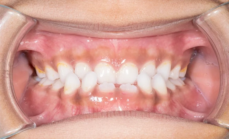
Gingival depigmentation, often referred to as gum bleaching, is a cosmetic dental procedure aimed at removing or reducing dark pigmentation on the gums. While dark gums are typically harmless and result from melanin deposits, they can be a cosmetic concern for some individuals. This comprehensive article delves into the causes of gingival pigmentation, various treatment options, and considerations for those seeking a brighter smile.
Table of Contents
ToggleUnderstanding Gingival Pigmentation
What Causes Dark Gums?
Gingival pigmentation is primarily due to melanin, the natural pigment responsible for the color of our skin, hair, and eyes. In the gums, melanin is produced by melanocytes located in the basal and suprabasal layers of the epithelium. An increased activity or number of these melanocytes leads to darker gum coloration. This condition, known as gingival hyperpigmentation, is benign and varies among individuals based on genetic factors.
Factors Influencing Gum Pigmentation
Several factors can contribute to increased pigmentation of the gums:
- Genetics: Individuals with darker skin tones naturally have more melanin, which can extend to the gums.
- Smoking: Tobacco use stimulates melanin production, leading to smoker’s melanosis, a condition characterized by darkened oral tissues.
- Medications: Certain drugs, such as antimalarials and minocycline, can cause pigmentation changes in the oral mucosa.
- Systemic Conditions: Diseases like Addison’s disease and Peutz-Jeghers syndrome can manifest as pigmentation in the oral cavity.
- Amalgam Tattoos: Particles from dental amalgam fillings can embed in the gum tissue, leading to localized dark spots.
Treatment Options for Gingival Depigmentation
Although gingival pigmentation is not harmful and does not require medical treatment, cosmetic concerns lead many individuals to seek interventions. Today, a variety of methods are available for depigmenting gums, each with distinct benefits, mechanisms, recovery timelines, and effectiveness. The selection depends on factors like the extent of pigmentation, patient preferences, clinician expertise, cost, and potential for recurrence.
1. Laser Depigmentation
Laser treatment is one of the most modern, efficient, and minimally invasive approaches for gingival depigmentation. Lasers work by vaporizing and removing the pigmented epithelial tissue and allowing healthy, lighter-colored tissue to regenerate in its place.
Common Laser Types:
- Diode Lasers (810–980 nm) – Highly effective, particularly in soft tissue surgeries, offering precision and faster healing.
- Er:YAG and Er,Cr:YSGG Lasers – Used for superficial ablation with less thermal damage, suitable for patients with sensitive gums.
- CO₂ Lasers – Offer deep penetration and coagulation but may cause more thermal injury compared to diode or Er:YAG lasers.
Advantages:
- Minimal bleeding due to coagulative properties
- Faster healing time (1–2 weeks)
- Reduced need for sutures or dressings
- Lower postoperative discomfort
- Precision and selective tissue targeting
Limitations:
- Costlier than traditional techniques
- May require more than one session depending on pigmentation depth
- Requires advanced equipment and operator training
2. Surgical Depigmentation (Scalpel Technique)
This is one of the oldest and most frequently used methods, involving the physical excision of the pigmented epithelium using a scalpel.
Procedure:
- Local anesthesia is administered.
- A partial-thickness flap is raised or the outer pigmented epithelium is scraped off.
- The area is then left to heal by secondary intention (open healing without sutures).
Advantages:
- Simple and cost-effective
- No requirement for complex equipment
- Immediate visible results
Limitations:
- Increased bleeding during the procedure
- Slower healing (7–14 days)
- Postoperative discomfort and sensitivity
- Risk of infection if oral hygiene is poor
- Possibility of repigmentation over time
3. Cryosurgery (Cryotherapy)
Cryotherapy involves applying extreme cold (usually via liquid nitrogen or nitrous oxide) to destroy melanocytes responsible for pigmentation.
Mechanism:
- Freezing causes cell lysis and death of pigmented epithelial cells.
- The treated area sloughs off and regenerates with new, lighter tissue.
Advantages:
- Non-invasive with minimal bleeding
- Usually painless, anesthesia often not required
- Good esthetic results
- Short procedure time
Limitations:
- Specialized equipment needed
- Less control over depth of tissue destruction
- Risk of damage to adjacent tissues
- Healing time can vary significantly
- May cause temporary discoloration or edema
4. Chemical Peeling (Chemical Cauterization)
This method uses chemical agents, like phenol and alcohol, to cauterize and peel off the pigmented gingival layer.
Procedure:
- The depigmentation agent is applied using a cotton swab or applicator.
- After a short contact time, the chemicals destroy the superficial pigmented layer.
- The tissue sloughs off in a few days, followed by regeneration.
Advantages:
- Simple, quick, and inexpensive
- Can be performed without high-end tools
- Suitable for small, localized pigmentation
Limitations:
- Chemical burns or tissue necrosis if improperly applied
- Potential for significant discomfort or pain
- Healing may take longer
- Risk of uneven pigmentation
- Not widely practiced due to safety concerns
5. Electrosurgery
Electrosurgery involves using a high-frequency electric current to burn off the pigmented tissue.
Procedure:
- Local anesthesia is applied.
- An electrode tip is used to “paint” over the pigmented area.
- The current generates heat that removes the epithelial layer.
Advantages:
- Good control over tissue removal
- Coagulates blood vessels, minimizing bleeding
- Cost-effective compared to laser
Limitations:
- Generates significant heat, potentially damaging deeper tissues
- Healing is slower than laser
- May require dressing and postoperative care
- Risk of scarring and patient discomfort
6. Gingival Grafting (Free Gingival Graft)
This advanced technique is used when the pigmented area is large or when other techniques are contraindicated. Gingival grafting involves harvesting a section of non-pigmented tissue (often from the palate) and transplanting it to the affected area.
Procedure:
- Pigmented gingiva is excised.
- Graft tissue from the donor site (palate) is prepared and sutured onto the exposed area.
- Graft integrates over time, replacing pigmented tissue.
Advantages:
- Provides long-lasting and often permanent depigmentation
- Reduces risk of repigmentation
- Natural color match when done precisely
Limitations:
- Invasive and technique-sensitive
- More expensive
- Requires healing of both donor and recipient sites
- Risk of graft failure or infection
7. Abrasive Techniques (Gingival Abrasion)
Gingival abrasion involves mechanically abrading the pigmented layer using rotary instruments like diamond burs or sanding disks.
Procedure:
- Under anesthesia, the surface of the gingiva is gently abraded until the pigmented layer is removed.
- The area is allowed to heal naturally.
Advantages:
- Cost-effective
- Immediate visual results
- Requires basic dental tools
Limitations:
- Risk of damaging deeper tissues
- Bleeding and discomfort during the procedure
- Possible uneven results and risk of recurrence
Combination Techniques
In some cases, clinicians combine techniques for optimal results. For example, laser treatment may be used after scalpel excision to ensure complete removal of residual pigmented cells, or cryotherapy might follow abrasion for enhanced depth.
Choosing the Right Method
The choice of treatment should be guided by:
| Factor | Recommendation |
|---|---|
| Extent of Pigmentation | Laser, scalpel, or grafting |
| Patient Budget | Scalpel or abrasion (cost-effective) |
| Desire for Minimally Invasive Treatment | Laser or cryotherapy |
| Recurrence Risk Concerns | Grafting or laser with good postoperative care |
| Sensitivity to Heat/Chemicals | Cryotherapy or mechanical abrasion |
Summary Table of Techniques
| Method | Invasiveness | Healing Time | Pain | Cost | Recurrence Risk |
|---|---|---|---|---|---|
| Laser | Low | 1–2 weeks | Low | High | Low |
| Scalpel | Moderate | 1–2 weeks | Medium | Low | Medium |
| Cryotherapy | Low | 1–3 weeks | Low | Medium | Low |
| Chemical Peeling | Low | 1–2 weeks | High | Low | High |
| Electrosurgery | Moderate | 1–2 weeks | Medium | Medium | Medium |
| Grafting | High | 2–4 weeks | High | High | Very Low |
| Abrasion | Moderate | 1–2 weeks | Medium | Low | Medium |
Considerations and Post-Treatment Care
Potential for Repigmentation
One of the challenges with gingival depigmentation is the possibility of repigmentation over time. Factors influencing this include the individual’s genetic predisposition and habits like smoking. Regular follow-ups with a dental professional can help monitor and manage any recurrence.
Post-Treatment Care
After undergoing depigmentation:
- Maintain Oral Hygiene: Proper brushing and flossing are essential to prevent infections and promote healing.
- Avoid Irritants: Refrain from smoking and consuming spicy foods during the healing period.
- Follow-Up Visits: Regular dental check-ups ensure the gums are healing properly and help detect any signs of repigmentation early.
Conclusion
Gingival depigmentation offers a solution for individuals seeking to enhance the aesthetics of their smile by addressing dark gum pigmentation. With various treatment options available, from advanced laser therapies to traditional surgical methods, patients can choose the approach that best suits their needs. Consulting with a qualified dental professional is crucial to determine the most appropriate treatment and ensure optimal results.

