
The head and neck examination is an indispensable aspect of dental practice, forming the cornerstone of accurate diagnosis and effective treatment planning. While the focus of dentistry often seems limited to the oral cavity, the surrounding structures of the head and neck—including the skin, eyes, ears, nose, lymphatic nodes, cranial nerves, and musculoskeletal framework—play a vital role in overall health and function. Dentists are frequently the first health professionals to identify abnormalities in these regions. Careful examination not only aids in diagnosing dental and maxillofacial conditions but also assists in detecting systemic diseases, infections, neoplasms, or neurological deficits at an early stage.
Table of Contents
ToggleImportance of Head and Neck Examination in Dentistry
1. Early Detection of Pathology
Many systemic diseases manifest initially in the head and neck region. For example, oral cancers, salivary gland tumors, and lymphomas may first present as swellings, ulcers, or asymmetries that can be identified during a dental examination.
2. Identifying Risk Factors
Certain risk factors such as tobacco use, alcohol consumption, and genetic predispositions may predispose patients to malignancies or precancerous lesions. Head and neck assessment allows dentists to document baseline findings and monitor changes over time.
3. Assessment of Trauma and Functional Disorders
Dentists frequently manage patients with maxillofacial trauma, temporomandibular joint (TMJ) dysfunction, or craniofacial anomalies. A structured examination facilitates diagnosis and helps in planning surgical or rehabilitative treatment.
4. Interdisciplinary Collaboration
Findings from the head and neck examination often necessitate referral to other medical specialties, such as otolaryngology, dermatology, neurology, or oncology. Dentists thus play a pivotal role in integrated healthcare.
Principles of Examination
The examination should be systematic, comprehensive, and tailored to the patient’s presenting complaint. It generally follows the principle of “look, feel, move, and listen”:
- Inspection (Look): Visual observation of the face, skin, eyes, and oral cavity.
- Palpation (Feel): Manual assessment of lymph nodes, muscles, TMJ, and other structures.
- Movement (Move): Functional evaluation, including TMJ range of motion, pupillary reflexes, or cranial nerve testing.
- Auscultation (Listen): Less frequently used in dentistry, but valuable in detecting bruits over carotid arteries or joint sounds in TMJ disorders.
In dentistry, the head and neck examination is often divided into extraoral (structures outside the mouth) and intraoral (structures inside the mouth) assessments. This article focuses primarily on the extraoral or head-and-neck aspects.
Components of the Head and Neck Examination
1. Head and Facial Appearance
Facial symmetry and contour provide important diagnostic clues. The dentist should look for:
- Congenital deformities: e.g., cleft lip and palate, craniofacial syndromes.
- Trauma-related changes: malar fractures, mandibular asymmetry, nasal bone injuries.
- Neurological disorders: facial palsy, Bell’s palsy, or post-stroke weakness.
- Orthodontic disharmony: skeletal malocclusions or disproportionate facial growth.
Observation should also extend to subtle abnormalities such as drooping eyelids, abnormal movements, or swelling. Documenting these findings establishes a baseline for future comparison.
2. Skin Examination
The skin of the head and neck is susceptible to numerous conditions:
- Colour changes: Pallor (anemia), cyanosis (hypoxia), jaundice (hepatic disease).
- Lesions: Ulcers, nodules, plaques, or pigmented spots, which may suggest infections, autoimmune conditions, or malignancy (e.g., melanoma, basal cell carcinoma).
- Texture and consistency: Hard, fixed lesions may indicate malignancy, while softer, fluctuant lesions may suggest cystic or infectious processes.
- Radiotherapy changes: Patients who have undergone head and neck cancer treatment may present with alopecia patches, fibrosis, or ulcerated skin within irradiated areas.
Careful inspection and palpation are essential to detect premalignant or malignant changes early.
3. Eyes
The eyes not only reflect systemic health but also serve as indicators of local head and neck pathology. Dentists should observe for:
Proptosis: Forward displacement of the eyeball (e.g., thyroid eye disease, orbital tumors).
Ptosis: Drooping eyelid, often associated with Horner’s syndrome or cranial nerve III palsy.
Conjunctiva: Pallor (anemia), chemosis (swelling, allergic or infective causes), or jaundice (yellow discoloration).
Pupils: Assess symmetry, size, and reactivity to light.
Unequal pupils (anisocoria) may indicate neurological disease.
Loss of pupillary reflex could point to head trauma.
Ophthalmoscopy: Although usually reserved for ophthalmologists, dentists should recognize when referral is needed for suspected optic disc swelling (papilledema) or retinal hemorrhages.
The eyes can also reflect systemic conditions relevant to dentistry, such as diabetes (retinopathy) or hypertension (retinal vascular changes).
4. Ears
While the ears are not the primary focus in dental practice, abnormalities may still be significant:
- External ear: Observe for deformities, skin lesions, or infections.
- Hearing assessment: Gross hearing loss may suggest conductive or sensorineural deficits.
- Otoscopy: Useful in detecting impacted wax, tympanic membrane perforations, or middle ear infections.
- Relevance to dentistry: Ear pain can sometimes be referred pain from TMJ disorders, impacted molars, or oropharyngeal conditions.
5. Mouth and Oropharynx
While intraoral examination is covered separately, extraoral assessment includes the oropharynx and tonsillar regions:
- Tonsils: Inspect for hypertrophy, exudates, or asymmetry. Persistent unilateral enlargement warrants suspicion of malignancy.
- Pharynx and hypopharynx: Examined using indirect laryngoscopy or flexible nasoendoscopy in specialist settings. Dentists should at least recognize abnormal appearances and refer appropriately.
- Speech quality: Nasal or muffled speech may indicate palatal defects or tonsillar hypertrophy.
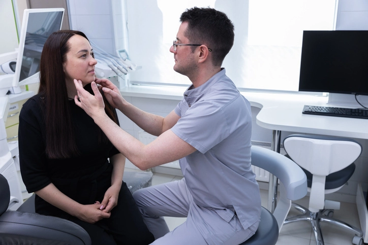
6. The Neck
A structured neck examination is crucial:
Inspection
- Observe for scars (previous surgery), swellings, asymmetry, or skin changes.
- Prominent pulsations may indicate vascular anomalies.
Palpation
Systematic palpation of lymph nodes, beginning under the chin and progressing along the cervical chain to the supraclavicular region.
Nodes are assessed for size, mobility, tenderness, and consistency:
Tender, soft nodes: usually reactive to infection.
Hard, fixed nodes: may indicate malignancy.
The thyroid gland is palpated for enlargement, nodules, or tenderness. Swelling that moves with swallowing suggests thyroid pathology.
Auscultation
Listen for bruits over the carotid arteries, which may indicate atherosclerosis.
7. Temporomandibular Joint (TMJ)
The TMJ is of paramount importance in dentistry due to its role in mastication and speech.
Examination involves:
- Palpation: Assess for tenderness, crepitus, or clicking sounds during opening and closing.
- Movement: Measure maximum opening, lateral excursions, and protrusion. Restricted movement may suggest joint pathology or muscular disorders.
- Observation: Deviation of the mandible on opening may indicate joint dysfunction.
Clinical relevance:
TMJ disorders are common in dental practice, associated with bruxism, stress, trauma, or degenerative joint disease. Differentiating between physiological clicking and pathological signs is essential to avoid unnecessary interventions.
Special Considerations in Dental Practice
1. Cranial Nerve Examination
Cranial nerves are particularly relevant in maxillofacial surgery and advanced dental practice. Assessment includes:
- CN V (Trigeminal): Sensory supply to face and motor supply to muscles of mastication.
- CN VII (Facial): Motor control of facial expression.
- CN IX, X, XII: Important for swallowing and tongue movements.
2. Oncological Screening
Dentists are at the forefront of oral cancer detection. Comprehensive head and neck examination should include vigilance for suspicious lesions, lymphadenopathy, or non-healing ulcers.
3. Impact of Systemic Conditions
Systemic diseases such as diabetes, hypertension, and autoimmune disorders often manifest in the head and neck region. Dentists should be adept at recognizing these signs and correlating them with oral findings.
Documentation and Communication
Findings from the examination should be meticulously documented, including negative findings. Clear communication with patients about the significance of abnormalities, as well as timely referral to specialists, ensures holistic patient care.
Conclusion
The head and neck examination is an essential component of dental practice, extending beyond the oral cavity to include structures that reveal both local and systemic pathology. A systematic approach—encompassing inspection, palpation, movement assessment, and occasionally auscultation—enables dentists to identify abnormalities, provide early interventions, and collaborate effectively with other healthcare professionals.
Dentists must cultivate not only technical skill in examination but also clinical judgment to discern when findings are significant and warrant referral. Given the increasing incidence of oral and head-and-neck cancers worldwide, the role of dentists in early detection cannot be overstated. A thorough, routine head and neck examination should thus be integral to every dental assessment.