
Endodontics is a critical component of restorative dentistry, focusing on the prevention, diagnosis, and treatment of diseases of the dental pulp and periradicular tissues. Despite advances in technology, materials, and clinical techniques, endodontic treatment continues to present a range of biological and technical challenges. These challenges may arise before treatment, during canal instrumentation, or after obturation, and they require sound biological understanding combined with meticulous clinical execution.
Common endodontic problems include acute infections such as periapical abscesses, post-operative pain, difficulties in canal negotiation due to sclerosis or calcification, fractured instruments, immature teeth with open apices, perforations, ledge formation, and combined periodontal–endodontic lesions. Successful management of these problems depends on accurate diagnosis, timely intervention, and the appropriate use of modern endodontic materials and techniques.
Table of Contents
ToggleAcute Periapical Abscess
Definition and Etiology
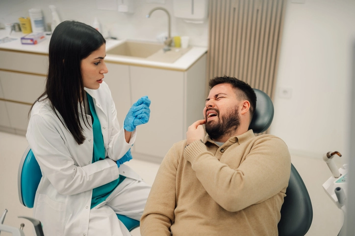
An acute periapical abscess is a localized accumulation of pus at the apex of a tooth root, usually resulting from pulpal necrosis and bacterial infection spreading into the periradicular tissues. It is often associated with severe pain, swelling, tenderness to percussion, and sometimes systemic signs such as fever and malaise.
Clinical Management
The primary goal in managing an acute periapical abscess is drainage, which provides immediate relief of symptoms by reducing tissue pressure and bacterial load. Whenever possible, drainage should be achieved through the tooth under rubber dam isolation. This approach minimizes contamination and maintains asepsis.
The pulp chamber is opened using a diamond bur in a high-speed turbine handpiece. Supporting the tooth during access preparation helps reduce vibration and patient discomfort. Local or regional anesthesia is often required, although anesthesia may be less effective in acutely inflamed tissues. In some cases, conscious sedation may be necessary.
Once access is gained, the root canal should be gently irrigated with sodium hypochlorite, which provides antimicrobial action and dissolves necrotic tissue. Importantly, the tooth should ideally be resealed after drainage rather than left open, as open drainage increases the risk of secondary infection. A review appointment within 24 hours is preferable if symptoms persist.
If a fluctuant soft tissue swelling is present, incision and drainage of the abscess through the mucosa may be required. Antibiotics are not routinely indicated unless there is evidence of spreading infection (cellulitis), systemic involvement, or compromised host immunity. Occlusal adjustment may also be necessary to eliminate traumatic occlusion and reduce post-operative discomfort.
Pain Following Instrumentation
Causes
Post-instrumentation pain is a relatively common occurrence in endodontics. It may result from:
- Mechanical irritation caused by over-instrumentation
- Chemical irritation from irrigants extruded beyond the apex
- Apical extrusion of debris, bacteria, or necrotic tissue
In some cases, a previously asymptomatic tooth may develop an acute flare-up following initial instrumentation, especially in teeth with chronic periapical pathology.
Management and Prevention
Prevention is key and includes:
- Accurate working length determination
- Use of crown-down instrumentation techniques
- Gentle irrigation with side-vented needles
- Avoiding apical over-enlargement
Management typically involves reassurance, analgesics, occlusal adjustment if indicated, and sometimes short-term intracanal medicaments such as calcium hydroxide to reduce inflammation.
Sclerosed and Calcified Canals
Clinical Significance
Canal sclerosis or obliteration is commonly associated with aging, trauma, or long-standing irritation. Although the incidence of pulp necrosis in sclerosed canals is relatively low (13–16%), pulp death can still occur, necessitating root canal treatment.
Challenges in Management
Locating the canal orifice can be extremely challenging due to calcification. Initial exploration with small stainless steel files may be unsuccessful. In such cases, the clinician should carefully assess radiographs and explore the expected anatomical location of the canal.
Techniques for Canal Location
- Use of magnification and illumination (loupes or operating microscope)
- Small round burs or ultrasonic tips to trough dentin
- Use of chelating agents such as EDTA-containing lubricants (e.g., RC Prep™, Glyde™, File-Eze®)
Once the canal is located, negotiation should begin with size 8 or 10 files using gentle watch-winding motions. Studies have shown success rates of approximately 80% even in canals that are radiographically indistinguishable. If complete blockage is encountered, obturation up to the level of blockage or periradicular surgery may be considered.
Pulp Stones
Pulp stones are calcified masses found within the pulp chamber or root canals. They may be free, attached, or embedded within dentin.
Management
- Pulp stones in the chamber can often be dislodged using an excavator or ultrasonic scaler.
- Stones within canals require the use of EDTA and small files to gently loosen and remove them.
Care must be taken to avoid excessive dentin removal or perforation.
Fractured Instruments
Etiology
Instrument separation may occur due to metal fatigue, excessive force, canal curvature, or improper use of rotary instruments.
Factors Affecting Prognosis
- Location of the fragment (coronal, middle, or apical third)
- Canal anatomy and curvature
- Length and type of fractured instrument
Management Options
- Bypassing the fragment using small files
- Removal using ultrasonics to create a staging platform
- Use of fine forceps such as Steiglitz forceps if accessible
- Use of specialized devices such as the Masserann kit, although this may weaken root dentin
If the fragment lies in the apical third and removal risks excessive dentin loss, obturating the canal coronal to the fragment and monitoring may be preferable. Periradicular surgery is considered a last resort.
Immature Teeth with Incomplete Root Formation
Clinical Challenges
Non-vital teeth with open apices present a unique challenge due to:
- Lack of apical constriction
- Thin dentinal walls
- Difficulty achieving an apical seal
Management Strategies
Working length determination can be achieved using radiographs, electronic apex locators, or modified K-files. Canal preparation emphasizes irrigation rather than aggressive instrumentation.
Apexification using materials such as mineral trioxide aggregate (MTA) or bioceramic cements has largely replaced traditional long-term calcium hydroxide therapy. Following apical barrier formation, obturation is completed using warm gutta-percha.
Revascularization (regenerative endodontics) is an alternative approach that promotes continued root development in immature non-vital teeth and is increasingly supported by clinical evidence.
Removal of Old Root Fillings
Indications
Removal of existing root fillings is often required during retreatment due to persistent infection, inadequate obturation, or procedural errors.
Techniques
- Hedström files for single gutta-percha cones
- Thermal softening using Gates-Glidden drills
- Rotary NiTi retreatment systems (e.g., ProTaper Universal Retreatment, Mtwo Retreatment files)
Chemical solvents such as chloroform or eucalyptus oil may assist but can leave a smear layer on canal walls. Complete removal is difficult, and remnants should be assessed under magnification.
Perforations
Causes
Perforations may be iatrogenic or result from pathological resorption. They represent a serious complication due to communication between the root canal system and periodontal tissues.
Management Principles
- Immediate sealing improves prognosis
- Control of bleeding and contamination is essential
Materials
MTA and bioceramic materials are the materials of choice due to their biocompatibility, sealing ability, and ability to promote hard tissue formation.
Management by Location
- Pulp chamber floor: Small perforations may be repaired; large ones may require hemisection or extraction.
- Lateral perforations: Management depends on proximity to the gingival margin and size; surgical repair may be required.
- Apical perforations: Vertical compaction techniques may succeed; otherwise, periradicular surgery is indicated.
Ledge Formation
A ledge is an artificial irregularity in the canal wall that prevents instruments from reaching the apex.
Management
Correction involves returning to a small, pre-curved file and carefully negotiating past the ledge using chelating lubricants such as EDTA or RC Prep™. Patience and tactile sensitivity are critical.
Periodontal–Endodontic Lesions
These lesions involve both pulpal and periodontal tissues and require careful diagnosis to determine the primary origin. Management may involve endodontic therapy alone or combined endodontic–periodontal treatment depending on lesion type.
Partial (Cvek) Pulpotomy
A partial pulpotomy is indicated primarily in traumatic crown fractures involving young permanent teeth. The procedure preserves pulp vitality and promotes continued root development when performed promptly under aseptic conditions.
Conclusion
Endodontic problems are diverse and often complex, requiring a deep understanding of pulp biology, pathology, and technical principles. Advances in magnification, materials such as MTA and bioceramics, and improved instrumentation techniques have significantly enhanced treatment outcomes. However, prevention through careful planning, meticulous technique, and sound clinical judgment remains paramount. A systematic and evidence-based approach allows clinicians to manage even the most challenging endodontic complications successfully.
References
- Andreasen JO, Andreasen FM, Andersson L. Textbook and Color Atlas of Traumatic Injuries to the Teeth. 4th ed. Oxford: Wiley-Blackwell; 2007.
- Torabinejad M, Chivian N. Clinical applications of mineral trioxide aggregate. J Endod. 1999;25(3):197–205.
- Ingle JI, Bakland LK, Baumgartner JC. Ingle’s Endodontics. 6th ed. Hamilton: BC Decker; 2008.
- Cohen S, Hargreaves KM. Pathways of the Pulp. 11th ed. St. Louis: Elsevier; 2016.
- European Society of Endodontology. Quality guidelines for endodontic treatment: consensus report of the European Society of Endodontology. Int Endod J. 2006;39(12):921–930.
- Siqueira JF Jr, Rôças IN. Clinical implications and microbiology of bacterial persistence after treatment procedures. J Endod. 2008;34(11):1291–1301.
- Kim S, Kratchman S. Modern endodontic surgery concepts and practice: a review. J Endod. 2006;32(7):601–623.
- Schilder H. Filling root canals in three dimensions. Dent Clin North Am. 1967;11(2):723–744.
- Peters OA. Current challenges and concepts in the preparation of root canal systems: a review. J Endod. 2004;30(8):559–567.
- Ricucci D, Siqueira JF Jr. Biofilms and apical periodontitis: study of prevalence and association with clinical and histopathologic findings. J Endod. 2010;36(8):1277–1288.
- Cvek M. A clinical report on partial pulpotomy and capping with calcium hydroxide in permanent incisors with complicated crown fracture. J Endod. 1978;4(8):232–237.
- Fuss Z, Tsesis I, Lin S. Root perforations: classification and treatment choices based on prognostic factors. Endod Topics. 2006;13(1):56–70.
- American Association of Endodontists. Guide to Clinical Endodontics. 6th ed. Chicago: AAE; 2013.
- Ng YL, Mann V, Gulabivala K. Outcome of secondary root canal treatment: a systematic review of the literature. Int Endod J. 2008;41(12):1026–1046.
- Banchs F, Trope M. Revascularization of immature permanent teeth with apical periodontitis: new treatment protocol? J Endod. 2004;30(4):196–200.