
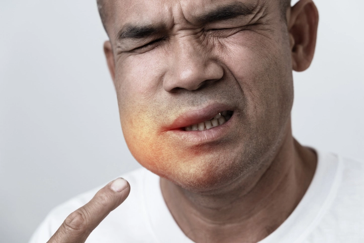
Dento-facial infections are among the most common yet potentially dangerous infections encountered in dental and oral surgery practice. Although many begin as localized odontogenic problems, they can rapidly progress into severe, life-threatening conditions if not recognized and managed promptly. A critical principle in managing dento-facial infections is understanding that antibiotics alone are rarely definitive treatment; instead, elimination of the source and appropriate surgical drainage are central to care.
Table of Contents
ToggleGeneral Principles of Dento-Facial Infections
Infections associated with teeth typically arise from:
- Necrotic dental pulps
- Periodontal pockets
- Pericoronitis around partially erupted teeth
While analgesics and antibiotics may provide temporary symptomatic relief, they do not eliminate the underlying cause. Failure to address the source can allow infections to spread into deeper anatomical spaces, leading to severe morbidity or even mortality.
Airway Risk: A Priority Assessment
One of the most critical aspects of managing dento-facial infections is airway assessment. Any patient who has difficulty swallowing their own saliva, exhibits drooling, voice changes, or shows signs of tongue elevation must be treated as an emergency. Such patients require urgent hospital admission, as airway obstruction may develop rapidly.
Deaths from odontogenic infections, though uncommon, continue to occur, underscoring the importance of vigilance.
Microbiology of Dento-Facial Infections
Dento-facial infections are typically polymicrobial, involving organisms that are part of the normal oral flora.
Key Microbial Groups
Anaerobes (most important)
Bacteroides species
Aerobic and facultative anaerobic streptococci
Occasionally:
Staphylococcus aureus
Haemophilus species (especially near the maxillary antrum)
Streptococcus milleri group is particularly significant due to its association with aggressive and potentially life-threatening infections.
Antibiotic Sensitivity
- Most organisms are sensitive to penicillins
- Bacteroides species are almost always sensitive to metronidazole
- Resistance is uncommon but increasing
Importantly, clinicians should not rely solely on metronidazole, as established infections often contain aerobic bacteria that require broader coverage.
Clinical Diagnosis
Diagnosis of dento-facial infection is usually clinical and based on:
- Pain
- Swelling
- Fever or systemic upset
- Presence of pus or discharge
- Tooth vitality and tenderness to percussion (TTP)
Imaging may be helpful in selected cases, but treatment should not be delayed when clinical signs are clear.
Apical Abscess
Definition and Pathogenesis
An apical abscess results from infection spreading beyond the apex of a tooth root, usually following pulp necrosis. The tooth is typically:
- Non-vital
- Tender to percussion
- Discoloured or previously restored
- Associated with trauma or root canal treatment history
Clinical Features
Severe localized pain initially
Pain may reduce once pus tracks into soft tissues
Discharge commonly occurs into the buccal sulcus
Exceptions:
Upper lateral incisors (palatal drainage)
Palatal roots of maxillary molars
Lower canines or incisors draining to the chin
Management
The guiding principle is drainage of pus:
- Through the root canal
- By incision and drainage of fluctuant swelling
- Or by extraction of the tooth
Local anesthesia is usually sufficient. Incision should be followed by blunt exploration and maintenance of drainage, often with:
- Tissue excision
- Placement of a small rubber drain (especially important in palatal abscesses)
Antibiotics
Used as adjuncts, not substitutes:
- Amoxicillin 500 mg three times daily for 5–7 days
- Metronidazole 400 mg three times daily for 5–7 days
- Combination therapy for more severe infections
Hospital admission is required for spreading infection or systemic involvement.
Periodontal Abscess
Periodontal abscesses arise from infection within a pre-existing periodontal pocket.
Clinical Features
- Localized swelling adjacent to a tooth
- Deep periodontal pocket
- Tooth often remains vital
Management
- Incision and drainage
- Elimination of the periodontal pocket
- Extraction if the tooth has a hopeless prognosis
Prompt intervention prevents progression into deeper fascial spaces.
Pericoronitis
Overview
Pericoronitis is inflammation and infection of the soft tissue flap (operculum) overlying a partially erupted tooth, most commonly a mandibular third molar.
Predisposing Factors
- Poor oral hygiene
- Trauma from opposing teeth
- Food stagnation under the operculum
Clinical Features
- Localized pain and swelling
- Trismus
- Bad taste or discharge
- Systemic symptoms in severe cases
Management
- Irrigation under the operculum with saline or chlorhexidine
- Removal of opposing traumatizing tooth if necessary
- Antibiotics if infection is spreading
- Definitive treatment is removal of the impacted tooth, as recurrence is common
Dry Socket (Alveolar Osteitis)
Definition
Dry socket is an inflammatory condition of the extraction socket caused by loss of the blood clot, leading to exposed bone.
Risk Factors
- Mandibular molar extractions
- Smoking
- Traumatic surgery
- Oral contraceptive use
- Immunocompromise
Clinical Presentation
- Severe pain starting 2–4 days post-extraction
- Pain worse than original toothache
- Inflamed socket with visible bone
- Halitosis
Management
- Gentle irrigation of the socket
- Placement of medicated dressings (e.g. Alvogyl®, BIPP, ZOE)
- Analgesia (NSAIDs preferred)
- Chlorhexidine or warm saline mouthwashes
Routine antibiotics are not indicated, but prophylactic anaerobic coverage may reduce incidence.
Actinomycosis
Actinomycosis is a chronic, low-grade infection caused by Actinomyces israelii.
Features
- Multiple draining sinuses
- Firm swelling
- Slow progression
Management
Surgical drainage
Prolonged antibiotics:
Amoxicillin 500 mg three times daily for up to 6 weeks
Doxycycline as an alternative
Staphylococcal Lymphadenitis
Most commonly seen in children, this condition arises from minor skin or mucosal breaches.
Clinical Features
- Enlarged tender lymph nodes
- May mimic viral exanthems (e.g. “slapped cheek” appearance)
Management
- Drainage if suppurative
- Flucloxacillin (dose adjusted for age)
Atypical Mycobacterial Infections
These present as cold, non-tender lymphadenopathy without systemic illness.
Key Points
- Slow-growing organisms
- Culture may take up to 12 weeks
- Standard anti-tuberculous therapy is inappropriate
Management
- Surgical excision is definitive
- Clarithromycin is the most useful antibiotic if required
Ludwig’s Angina
Definition
Ludwig’s angina is a rapidly spreading cellulitis involving:
- Submandibular spaces
- Sublingual spaces
- Typically bilateral
Clinical Features
- Board-hard swelling of the floor of mouth
- Tongue elevation and posterior displacement
- Dysphagia and drooling
- Systemic toxicity
Clinical Significance
This condition is a medical and surgical emergency. The airway is at immediate risk.
Management
- Immediate hospital admission
- Airway protection
- IV antibiotics
- Surgical drainage
Necrotizing Fasciitis
A rare but devastating infection caused by highly virulent bacteria, often streptococci.
Features
- Rapid tissue destruction
- Severe systemic illness
- High mortality if untreated
Management
- Aggressive surgical debridement
- IV antibiotics
- Intensive supportive care
Abscess vs Cellulitis
Abscess
- Localized pus collection
- Poor antibiotic penetration
- Requires drainage
Cellulitis
- Diffuse inflammatory spread
- Good blood supply
- Responds well to high-dose antibiotics
Many head and neck infections exhibit features of both.
Conclusion
Dento-facial infections represent a spectrum ranging from minor localized abscesses to life-threatening deep neck space infections. The cornerstone of management is early recognition, source control, and appropriate surgical drainage, supported by antibiotics rather than replaced by them.
A thorough understanding of anatomy, microbiology, and clinical presentation allows clinicians to intervene early, prevent complications, and save lives. For dental and medical practitioners alike, vigilance, sound clinical judgment, and respect for the potential severity of these infections remain essential.
References
- Peterson LJ, Ellis E, Hupp JR, Tucker MR. Contemporary Oral and Maxillofacial Surgery. 7th ed. St. Louis: Elsevier; 2019.
- Flynn TR. Principles and surgical management of head and neck infections. Oral Maxillofac Surg Clin North Am. 2011;23(3):327–337.
- Hupp JR, Ellis E, Tucker MR. Contemporary Oral and Maxillofacial Surgery. 6th ed. St. Louis: Mosby Elsevier; 2014.
- Topazian RG, Goldberg MH, Hupp JR. Oral and Maxillofacial Infections. 4th ed. Philadelphia: Saunders; 2002.
- Brook I. Microbiology and management of deep facial infections and Ludwig’s angina. J Oral Maxillofac Surg. 2004;62(9):1041–1048.
- Flynn TR, Shanti RM, Levi MH, et al. Severe odontogenic infections, part 1: Prospective report. J Oral Maxillofac Surg. 2006;64(7):1093–1103.
- Flynn TR, Shanti RM, Hayes C. Severe odontogenic infections, part 2: Prospective outcomes study. J Oral Maxillofac Surg. 2006;64(7):1104–1113.
- Bahl R, Sandhu S, Singh K, Sahai N, Gupta M. Odontogenic infections: Microbiology and management. Contemp Clin Dent. 2014;5(3):307–311.
- Bakathir AA, Moos KF, Ayoub AF. Management of odontogenic infections. Br J Oral Maxillofac Surg. 2006;44(4):331–336.
- Koorbusch GF, Fotos P, Goll KT. Retrospective assessment of antibiotic prescribing for odontogenic infections. J Oral Maxillofac Surg. 1992;50(11):1218–1222.
- Pogrel MA, Schmidt BL, Ammar A. Necrotizing fasciitis of the head and neck. J Oral Maxillofac Surg. 2003;61(4):455–460.
- Britt JC, Josephson GD, Gross CW. Ludwig’s angina in the pediatric population. Laryngoscope. 2000;110(5):821–826.
- Sharma M, Gupta R, Choudhary A. Ludwig’s angina: A case report and review. Indian J Otolaryngol Head Neck Surg. 2012;64(2):197–200.
- Seppänen L, Rautemaa R, Lindqvist C, Lauhio A. Changing clinical features of odontogenic maxillofacial infections. Clin Oral Investig. 2010;14(4):459–465.
- Cope AL, Francis NA, Wood F, Chestnutt IG. Antibiotic prescribing in UK general dental practice. Br Dent J. 2016;220(1):25–29.
- Scottish Dental Clinical Effectiveness Programme (SDCEP). Drug Prescribing for Dentistry. 3rd ed. Dundee: SDCEP; 2016.
- National Institute for Health and Care Excellence (NICE). Antimicrobial prescribing: dental abscess. NICE guideline NG187. London: NICE; 2020.
- Brook I. Actinomycosis: diagnosis and management. South Med J. 2008;101(10):1019–1023.
- Goldenberg D, Golz A, Netzer A, et al. Atypical mycobacterial infection of the head and neck. Otolaryngol Head Neck Surg. 2001;125(3):293–299.
- Thomas DW, Hill CM, Lewis MAO, et al. Guidelines for the management of acute dental pain and infection. Br Dent J. 1998;184(1):44–47.