
Cysts of the jaws represent a significant group of pathological lesions commonly encountered in dental and maxillofacial practice. They are often discovered incidentally on routine radiographic examination, yet they may also present with symptoms ranging from painless swelling to infection, pain, and pathological fracture. Understanding the origin, diagnosis, classification, and management of jaw cysts is essential for dental students and clinicians, as early recognition and appropriate treatment can prevent complications such as bone destruction, tooth displacement, recurrence, and facial deformity.
A cyst is defined as a pathological cavity that is lined by epithelium and typically contains fluid or semi-fluid material. In the jaws, most cysts are odontogenic in origin, meaning they arise from tissues involved in tooth development. Although many jaw cysts grow slowly and remain asymptomatic for long periods, some may behave aggressively, expanding rapidly and invading surrounding bone. The complexity of jaw cysts lies not only in their varied origins but also in their overlapping clinical and radiographic features, which can make diagnosis challenging.
Table of Contents
ToggleDefinition and General Characteristics of Jaw Cysts
Jaw cysts are abnormal cavities lined by epithelium and enclosed within bone or soft tissue. They commonly contain clear or straw-coloured fluid but may contain pus if secondary infection occurs. The epithelial lining distinguishes true cysts from pseudocysts, which lack an epithelial lining.
Most jaw cysts are derived from odontogenic epithelium, including remnants of the dental lamina, reduced enamel epithelium, or cell rests of Malassez. These epithelial remnants normally persist within the jaws after tooth development and may proliferate under certain pathological conditions, giving rise to cyst formation.
Cyst enlargement occurs through several mechanisms, including:
- Epithelial proliferation of the cyst lining
- Increased intracystic osmotic pressure, drawing fluid into the cavity
- Bone resorption, mediated by inflammatory cytokines and prostaglandins
- Hydrostatic pressure, leading to gradual expansion
As cysts enlarge, they may cause cortical bone thinning, displacement of teeth, root resorption, and, in severe cases, pathological fracture.
Pathogenesis of Jaw Cysts
The precise mechanisms underlying cyst development and growth are not fully understood, but several contributing factors have been identified.
Epithelial Proliferation
Epithelial remnants in the jaws may proliferate in response to inflammatory stimuli, trauma, or developmental abnormalities. This proliferation forms an epithelial-lined cavity that gradually enlarges.
Osmotic Pressure
The breakdown of epithelial cells within the cyst lumen increases protein concentration, raising osmotic pressure. Fluid is drawn into the cyst, increasing its size and exerting pressure on surrounding bone.
Bone Resorption
Prostaglandins and other inflammatory mediators stimulate osteoclastic activity, leading to bone resorption and cyst expansion.
Inflammation
In inflammatory cysts, necrotic pulp tissue or chronic periapical infection provides a persistent stimulus for epithelial proliferation and cyst growth.
Clinical Presentation
The clinical presentation of jaw cysts varies depending on size, location, and whether infection is present.
Asymptomatic Presentation
Many cysts are asymptomatic and discovered incidentally on routine radiographs. These lesions often grow slowly and may reach considerable size before producing symptoms.
Swelling
A painless swelling is a common presenting feature. The swelling is typically bony hard initially but may become fluctuant as cortical bone thins.
Pain and Infection
Infected cysts may present with pain, tenderness, swelling, erythema, and purulent discharge. Systemic signs such as fever are less common but may occur.
Tooth-Related Symptoms
Cysts may cause displacement of teeth, delayed eruption, or mobility. Teeth associated with inflammatory cysts are usually non-vital.
Pathological Fracture
Large cysts, especially in the mandible, may weaken bone sufficiently to result in pathological fracture.
Diagnosis
Accurate diagnosis of jaw cysts relies on a combination of clinical examination, radiographic assessment, and histopathological evaluation.
Clinical Examination
The clinician should assess:
- Presence of swelling
- Consistency (bony hard vs. fluctuant)
- Tenderness
- Relationship to teeth
- Signs of infection
Vitality testing of associated teeth is essential, as non-vital teeth suggest inflammatory odontogenic cysts.
Radiographic Assessment
Radiographic examination is crucial in the diagnosis of jaw cysts.
Common Imaging Modalities
- Dental panoramic tomography (DPT): Provides an overview of lesion size, location, and relation to adjacent structures.
- Periapical radiographs: Useful for assessing tooth involvement and periapical pathology.
- Occlusal radiographs: May demonstrate cortical expansion.
- CBCT (in selected cases): Provides detailed three-dimensional assessment.
Radiographic Features
- Well-defined radiolucency
- Corticated margins (unless infected)
- Unilocular or multilocular appearance
- Tooth displacement or root resorption
Aspiration
Aspiration of cyst contents may assist diagnosis. Straw-coloured fluid suggests a benign cyst, while thick keratinous material may indicate an odontogenic keratocyst.
Histopathology
Histological examination of the cyst lining is essential for definitive diagnosis. All excised cyst linings should be submitted for histopathological analysis to exclude neoplasia.
Principles of Treatment
The management of jaw cysts depends on the type, size, location, and patient factors.
Enucleation with Primary Closure
This is the most common and preferred treatment for most jaw cysts. It involves complete removal of the cyst lining from the bony cavity, followed by repositioning and suturing of the mucoperiosteal flap.
Advantages include:
- Definitive removal
- Rapid healing
- Low recurrence (for most cyst types)
Associated dental pathology, such as non-vital teeth, may be treated concurrently with endodontic therapy or extraction.
Enucleation with Packing and Delayed Closure
This approach is used for large or infected cysts where primary closure is unsuitable. The cavity is packed with antiseptic dressings such as Whitehead’s varnish or BIPP, allowing gradual healing.
Enucleation with Bone Grafting
Rarely indicated, this technique may be used in selected cases to restore bone volume but is generally unnecessary due to natural bone regeneration.
Marsupialization
Marsupialization involves creating a surgical window in the cyst wall and suturing it to the oral mucosa, allowing continuous drainage.
Indications include:
- Large cysts
- Cysts in medically compromised patients
- Facilitation of tooth eruption
- Cases where enucleation is contraindicated
Disadvantages include prolonged healing time and persistence of a residual cavity.
Classification of Jaw Cysts
Although many classifications exist, a practical approach divides cysts into odontogenic and non-odontogenic categories.
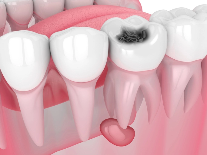
Inflammatory Odontogenic Cysts (Radicular Cysts)
Radicular cysts are the most common jaw cysts. They arise from chronic periapical inflammation associated with a non-vital tooth.
Pathogenesis
Necrotic pulp tissue stimulates epithelial proliferation from the cell rests of Malassez, leading to cyst formation.
Clinical Features
- Associated with non-vital tooth
- Often asymptomatic
- May present as swelling or infection
Radiographic Features
- Well-defined radiolucency at tooth apex
- May be lateral or residual (post-extraction)
Treatment
Enucleation combined with endodontic therapy or extraction of the offending tooth.
Dentigerous Cysts
Dentigerous cysts develop around the crown of an unerupted permanent tooth.
Pathogenesis
They arise from the reduced enamel epithelium after enamel formation.
Clinical Features
- Often associated with impacted third molars or canines
- May delay tooth eruption
Radiographic Features
- Radiolucency attached at the cemento-enamel junction
- Surrounds tooth crown
Treatment
Marsupialization or enucleation depending on tooth position and treatment goals.
Odontogenic Keratocysts
Odontogenic keratocysts are distinctive due to their aggressive behavior and high recurrence rate.
Biological Behavior
Previously classified as keratocystic odontogenic tumors, they demonstrate rapid growth and tendency to recur.
Histology
- Parakeratinized epithelial lining
- Thin, uniform epithelium
- Satellite cysts common
Clinical and Radiographic Features
- Often asymptomatic
- May appear multilocular
- Commonly in posterior mandible
Treatment
Careful enucleation with adjunctive measures such as Carnoy’s solution or cryotherapy to reduce recurrence.
Calcifying Epithelial Odontogenic Cysts
These rare cysts are characterized histologically by calcifications and ghost cells.
Diagnosis
Based primarily on histological findings.
Treatment
Simple enucleation is usually curative.
Solitary Bone Cysts
Solitary bone cysts are pseudocysts lacking epithelial lining.
Etiology
Believed to arise from unresolved intraosseous hematoma.
Clinical Features
- Usually asymptomatic
- Often discovered incidentally
Radiographic Features
- Scalloped margins between tooth roots
- Lamina dura preserved
Treatment
Surgical exploration and curettage stimulate healing; no further treatment required.
Aneurysmal Bone Cysts
These lesions consist of blood-filled spaces within bone.
Clinical Features
- Rapid expansion
- Pain if traumatized
Treatment
Small lesions may be enucleated; large lesions require excision and possible reconstruction due to recurrence risk.
Fissural Cysts
Fissural cysts arise from embryonic epithelial remnants unrelated to tooth development.
Examples
- Incisive canal cyst
- Nasolabial cyst
Treatment
Enucleation is curative.
Importance of Histopathological Examination
Submission of all cyst linings for histopathological examination is mandatory. This ensures accurate diagnosis, excludes neoplastic transformation, and guides further management.
Conclusion
Cysts of the jaws represent a diverse group of lesions with varying origins, behaviors, and treatment requirements. While many are benign and slow-growing, others may exhibit aggressive features and a high tendency to recur. Accurate diagnosis through careful clinical assessment, radiographic evaluation, and histopathological confirmation is essential.
A thorough understanding of jaw cysts enables clinicians to select appropriate treatment strategies, minimize complications, and ensure optimal patient outcomes. For dental students and practitioners, mastery of this topic is fundamental to safe and effective oral surgical practice.
References
- Cawson, R. A., Odell, E. W.
Cawson’s Essentials of Oral Pathology and Oral Medicine.
9th ed. Edinburgh: Churchill Livingstone Elsevier; 2017. - Neville, B. W., Damm, D. D., Allen, C. M., Chi, A. C.
Oral and Maxillofacial Pathology.
4th ed. St. Louis: Elsevier; 2016. - Shear, M., Speight, P.
Cysts of the Oral and Maxillofacial Regions.
4th ed. Oxford: Blackwell Munksgaard; 2007. - Shafer, W. G., Hine, M. K., Levy, B. M.
Shafer’s Textbook of Oral Pathology.
8th ed. New Delhi: Elsevier India; 2016. - Peterson, L. J., Ellis, E., Hupp, J. R., Tucker, M. R.
Contemporary Oral and Maxillofacial Surgery.
7th ed. St. Louis: Elsevier; 2019. - Regezi, J. A., Sciubba, J. J., Jordan, R. C. K.
Oral Pathology: Clinical Pathologic Correlations.
7th ed. St. Louis: Elsevier; 2017. - WHO Classification of Head and Neck Tumours.
Odontogenic and Maxillofacial Bone Tumours.
4th ed. Lyon: IARC Press; 2017. - Pogrel, M. A.
The Keratocystic Odontogenic Tumor.
Oral and Maxillofacial Surgery Clinics of North America. 2013;25(1):21–30. - MacDonald-Jankowski, D. S.
Odontogenic cysts: a review of the literature.
Dentomaxillofacial Radiology. 2011;40(1):1–14. - Johnson, N. R., Batstone, M. D., Savage, N. W.
Management and recurrence of keratocystic odontogenic tumor: a systematic review.
Oral Surgery, Oral Medicine, Oral Pathology, Oral Radiology. 2013;116(4):e271–e276.