
Benign tumours of the mouth represent a diverse group of lesions that arise from the various tissues found within the oral cavity and jaws. Although benign, these tumours can cause significant morbidity due to local invasion, facial deformity, interference with mastication, speech, and aesthetics, and in some cases, difficulty in diagnosis due to overlapping clinical and radiographic features with malignant lesions.
Understanding benign oral tumours is essential for dental practitioners, oral and maxillofacial surgeons, and clinicians involved in head and neck care. Accurate diagnosis relies on a combination of clinical examination, radiographic assessment, histopathological evaluation, and appropriate surgical management.
Table of Contents
ToggleClassification of Benign Oral Tumours
Benign tumours of the mouth can be broadly classified based on their tissue of origin:
Non-odontogenic tumours
AdvertisementsEpithelial tumours
Connective tissue tumours
Odontogenic tumours
Tumours derived from odontogenic epithelium
Tumours derived from odontogenic mesenchyme
Mixed odontogenic tumours
Odontogenic hamartomas
This classification is clinically relevant, as it guides diagnostic approaches and treatment planning.
Non-Odontogenic Tumours
1. Epithelial Tumours
Squamous Cell Papilloma
Squamous cell papilloma is one of the most common benign epithelial tumours of the oral cavity. It is caused by infection with the human papillomavirus (HPV), most commonly HPV types 6 and 11.
Clinical Features:
- Appears as a white or pink, exophytic, cauliflower-like lesion
- May be pedunculated or sessile
- Common sites include the palate, tongue, lips, and buccal mucosa
- Typically small and asymptomatic
- Slow-growing
Pathology:
Composed of finger-like projections of stratified squamous epithelium supported by fibrovascular connective tissue cores
Malignant Potential:
Does not undergo malignant transformation, distinguishing it from premalignant epithelial lesions
Management:
- Simple surgical excision is curative
- Recurrence is rare if completely excised
2. Connective Tissue Tumours
Connective tissue tumours arise from mesenchymal elements such as fibrous tissue, adipose tissue, nerve tissue, bone, and histiocytic cells.
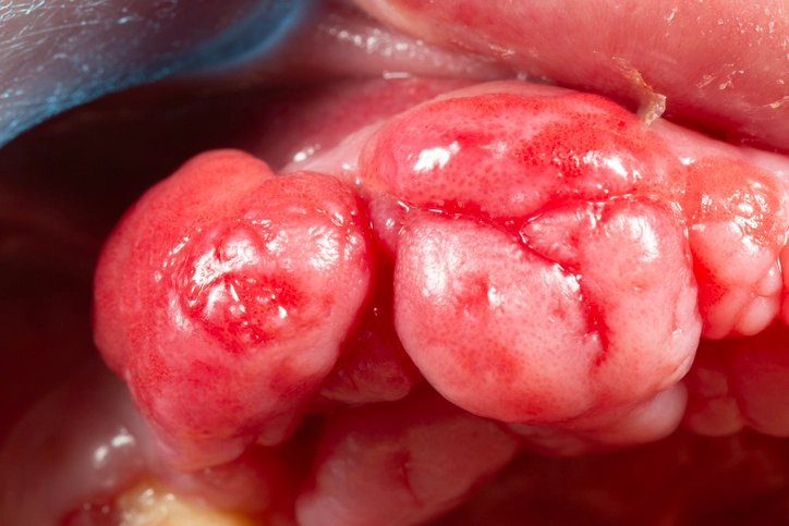
Fibroma
Fibroma is a benign fibrous tumour of the oral cavity, often representing a reactive lesion rather than a true neoplasm.
Clinical Features:
- Usually pink, firm, and pedunculated
- Commonly found on the buccal mucosa, tongue, or gingiva
- Often associated with chronic irritation or trauma
- Slow-growing and painless
Pathology:
- Dense collagen fibers with few cells
- Covered by normal stratified squamous epithelium
Management:
- Excision with a narrow margin
- Excellent prognosis with minimal recurrence
Lipoma
Lipoma is a benign tumour composed of mature adipose tissue and is relatively uncommon in the oral cavity.
Clinical Features:
- Soft, smooth, doughy consistency
- Yellowish hue may be visible through the mucosa
- Common sites include the buccal mucosa, floor of mouth, and tongue
- Typically slow-growing and asymptomatic
Pathology:
- Mature fat cells arranged in lobules
- Encapsulated
Management:
- Enucleation or conservative excision
- Recurrence is rare
Osteoma
Osteoma is a benign tumour of mature bone.
Clinical Features:
- Hard, well-circumscribed swelling
- Usually unilateral
- Covered by normal oral mucosa
- Most commonly affects the mandible and paranasal sinuses
Clinical Significance:
Osteomas may be associated with Gardner syndrome, a variant of familial adenomatous polyposis, characterized by:
Multiple osteomas
Impacted supernumerary teeth
Intestinal polyps with malignant potential
Management:
- Surgical excision if symptomatic or cosmetically concerning
- Asymptomatic lesions may be observed
Neurofibroma
Neurofibroma is a benign tumour arising from peripheral nerve fibroblasts.
Clinical Features:
- May occur as a solitary lesion or as part of neurofibromatosis type 1 (von Recklinghausen disease)
- Commonly affects the tongue
- Soft to firm, non-encapsulated lesion
Pathology:
Mixed population of Schwann cells, fibroblasts, and perineural cells
Malignant Potential:
May undergo sarcomatous transformation, particularly in patients with neurofibromatosis
Management:
- Excision with a small margin
- Long-term follow-up is advised in syndromic cases
Neurilemmoma (Schwannoma)
Neurilemmoma, also known as schwannoma, is a benign nerve sheath tumour composed exclusively of Schwann cells.
Clinical Features:
- Slow-growing, encapsulated mass
- May cause nerve-related symptoms if large
- Common oral sites include the tongue and floor of mouth
Pathology:
- Antoni A and Antoni B patterns
- Verocay bodies are characteristic
Management:
- Surgical excision
- Nerve fibers may often be preserved due to eccentric tumour growth
Granular Cell Myoblastoma
Granular cell myoblastoma is a rare benign tumour thought to arise from histiocytic or Schwann cell origin.
Clinical Features:
- Firm nodule, commonly on the tongue
- May mimic malignancy clinically due to firmness
Pathology:
Large polygonal cells with granular cytoplasm
Management:
- Excision with a margin
- Excellent prognosis
Ossifying Fibroma
Ossifying fibroma is a benign fibro-osseous lesion of the jaws.
Clinical Features:
- Painless, slow-growing swelling
- Causes expansion of both buccal and lingual cortical plates
- Most commonly affects the mandible
Radiographic Features:
- Well-defined radiolucent lesion with a radio-opaque margin
- Progressive calcification over time
Histology:
- Fibrous tissue containing varying amounts of bone or cementum-like material
- Similar to fibrous dysplasia but well-demarcated
Management:
- Enucleation or conservative excision
- A more aggressive juvenile form occurs in children but remains benign
Odontogenic Tumours
Odontogenic tumours arise from tissues involved in tooth development and are unique to the jaws.
Ameloblastoma
Ameloblastoma is one of the most clinically significant odontogenic tumours due to its aggressive local behavior.
Epidemiology:
- Common in adults
- Higher prevalence in males and individuals of African descent
- Predominantly affects the posterior mandible
Clinical Features:
- Painless jaw swelling
- Facial asymmetry
- Loosening or displacement of teeth
Types:
- Unicystic
- Polycystic (multicystic)
- Peripheral
Radiographic Features:
Multilocular “soap bubble” or “honeycomb” appearance
Histological Patterns:
- Plexiform
- Follicular
Management:
- Unicystic ameloblastoma: enucleation with removal of a rim of bone
- Other types: surgical excision with a margin due to high recurrence risk
Adenoameloblastoma
Adenoameloblastoma typically occurs in young females and involves the anterior maxilla.
Clinical Features:
- Slow-growing, painless swelling
- Often associated with impacted teeth
Management:
- Conservative excision
- Recurrence is uncommon
Calcifying Epithelial Odontogenic Tumour (Pindborg Tumour)
This rare tumour is characterized by calcified deposits within the lesion.
Radiographic Features:
Radiolucent lesion with scattered radiopaque foci
Management:
- Excision with a margin
- Recurrence is possible but uncommon
Myxoma
Odontogenic myxoma arises from odontogenic mesenchyme.
Clinical Features:
- Occurs in young adults
- Slow-growing but locally invasive
- Causes painless jaw expansion
Radiographic Features:
Multilocular radiolucency with a “soap bubble” appearance
Histology:
Spindle-shaped cells in a loose mucoid stroma
Management:
- Wide excision with surrounding normal bone
- High recurrence if inadequately removed
Ameloblastic Fibroma
A rare mixed odontogenic tumour.
Clinical Features:
- Occurs in young adults
- Painless jaw expansion
Radiographic Features:
Unilocular radiolucency
Management:
- Enucleation
- Excellent prognosis
Odontomes
Odontomes are the most common odontogenic lesions and are considered hamartomas rather than true neoplasms.
Types:
- Compound odontome: multiple miniature teeth
- Complex odontome: irregular mass of dental tissue
Clinical Significance:
- Often associated with delayed eruption of teeth
- Usually asymptomatic
Management:
Surgical removal using standard dento-alveolar techniques
Conclusion
Benign tumours of the mouth encompass a wide spectrum of lesions with varying origins, clinical behavior, and management requirements. While benign, many of these tumours can cause significant local destruction and functional impairment if not diagnosed and treated early. A thorough understanding of their clinical presentation, radiographic features, and histopathological characteristics is essential for effective management.
Dental professionals must maintain a high index of suspicion when evaluating oral swellings and ensure timely referral and appropriate surgical intervention. With proper diagnosis and treatment, the prognosis for benign oral tumours is excellent.
References
- Neville BW, Damm DD, Allen CM, Chi AC.
Oral and Maxillofacial Pathology. 4th ed. St. Louis: Elsevier; 2016. - Shafer WG, Hine MK, Levy BM.
Shafer’s Textbook of Oral Pathology. 8th ed. New Delhi: Elsevier India; 2016. - Regezi JA, Sciubba JJ, Jordan RCK.
Oral Pathology: Clinical Pathologic Correlations. 7th ed. St. Louis: Elsevier; 2017. - Cawson RA, Odell EW.
Cawson’s Essentials of Oral Pathology and Oral Medicine. 9th ed. London: Churchill Livingstone; 2017. - Marx RE, Stern D.
Oral and Maxillofacial Pathology: A Rationale for Diagnosis and Treatment. 2nd ed. Hanover Park: Quintessence Publishing; 2012. - Pogrel MA, Schmidt BL, Ammar A.
Oral and Maxillofacial Surgery: Essentials for Students and Practitioners. Oxford: Oxford University Press; 2014. - Kramer IRH, Pindborg JJ, Shear M.
Histological Typing of Odontogenic Tumours. 2nd ed. World Health Organization; 1992. - Barnes L, Eveson JW, Reichart P, Sidransky D (eds).
World Health Organization Classification of Tumours: Pathology and Genetics of Head and Neck Tumours. Lyon: IARC Press; 2005. - Sapp JP, Eversole LR, Wysocki GP.
Contemporary Oral and Maxillofacial Pathology. 2nd ed. St. Louis: Mosby; 2004. - Rajendran R, Sivapathasundharam B.
Shafer’s Textbook of Oral Pathology. 7th ed. New Delhi: Elsevier India; 2012.