
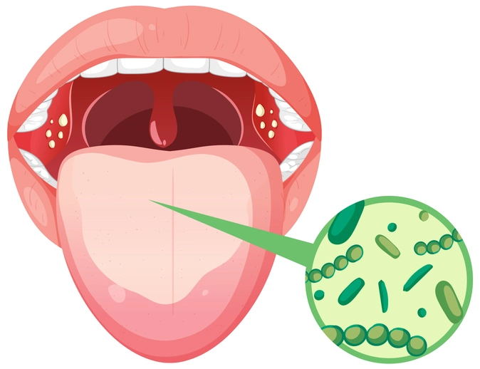
The oral cavity is a complex anatomical and ecological environment that hosts a diverse microbiota. While many microorganisms exist in a commensal or symbiotic relationship with the host, disruptions in local or systemic immunity can allow pathogenic bacteria to cause disease. Bacterial infections of the mouth represent a significant component of oral medicine and are encountered frequently in dental and medical practice. These infections may be localized to the oral mucosa or may reflect manifestations of systemic disease.
Oral bacterial infections can arise from endogenous flora, as seen in dental caries and periodontal disease, or from exogenous pathogens transmitted through respiratory droplets, sexual contact, or hematogenous spread. Importantly, the mouth may be the first site where systemic infections present, making oral examination a critical component of early diagnosis.
Bacterial infections of the mouth can be broadly categorized into:
- Odontogenic infections – such as dental caries, pulpitis, periapical abscesses, and periodontal disease.
- Non-odontogenic mucosal infections – caused by specific bacterial pathogens with characteristic oral manifestations.
- Systemic infections with oral signs – where the oral cavity reflects an underlying systemic disease.
This review focuses primarily on mucosal infections, particularly those with distinctive oral features that aid in diagnosis.
Table of Contents
ToggleScarlet Fever
Etiology and Pathogenesis
Scarlet fever is an acute infectious disease most commonly affecting children aged 4 to 8 years. It is caused by infection with Group A beta-hemolytic Streptococcus (Streptococcus pyogenes), specifically strains that produce erythrogenic (pyrogenic) exotoxins. The disease results from a delayed-type hypersensitivity reaction to these toxins rather than from direct bacterial invasion.
The exotoxins act as superantigens, stimulating massive T-cell activation and cytokine release, which leads to the systemic and mucocutaneous manifestations characteristic of scarlet fever.
Clinical Features
The disease typically follows streptococcal pharyngitis and presents with:
- Sudden onset sore throat
- Fever
- General malaise
- Headache
- Cervical lymphadenopathy
Within 24–48 hours, a diffuse erythematous rash develops. This rash has a fine, sandpaper-like texture and typically spares the perioral region, producing circumoral pallor.
Oral Manifestations
The oral cavity exhibits some of the most pathognomonic features of scarlet fever:
- Erythematous oral mucosa – due to increased vascular permeability and inflammation.
- Strawberry tongue – initially, the dorsum of the tongue becomes coated with a white exudate through which enlarged, edematous fungiform papillae protrude.
- Raspberry tongue – later, as the white coating desquamates, the tongue becomes bright red, smooth, and glossy, with prominent papillae.
These changes are diagnostically significant and are often emphasized in clinical teaching.
Diagnosis and Management
Diagnosis is primarily clinical, supported by:
- Throat swab and culture
- Rapid antigen detection tests
Treatment consists of systemic antibiotic therapy, usually penicillin. Early treatment prevents complications such as rheumatic fever and post-streptococcal glomerulonephritis. Oral manifestations typically resolve completely within two weeks.
Tuberculosis (TB)
Epidemiology and Etiology
Tuberculosis is a chronic infectious disease caused by Mycobacterium tuberculosis. Although TB incidence declined significantly in the mid-20th century in many Western countries, it has re-emerged due to factors such as:
- Increased global migration
- HIV/AIDS pandemic
- Immunosuppressive therapies
- Socioeconomic disparities
While approximately one-third of the world’s population is infected with TB, oral involvement is rare.
Pathogenesis of Oral TB
Oral tuberculosis usually represents secondary involvement, arising from:
- Autoinoculation from infected sputum in pulmonary TB
- Hematogenous spread
- Coexisting immunosuppression, particularly HIV infection
The intact oral mucosa is relatively resistant to TB infection. Breaches such as trauma, ulcers, or inflammation increase susceptibility.
Clinical Presentation
Oral TB commonly presents as:
- A deep, painful ulcer
- Irregular or raised margins
- Gradual enlargement
- Induration at the base
The most frequently affected site is the posterior dorsum of the tongue, though any oral mucosal surface may be involved.
Histopathology and Diagnosis
Histological examination typically reveals:
- Necrotizing granulomatous inflammation
- Langhans giant cells
- Epithelioid histiocytes
Special staining with Ziehl–Neelsen stain demonstrates acid-fast bacilli. Polymerase chain reaction (PCR) testing may aid diagnosis, particularly in atypical cases.
Radiographic findings may include calcified lymph nodes visible as facial radio-opacities, indicating prior infection.
Management
Management requires referral to a chest physician. Treatment involves combination anti-tubercular chemotherapy for several months. Surgical intervention, such as lymph node excision, may be required in pediatric cases.
Syphilis
Etiology and Transmission
Syphilis is a chronic, multisystem sexually transmitted infection caused by the spirochete Treponema pallidum. Transmission occurs primarily through sexual contact, but vertical transmission from mother to fetus is also possible.
Syphilis progresses through distinct clinical stages, each with characteristic oral manifestations.
Primary Syphilis
Clinical Features
The primary lesion, known as a chancre, develops at the site of inoculation approximately 3 weeks after exposure. In the oral cavity, chancres may occur on the lips, tongue, or oral mucosa.
Key features include:
- Firm, indurated ulcer
- Painless
- Clean base
- Highly infectious
Regional lymphadenopathy, particularly cervical nodes in oral lesions, is common.
Natural History
The chancre heals spontaneously within 1–2 months, even without treatment, which may lead to false reassurance and disease progression.
Secondary Syphilis
Systemic Features
Secondary syphilis develops 2–4 months after the primary stage and reflects widespread hematogenous dissemination. Features include:
- Generalized rash
- Fever
- Malaise
- Headache
- Weight loss
- Lymphadenopathy
Oral Manifestations
Oral lesions are highly characteristic and include:
- Mucous patches – shallow, grayish-white lesions
- Snail-track ulcers – serpiginous, sloughy ulcerations
These lesions are extremely infectious and often painful, interfering with eating and speaking.
Diagnosis
Serological tests are reliably positive at this stage. Dark-field microscopy can identify spirochetes from lesions.
Tertiary Syphilis
Pathogenesis
Tertiary syphilis develops years later in approximately 30% of untreated patients. It is characterized by immune-mediated tissue destruction rather than active infection.
Oral Features
The hallmark lesion is the gumma, a necrotic granulomatous mass that commonly affects:
- Palate
- Tongue
Gummas may enlarge, ulcerate, and cause palatal perforation, leading to oronasal communication.
Lesions are non-infectious but destructive.
Systemic Involvement
Tertiary syphilis is a multisystem disorder, potentially causing:
- Neurosyphilis
- Cardiovascular syphilis
- Vasculitis
Congenital Syphilis
Pathogenesis
Congenital syphilis results from transplacental transmission of T. pallidum. The severity depends on the stage of maternal infection and adequacy of treatment.
Classical Features
Late congenital syphilis is associated with a characteristic triad and additional features:
- Saddle nose deformity
- Frontal bossing
- Sensorineural deafness
- Hutchinson incisors (peg-shaped, notched)
- Mulberry (Moon) molars
These dental anomalies are of particular relevance to dental professionals, as they may provide the first clue to diagnosis.
Gonorrhoea
Etiology and Epidemiology
Gonorrhoea is caused by Neisseria gonorrhoeae and is significantly more common than syphilis. Oral infection occurs through oro-genital contact.
Oral and Pharyngeal Manifestations
Oral gonorrhoea often presents non-specifically as:
- Stomatitis
- Pharyngitis
- Persistent superficial ulcers
- Purulent gingivitis
Because symptoms may be mild or absent, oral gonorrhoea is frequently underdiagnosed.
Diagnosis
Diagnosis is confirmed by microbiological analysis:
- Swabs reveal Gram-negative intracellular diplococci
- Culture or nucleic acid amplification tests may be used
Management
Treatment involves high-dose antibiotic therapy, traditionally penicillin, though resistance patterns must be considered. Patients should be referred to genitourinary medicine specialists, and contact tracing is essential.
Clinical Importance of Oral Bacterial Infections
The oral cavity serves as a window to systemic health. Recognition of bacterial infections with oral manifestations is crucial for:
- Early diagnosis of systemic disease
- Prevention of complications
- Appropriate referral and multidisciplinary care
Dental and medical professionals must maintain a high index of suspicion when encountering unusual oral ulcers, mucosal changes, or persistent lesions.
Conclusion
Bacterial infections of the mouth encompass a wide spectrum of diseases, ranging from common childhood illnesses such as scarlet fever to chronic systemic infections like tuberculosis and syphilis. While some conditions present with distinctive oral signs, others may be subtle or mimic benign lesions. A thorough understanding of their pathogenesis, clinical features, and management is essential for effective diagnosis and patient care.
The oral cavity often provides the first visible evidence of systemic disease. Consequently, clinicians who are skilled in recognizing these manifestations play a vital role in early detection, appropriate referral, and improved patient outcomes.