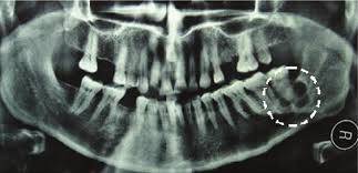
Ameloblastoma is a rare, benign but locally aggressive odontogenic tumor that predominantly affects the jawbones. It originates from the remnants of the dental lamina and is associated with the enamel-forming cells known as ameloblasts. This tumor is characterized by its slow growth and high recurrence rates if not appropriately treated. In this article, we will delve into the various aspects of ameloblastoma, including its etiology, classification, clinical presentation, diagnosis, treatment options, and prognosis.
Table of Contents
ToggleEtiology and Pathogenesis of Ameloblastoma
The exact cause of ameloblastoma remains unclear. However, it is believed to stem from the epithelial tissue associated with tooth development, particularly from the remnants of the dental lamina, enamel organ, or developing enamel. Genetic mutations and alterations in certain signaling pathways, such as the Wnt/β-catenin pathway, have been implicated in the pathogenesis of ameloblastoma.
Classification of Ameloblastoma
Ameloblastomas are typically classified into several subtypes, each with distinct clinical and histological features. The main subtypes include:
- Conventional/Classic Ameloblastoma
- Unicystic Ameloblastoma
- Extraosseous/Peripheral Ameloblastoma
- Mural Ameloblastoma
Conventional/Classic Ameloblastoma
This subtype constitutes the majority of cases and presents with well-defined epithelial islands resembling the dental enamel organ.
Unicystic Ameloblastoma
A less aggressive variant that manifests as a unilocular radiolucency and shows cystic changes histologically.
Extraosseous/Peripheral Ameloblastoma
A rare variant that occurs in the soft tissues outside the bone, often associated with the gingiva.
Mural Ameloblastoma
It is characterized by the tumor invading the cystic wall and displaying a distinctive mural pattern.
Symptoms of Ameloblastoma
Ameloblastoma predominantly occurs in the mandible (lower jaw) but can also affect the maxilla (upper jaw). Common clinical features include:
- Asymptomatic swelling
- Paresthesia
- Tooth displacement or mobility
- Facial deformity
Asymptomatic swelling
Often painless, slow-growing swelling or mass in the jawbone, particularly the mandible.
Paresthesia
Numbness or altered sensation in the affected area due to pressure on nerves.
Tooth displacement or mobility
Due to the tumor’s growth and expansion, adjacent teeth may be pushed or displaced.
Facial deformity
In advanced cases, the tumor can cause noticeable facial asymmetry and distortion.
Diagnosis of Ameloblastoma
Accurate diagnosis of ameloblastoma involves a combination of clinical, radiological, and histopathological assessments.
- Clinical Examination
- Radiographic Studies
- MRI (Magnetic Resonance Imaging)
- Histopathological Examination
Clinical Examination
Detailed examination of the oral cavity and jaw, assessing for signs of swelling, tenderness, tooth mobility, and nerve involvement.
Radiographic Studies
X-rays
Initial screening to visualize radiolucencies or radiopacities in the jawbone.
CT (Computed Tomography) Scan
Provides detailed 3D imaging, aiding in evaluating the extent and invasion of the tumor.
MRI (Magnetic Resonance Imaging)
Helpful in determining soft tissue involvement and relationship with adjacent structures.
Histopathological Examination
Biopsy
Tissue sample obtained through a surgical procedure for microscopic evaluation by a pathologist to confirm the diagnosis.
Treatment of Ameloblastoma
Treatment of ameloblastoma involves a multidisciplinary approach, including surgery, with the goal of complete removal of the tumor and reconstruction of the affected area.
- Surgical Resection
- Reconstruction
- Adjuvant Therapy
Surgical Resection
- Enucleation and Curettage
- Segmental Resection
- Marginal Resection
Enucleation and Curettage
Commonly used for unicystic ameloblastoma. The cystic lining is removed, and the cavity is thoroughly scraped and cleaned.
Segmental Resection
Involves removal of the affected portion of the jawbone, followed by reconstruction using bone grafts or other surgical techniques.
Marginal Resection
Removal of the tumor with a margin of normal tissue to reduce the risk of recurrence.
Reconstruction
- Bone Grafts
- Free Flap Reconstruction
Bone Grafts
Autografts, allografts, or synthetic grafts may be used to restore the shape and function of the jaw.
Free Flap Reconstruction
Involves transferring tissue from another part of the body, such as fibula or iliac crest, to reconstruct the jaw.
Adjuvant Therapy
- Radiation Therapy
- Chemotherapy
Radiation Therapy
Sometimes used postoperatively to reduce the risk of recurrence, especially in cases where complete resection is challenging.
Chemotherapy
Limited effectiveness and mainly reserved for aggressive or recurrent cases.
Prognosis for Ameloblastoma
The prognosis for ameloblastoma is generally good, but it depends on various factors, including the subtype, size, location, and surgical margins achieved during resection. Recurrence rates can range from 15% to 90%, emphasizing the importance of thorough surgical excision and long-term follow-up. Regular monitoring and prompt intervention in case of recurrence are essential to manage the disease effectively.
Emerging Therapeutic Approaches and Research
Continued research and advancements in the field of oncology have paved the way for exploring new therapeutic approaches for ameloblastoma. These approaches aim to enhance treatment efficacy, reduce recurrence rates, and improve patients’ quality of life.
- Targeted Therapies
- Immunotherapies
- Gene Therapy
- Radiofrequency Ablation
- Adjuvant Therapies
Targeted Therapies
Targeted therapies involve drugs or other substances that specifically target molecular and genetic changes present in ameloblastoma cells. The inhibition of specific signaling pathways, such as the Wnt/β-catenin pathway, is a promising avenue for targeted therapy.
Immunotherapies
Immunotherapies harness the body’s immune system to recognize and destroy cancer cells. These treatments can potentially be used to augment the body’s natural defense mechanisms against ameloblastoma, particularly in cases of recurrence or advanced disease.
Gene Therapy
Gene therapy involves modifying or replacing genes within the tumor cells to inhibit their growth or induce cell death. Research is ongoing to develop gene-based treatment strategies to target the genetic alterations associated with ameloblastoma.
Radiofrequency Ablation
Radiofrequency ablation is a minimally invasive technique that uses heat generated by radio waves to destroy tumor cells. It has shown promise in the treatment of recurrent or unresectable ameloblastoma.
Adjuvant Therapies
Ongoing studies are investigating the role of various adjuvant therapies, including bisphosphonates, antiangiogenic agents, and epidermal growth factor receptor (EGFR) inhibitors, in preventing recurrence and improving outcomes in patients with ameloblastoma.
Conclusion
ameloblastoma is a complex odontogenic tumor that necessitates a multidisciplinary approach for proper diagnosis and management. Advances in surgical techniques, reconstruction options, and a deeper understanding of its pathogenesis continue to improve outcomes for individuals affected by this condition. Early detection, accurate diagnosis, and timely treatment are crucial in achieving a favorable prognosis and minimizing the risk of recurrence.

