
The structural integrity and appearance of human teeth depend on the harmonious development of enamel, dentin, and cementum. Any disruption during odontogenesis—whether genetic, systemic, or environmental—can result in developmental anomalies that compromise both function and esthetics. Such abnormalities are often evident during childhood and may influence oral health, mastication, and psychosocial well-being.
Table of Contents
ToggleDisturbances in the Structure of Enamel
Enamel, the hardest tissue in the human body, is entirely ectodermal in origin and lacks regenerative capacity. It develops through a highly regulated sequence of stages—matrix secretion, maturation, and mineralization—under the control of ameloblasts. Any deviation in this process leads to structural or compositional enamel defects.
Developmental enamel disturbances can broadly be divided into two categories:
- Hypoplasia – defective enamel matrix formation resulting in quantitative loss.
- Hypomineralization or Hypocalcification – defective enamel mineralization resulting in qualitative changes.
Most cases involve a combination of both, but typically one pattern predominates clinically.
Enamel Hypoplasia
Definition:
Enamel hypoplasia refers to a quantitative deficiency of enamel, wherein the matrix is inadequately formed. It results in enamel that is thinner than normal, pitted, grooved, or even absent in localized regions.
Pathogenesis:
Ameloblasts are highly sensitive cells. During enamel formation, systemic or local insults can impair their activity, resulting in reduced matrix secretion. The timing and duration of the insult determine the distribution and severity of the defect.
Clinical Features:
- Enamel surface appears rough, pitted, or grooved.
- The affected enamel may have normal hardness but insufficient thickness.
- Color may vary from chalky white to yellow-brown depending on the degree of mineralization.
- The teeth may show increased susceptibility to caries and wear due to the irregular surface.
Localized vs. Generalized Hypoplasia:
Localized hypoplasia typically results from trauma or infection of a primary predecessor (known as Turner’s tooth). Generalized hypoplasia often reflects systemic disturbances such as nutritional deficiency, prenatal infections, or hereditary conditions like Amelogenesis Imperfecta.
Chronological Hypoplasia:
When systemic factors such as fever, malnutrition, or neonatal stress affect enamel formation at a specific developmental stage, the defects appear in bands or lines corresponding to the period of enamel deposition. This can manifest as horizontal grooves on multiple teeth, reflecting the time of insult.
Enamel Hypomineralization (Hypocalcification)
Definition:
Hypomineralization refers to a qualitative defect of enamel, characterized by reduced mineral content and increased porosity, although the total enamel thickness may be normal.
Clinical Presentation:
- Enamel appears opaque, chalky white, yellow, or brown.
- Affected enamel is weak, easily discolored, and prone to post-eruptive breakdown.
- Sensitivity is often reported due to exposed dentinal tubules following enamel loss.
Pathogenesis:
During the mineralization phase, incomplete or defective crystal growth leads to reduced enamel hardness. This may occur due to transient systemic disturbances or exposure to high fluoride levels (fluorosis), leading to porous and brittle enamel.
Etiological Factors of Enamel Defects
The causes of enamel developmental defects are multifactorial, encompassing local, systemic, and hereditary influences.
Localized Causes:
- Infection: e.g., periapical inflammation from an infected deciduous tooth affecting the developing successor (Turner’s hypoplasia).
- Trauma: injury to primary teeth can disturb the enamel organ of the underlying permanent tooth.
- Irradiation: exposure to therapeutic radiation during tooth development.
- Idiopathic causes: unknown factors occasionally result in localized opacities.
Generalized Causes:
1. Environmental:
- Prenatal factors: maternal rubella, syphilis, or drug toxicity.
- Neonatal factors: premature birth, hypocalcemia, prolonged labor.
- Postnatal factors: systemic diseases (measles, rickets), malnutrition, or fluoride toxicity.
2. Hereditary Causes:
- Amelogenesis Imperfecta (AI) – a group of genetic conditions affecting enamel formation in both dentitions.
- Systemic Syndromes: Down syndrome, tuberous sclerosis, and others may involve enamel hypoplasia as part of broader developmental abnormalities.
Management of Enamel Defects
The treatment of enamel hypoplasia or hypomineralization depends on the extent and severity of the defect as well as the tooth type affected.
Posterior Teeth:
- Mild cases can be treated with fissure sealants or composite restorations to protect the enamel from further breakdown.
- Severe cases, particularly with post-eruptive breakdown, may require stainless steel crowns (SSCs) to maintain function and prevent sensitivity.
- In the permanent dentition, porcelain or metal-ceramic crowns may be used once growth is complete.
Anterior Teeth:
- Mild discolorations can be improved with microabrasion, resin infiltration, or composite veneers.
- Larger defects often require porcelain veneers or full-coverage crowns for esthetic and structural rehabilitation.
Molar Incisor Hypomineralization (MIH)
Definition:
MIH is a specific type of enamel hypomineralization affecting one or more first permanent molars (FPMs) and frequently associated with permanent incisors.
Epidemiology:
The prevalence of MIH has increased markedly in recent decades, now estimated at around 14% globally. The etiology remains uncertain but appears multifactorial.
Clinical Features:
- Affects first permanent molars and sometimes incisors (about 50% co-occurrence).
- Enamel defects range from minor discoloration to severe enamel loss with sensitivity and caries.
- Lesions often present as asymmetric yellow or brown opacities.
- Enamel is soft and prone to breakdown soon after eruption, complicating restorative procedures.
- Incisors, when affected, usually show aesthetic discolorations rather than structural breakdown.
Management:
- Mild MIH: Sealants, composite restorations, or microabrasion.
- Moderate to severe MIH: Stainless steel crowns for molars; extraction of poor-quality first permanent molars (ideally between ages 8–10 years) to allow for natural space closure by second molars.
- Severe aesthetic cases: Consider veneers or resin infiltration for affected incisors.
Clinical Note:
Recent studies suggest that hypomineralized second primary molars (HSPM) may predict MIH occurrence in the permanent dentition. Early detection is therefore crucial for preventive management.
Amelogenesis Imperfecta (AI)
Definition:
Amelogenesis Imperfecta represents a diverse group of inherited disorders affecting enamel formation. It involves both primary and permanent dentitions and results from mutations in one or more genes regulating enamel matrix secretion or mineralization (e.g., AMELX, ENAM, MMP20, KLK4, WDR72).
Classification:
AI is classified based on the stage of enamel formation affected:
- Hypoplastic Type – Defect in matrix formation (thin enamel).
- Hypocalcified Type – Defect in mineralization (soft enamel).
- Hypomaturation Type – Defect in crystal maturation (mottled enamel).
- Mixed Types – Overlapping features.
1. Hypoplastic Amelogenesis Imperfecta
Clinical Features:
- Enamel is thin and may appear smooth or pitted.
- Teeth have normal hardness but reduced thickness.
- Color varies from opaque white to yellow-brown.
- Margins often have a square shape with open contacts.
- In severe cases, enamel may be completely absent.
Inheritance:
Most commonly autosomal dominant, but recessive and X-linked forms also exist.
Associated Syndrome:
A rare variant known as Enamel-Renal Syndrome, caused by FAM20A mutations, involves hypoplastic enamel along with nephrocalcinosis. Other features include gingival hyperplasia, delayed eruption, and intra-pulpal calcifications. Genetic testing and renal evaluation are essential in suspected cases.
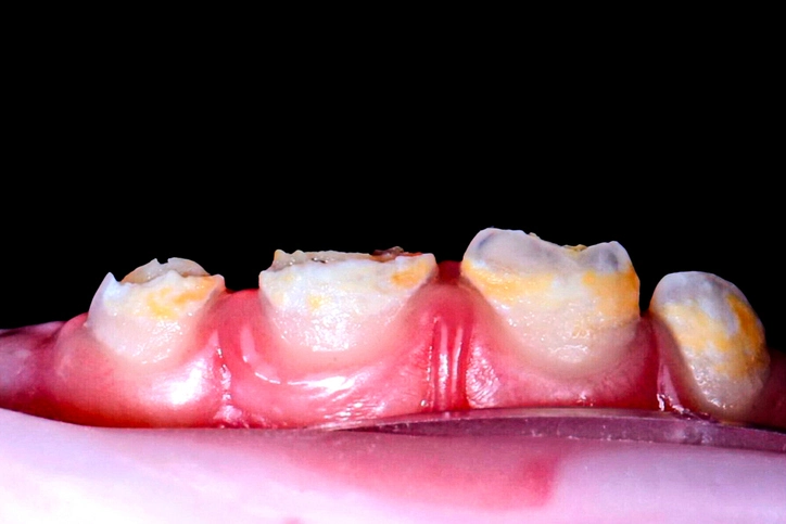
2. Hypocalcified Amelogenesis Imperfecta
Features:
- Enamel thickness is normal at eruption but soft and chalky.
- Color ranges from yellow-brown to honey-colored.
- Rapid post-eruptive wear and calculus accumulation are common.
- Teeth are extremely sensitive due to exposed dentin.
- Inheritance may be autosomal dominant or recessive.
Management:
Early coverage with stainless steel crowns in posterior teeth prevents sensitivity and attrition. In adolescents and adults, ceramic restorations or veneers can restore esthetics and function.
3. Hypomaturation Amelogenesis Imperfecta
Features:
- Enamel appears mottled, opaque white, or frosty.
- Hardness is reduced, but enamel is not as soft as in hypocalcified types.
- Often localized to the incisal third or cusp tips (“snow-capped teeth”).
- Increased sensitivity and chipping may occur.
Management:
- Conservative restoration with composite resin or veneers.
- In severe cases, full-coverage crowns may be necessary to protect dentin.
4. General Considerations in AI Management
The management of AI is complex and multidisciplinary. Since both primary and permanent dentitions are affected, the approach must evolve with the patient’s growth and development. The goals are to restore esthetics, maintain function, and prevent psychological distress.
Treatment strategies include:
- Primary dentition: Stainless steel crowns, glass ionomer restorations, or fluoride varnishes.
- Mixed dentition: Composite restorations or preformed crowns.
- Permanent dentition: Veneers, onlays, or full crowns depending on severity.
- Preventive care: Regular fluoride application, dietary counseling, and desensitizing agents.
Prognosis:
With early diagnosis and planned multidisciplinary care, affected children can maintain a functional and esthetically acceptable dentition throughout life.
Disturbances in the Structure of Dentine
Dentinogenesis Imperfecta (DI) is the principal hereditary dentin defect, also known as hereditary opalescent dentine. It affects both primary and permanent teeth and can occur in isolation or association with systemic disorders like osteogenesis imperfecta.
Prevalence: Approximately 1 in 8000 individuals.
Types of Dentinogenesis Imperfecta
- Type I: Associated with osteogenesis imperfecta (brittle bone disease).
- Type II: Isolated dentin defect without systemic involvement.
Clinical Features:
- Teeth exhibit an opalescent hue—blue-gray or amber-brown.
- Bulbous crowns with cervical constrictions.
- Short, narrow roots and obliterated pulp chambers on radiographs.
- The amelodentinal junction (ADJ) is abnormal, leading to enamel flaking and rapid wear of exposed dentin.
Histology:
The dentin shows irregular tubules and interglobular areas, accounting for its weak structure and opalescent appearance.
Management:
- Early protection of teeth using full-coverage crowns.
- Adhesive restorations to minimize wear.
- In severe cases, overlay dentures or implants may be required once growth is complete.
- Preventive care is vital to avoid secondary caries and further wear.
Association with Osteogenesis Imperfecta:
In type I cases, children may exhibit bone fragility, blue sclerae, and hearing loss. Collaboration with pediatricians and geneticists is essential.
Disturbances in Cementum Structure
Though less common than enamel and dentin defects, abnormalities in cementum can have significant clinical consequences.
Hypoplasia and Aplasia of Cementum
These are rare and usually observed in conditions such as hypophosphatasia, a metabolic disorder affecting bone and cementum mineralization. Affected children may experience premature exfoliation of primary teeth due to defective periodontal attachment.
Hypercementosis
This condition involves excessive deposition of cementum, often as a response to local irritation (e.g., periapical inflammation), occlusal trauma, or systemic diseases like Paget’s disease. It is typically asymptomatic but may complicate extraction procedures.
Concrescence
Concrescence refers to the pathological union of adjacent tooth roots through cementum deposition. It may be developmental or acquired following inflammation or trauma.
Multidisciplinary Management and Preventive Strategies
Children presenting with developmental dental anomalies often require the collaboration of multiple specialists including pediatric dentists, orthodontists, restorative dentists, and genetic counselors.
Goals of Management:
- Functional rehabilitation — ensuring mastication and speech are not compromised.
- Aesthetic improvement — to address psychological impacts, especially in visible anterior defects.
- Prevention of secondary complications — caries, sensitivity, and periodontal problems.
- Long-term maintenance — through fluoride therapy, regular reviews, and oral hygiene reinforcement.
Preventive Emphasis:
Because defective enamel and dentin are more susceptible to demineralization, prevention of secondary caries is paramount. Fluoride varnishes, desensitizing agents, dietary counseling, and meticulous plaque control are essential components of care.
Conclusion
Developmental abnormalities of enamel, dentin, and cementum represent a significant clinical challenge in pediatric dentistry. While some conditions like molar incisor hypomineralization have multifactorial etiologies, others, such as amelogenesis imperfecta and dentinogenesis imperfecta, are genetically determined.
Early diagnosis, preventive measures, and comprehensive multidisciplinary management can preserve function and appearance while minimizing long-term complications. Advances in genetic research and restorative materials continue to improve the prognosis and quality of life for affected individuals.
Understanding these conditions not only enhances clinical outcomes but also contributes to the broader understanding of dental tissue biology and the complex interplay of genetic and environmental factors during tooth development.
References
- Wright, J. T., & Hart, T. C. (2002). The Developmental Defects of Enamel: Genetic and Environmental Factors. Dental Clinics of North America, 46(1), 25–41.
- Roberts-Harry, D., & Curzon, M. E. J. (2021). Abnormalities of tooth structure. In W. R. Proffit & R. P. Vinson (Eds.), Handbook of Paediatric Dentistry (6th ed., pp. 73–83). Elsevier.
- Wright, J. T. (2019). Developmental Defects of the Teeth.
- Zhao, D., et al. (2018). Prevalence and clinical features of molar incisor hypomineralization in Chinese children: A systematic review. International Journal of Paediatric Dentistry, 28(2), 170–179.
- Alaluusua, S. (2010). Aetiology of Molar-Incisor Hypomineralisation: A systematic review. European Archives of Paediatric Dentistry, 11(2), 53–58.
- Suckling, G. W. (1989). Developmental defects of enamel—historical and present-day perspectives of their pathogenesis. Advances in Dental Research, 3(2), 87–94.
- Ten Cate, A. R., & Nanci, A. (2017). Ten Cate’s Oral Histology: Development, Structure, and Function (9th ed.). Elsevier.
- Neville, B. W., Damm, D. D., Allen, C. M., & Chi, A. C. (2016). Oral and Maxillofacial Pathology (4th ed.). Elsevier.
- Hillson, S. (2014). Tooth Development in Human Evolution and Bioarchaeology. Cambridge University Press.
- Crawford, P. J. M., Aldred, M., & Bloch-Zupan, A. (2007). Amelogenesis imperfecta. Orphanet Journal of Rare Diseases, 2, 17.
- Koch, G., Poulsen, S., Espelid, I., & Haubek, D. (2017). Pediatric Dentistry: A Clinical Approach (3rd ed.). Wiley Blackwell.
- Batra, P., Duggal, R., & Parkash, H. (2005). Amelogenesis imperfecta: An updated classification and treatment options in light of advances in genetic research. Oral Surgery, Oral Medicine, Oral Pathology, Oral Radiology, and Endodontology, 99(6), 662–672.
- Kim, J. W., & Simmer, J. P. (2007). Hereditary enamel defects. Journal of Dental Research, 86(5), 392–399.
- Aldred, M. J., Savarirayan, R., & Crawford, P. J. M. (2003). Amelogenesis imperfecta: A classification and catalogue for the 21st century. Oral Diseases, 9(1), 19–23.
- Rajendran, R., & Sivapathasundharam, B. (2020). Shafer’s Textbook of Oral Pathology (9th ed.). Elsevier.