
Variations in tooth morphology represent one of the most intriguing aspects of dental development and paediatric dentistry. Abnormalities of tooth form may arise due to disturbances during the morphodifferentiation and histodifferentiation stages of tooth development. These anomalies can affect a single tooth or multiple teeth and may involve alterations in size, shape, or internal structure. Although many of these anomalies are incidental findings, some can have significant clinical implications, including esthetic concerns, occlusal disturbances, caries susceptibility, and endodontic complications.
The normal mesiodistal width of the maxillary central incisor (1) is approximately 8.5 mm, while that of the lateral incisor (2) is about 6.5 mm. Deviations from these norms can indicate macrodontia, microdontia, or other developmental disturbances.
Table of Contents
ToggleDouble Teeth
The term “double teeth” refers to the presence of an abnormally large tooth structure that appears as if two teeth have been joined or partially split. It encompasses two distinct developmental anomalies—gemination and fusion—that can appear clinically similar.
Gemination
Gemination results from an attempted division of a single tooth germ. During the cap or bell stage of odontogenesis, the tooth germ partially splits, leading to the development of a single enlarged tooth with a bifid or notched crown. Despite the crown enlargement, the tooth typically possesses a single root and a single root canal system.
Clinical Features
- Appears as an enlarged or bifid crown with a groove or notch on the incisal edge.
- The total number of teeth in the arch remains normal, since no actual fusion occurs.
- Most commonly affects anterior teeth, particularly the maxillary incisors.
Radiographic Features
- A single root canal system is usually seen.
- The pulp chamber may appear partially divided.
Clinical Significance and Management
Geminated teeth may cause esthetic concerns, malalignment, and spacing issues. If the anomaly results in deep developmental grooves, it may predispose to caries and periodontal involvement.
Treatment depends on the degree of division and esthetic demands:
- Mild cases: reshaping and cosmetic contouring with composite resin.
- Severe cases: extraction followed by orthodontic closure or prosthetic replacement.
Fusion
Fusion is the union of two adjacent tooth germs, resulting in a single large tooth with a confluence of dentine and, in some cases, pulp tissues. The extent of union can vary from complete to partial fusion, depending on the developmental stage at which it occurs.
Clinical Features
- Appears as a broad tooth with an irregular or notched crown.
- The number of teeth in the arch is reduced by one, distinguishing it from gemination.
- Commonly seen in primary dentition, particularly in anterior segments.
Radiographic Features
- The fused teeth may have separate or fused pulp chambers and root canals.
- The crown morphology is irregular, and root length may vary.
Prevalence and Aetiology
Fusion occurs more frequently in the primary dentition (0.1–0.2%) and may have genetic or environmental influences. Trauma and local metabolic disturbances during odontogenesis have been suggested as contributing factors.
Management
Aesthetic correction should be delayed until root formation is complete. If separate pulp chambers and root canals exist, surgical separation may be considered. When fusion involves a supernumerary tooth, extraction of the accessory portion is recommended. Alternatively, the tooth can be contoured to simulate two separate crowns.
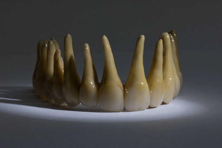
Macrodontia (Megalodontia)
Macrodontia refers to an increase in the size of teeth beyond normal limits, without alteration in tooth morphology. It may occur as a generalized, localized, or unilateral phenomenon.
Types and Associations
- True Generalized Macrodontia: Very rare and usually associated with conditions such as pituitary gigantism or hormonal disturbances during growth.
- Localized or Unilateral Macrodontia: Involves one or few teeth, often associated with hemifacial hypertrophy, a developmental condition characterized by unilateral overgrowth of the facial tissues, bones, and teeth.
- Isolated Macrodontia: Involves a single tooth without systemic involvement.
Clinical and Radiographic Features
- Affected tooth is significantly larger than normal.
- May cause crowding, displacement, and occlusal disturbances.
- Pulp chamber and root canal morphology are normal.
Management
Treatment is primarily esthetic or orthodontic. In cases with severe size discrepancy, reduction by enameloplasty, composite contouring, or extraction may be indicated.
Microdontia
Microdontia describes teeth that are smaller than the average size, with otherwise normal morphology. It can affect one or multiple teeth.
Prevalence and Distribution
- Occurs in less than 0.5% of the primary dentition.
- Approximately 2.5% prevalence in the permanent dentition.
- The most commonly affected teeth are the maxillary lateral incisors (“peg laterals”) and third molars.
Aetiology
- Hereditary factors play a significant role.
- Often seen in association with conditions such as pituitary dwarfism, Down syndrome, and ectodermal dysplasia.
Clinical Significance
- Peg-shaped lateral incisors are associated with spacing and esthetic issues.
- Short roots may contribute to increased tooth mobility.
- Microdontia of the lateral incisors is a predisposing factor for the palatal displacement of maxillary canines.
Management
- Restorative build-up with composite resin or porcelain veneer for esthetics.
- Orthodontic correction of spacing if required.
Dens in Dente (Dens Invaginatus)
Dens in dente, also known as dens invaginatus, is a developmental anomaly resulting from invagination of the enamel organ into the dental papilla during tooth formation. This creates a tooth-like structure within the crown, appearing as a “tooth within a tooth.”
Clinical Features
- Most frequently affects maxillary lateral incisors.
- May also occur in canines, premolars, and occasionally molars.
- Presents as a deep pit or fissure on the palatal surface.
- The invagination is lined by enamel and may extend into the root, sometimes reaching the pulp chamber.
Radiographic Features
The radiograph reveals an infolding of enamel and dentine that appears as a radiopaque pocket extending into the crown or root.
Complications
- The enamel lining the invagination may be thin and incomplete, allowing bacterial ingress and early pulpal involvement.
- Pulp necrosis and periapical pathology can occur even before eruption is complete.
Management
- Preventive approach: sealing of the invagination with flowable composite or resin after eruption.
- If pulp involvement occurs: endodontic treatment may be required, though it is often complex due to the aberrant canal anatomy.
- Extraction may be necessary in cases of severe infection or non-restorable morphology.
Dilaceration
Dilaceration is defined as a distortion or angulation of the crown or root of a tooth. The deformity results from a disturbance during root or crown development, leading to a sharp bend or curve.
Aetiology
Dilaceration can be of developmental or traumatic origin:
1. Developmental Type
- Caused by displacement of the developing tooth germ due to mechanical interference, such as pressure from adjacent teeth or cysts.
- Commonly affects the maxillary central incisors.
- The crown may be turned upward and labially.
2. Traumatic Type
- Usually results from trauma to the primary predecessor, particularly intrusion injuries.
- The developing permanent tooth germ becomes displaced, and subsequent calcification follows an altered path, producing an angulated root.
Epidemiology
- Most commonly affects permanent maxillary incisors.
- The prevalence varies but is higher in populations with a history of early dental trauma.
Radiographic Features
- Angulation between the crown and root axis.
- The degree of curvature may vary from mild to severe.
Clinical Implications
- Eruption disturbances or impaction.
- Complications during endodontic and orthodontic treatment.
- Esthetic problems due to abnormal crown orientation.
Management
- Mild cases: orthodontic traction can be attempted if the root apex is open.
- Severe cases with significant displacement: extraction may be indicated.
- Endodontic procedures should be approached with care to avoid perforation.
Turner Tooth
Turner tooth, or Turner hypoplasia, refers to a localized enamel and dentine defect in a permanent tooth caused by periapical infection or trauma of the overlying primary predecessor. The infection interferes with ameloblast activity during tooth formation.
Commonly Affected Teeth
Premolars are most commonly affected because their developing tooth germs are located near the roots of the primary molars.
Clinical Features
- Presents as localized enamel hypoplasia or hypomineralization.
- The surface may appear pitted, discolored, or irregular.
- The severity depends on the intensity and duration of the infection or trauma in the primary tooth.
Radiographic Features
May show reduced enamel thickness or localized radiolucency corresponding to hypoplastic areas.
Management
- Mild cases: cosmetic restoration using composite or microabrasion techniques.
- Severe cases: full coverage crowns may be required to restore function and esthetics.
Taurodontism
Taurodontism is a developmental anomaly characterized by an enlarged pulp chamber and apical displacement of the pulpal floor, giving the tooth a “bull-like” appearance radiographically.
Etymology and Pathogenesis
The term “taurodontism” derives from “tauros” (bull) and “odous” (tooth), describing the resemblance to bovine teeth. The anomaly arises due to a failure of Hertwig’s epithelial root sheath to invaginate at the proper level, resulting in delayed root formation and a vertically elongated pulp chamber.
Classification (Shaw, 1928)
- Hypotaurodont – Mild elongation of the pulp chamber.
- Mesotaurodont – Moderate elongation, with the pulp chamber extending further apically.
- Hypertaurodont – Extreme form, where the pulp floor is close to the root apex.
Prevalence and Associations
- Occurs more commonly in molars and premolars.
- Associated with conditions such as amelogenesis imperfecta, Klinefelter syndrome, Down syndrome, and tricho-dento-osseous syndrome.
Clinical Features
- Clinically, the crowns appear normal, and the condition is usually discovered radiographically.
- Radiographs show an elongated pulp chamber and short roots.
Clinical Significance
- Taurodontism can complicate endodontic therapy due to elongated pulp chambers and atypical canal morphology.
- Extraction may also be more difficult due to the altered root structure.
Management
- No specific treatment is required unless other pathologies are present.
- Careful endodontic planning and radiographic evaluation are essential when intervention is necessary.
Conclusion
Abnormalities of tooth form encompass a diverse range of developmental disturbances, each with unique etiological, morphological, and clinical implications. Understanding the nature and origin of these anomalies is essential for accurate diagnosis and appropriate management in paediatric and restorative dentistry.
While some anomalies such as microdontia or macrodontia may primarily present esthetic challenges, others like dens invaginatus or dilaceration can significantly complicate endodontic, orthodontic, and surgical procedures. Early recognition, preventive care, and timely intervention are crucial in minimizing long-term complications.
Radiographic examination remains an indispensable diagnostic tool, providing insight into the internal anatomy and developmental status of affected teeth. Interdisciplinary management involving paediatric dentists, orthodontists, and endodontists is often necessary to achieve optimal functional and esthetic outcomes.
References
- Wright, G.Z., Kupietzky, A. (2021). Behavior Management in Dentistry for Children. 3rd ed. Wiley-Blackwell.
→ Provides foundational principles of paediatric dental anomalies and their management. - W. M. Till and R. P. Hillson. (2018). Clinical Paediatric Dentistry. 5th ed. Elsevier.
→ Discusses developmental disturbances of teeth, including abnormalities of size and form. - Neville, B.W., Damm, D.D., Allen, C.M., Chi, A.C. (2016). Oral & Maxillofacial Pathology. 4th ed. Elsevier Saunders.
→ Comprehensive source for etiology, histopathology, and radiographic features of dental anomalies. - Cameron, A.C., Widmer, R.P. (2021). Handbook of Pediatric Dentistry. 6th ed. Elsevier.
→ Primary reference for paediatric oral development, including gemination, fusion, and dens invaginatus. - Ten Cate, A.R. (2019). Oral Histology: Development, Structure, and Function. 9th ed. Elsevier.
→ Authoritative text explaining embryologic development and disruptions leading to anomalies. - Hillson, S. (2014). Tooth Development in Human Evolution and Bioarchaeology. Cambridge University Press.
→ Provides insight into developmental mechanisms underlying variations in tooth form. - Proffit, W.R., Fields, H.W., Larson, B., Sarver, D.M. (2019). Contemporary Orthodontics. 6th ed. Elsevier.
→ Discusses clinical implications of anomalies like dilaceration and microdontia on orthodontic management. - Shafer, W.G., Hine, M.K., Levy, B.M. (2015). Shafer’s Textbook of Oral Pathology. 9th ed. Elsevier India.
→ Classic resource detailing pathology and classification of developmental disturbances in teeth. - Lovschall, H., and Andreasen, J.O. (2019). “Developmental disturbances following dental trauma in primary teeth.” Dental Traumatology, 35(3): 143–152.
→ Key paper linking traumatic injuries to anomalies such as dilaceration and Turner teeth. - White, S.C., Pharoah, M.J. (2014). Oral Radiology: Principles and Interpretation. 8th ed. Elsevier.
→ Describes radiographic appearance and differential diagnosis of anomalies like dens in dente and taurodontism.