
Restorative dentistry plays a critical role in preserving form, function, and aesthetics of natural teeth following disease or trauma. As restorative materials and techniques continue to evolve, patient expectations for longevity and aesthetics rise in parallel. However, despite advancements, restorations do not last forever; they are subject to mechanical, chemical, and biological challenges within the oral environment. Understanding the factors that influence the survival and failure of restorations is essential not only for clinicians but also for students, researchers, and patients seeking long-term oral health.
Table of Contents
ToggleThe Dynamics of Restoration Longevity
Dental restorations substitute or reinforce portions of natural tooth structure lost to caries, trauma, wear, or developmental defects. The success of a restoration depends on several interrelated factors, including:
- The patient’s oral hygiene habits
- The dentist’s technical skills
- Material properties
- The functional demands on the tooth
- Biological processes such as caries and periodontal disease
Interestingly, clinical research shows that patients who frequently switch dentists tend to have higher rates of restoration replacement than those who consistently see the same practitioner. Continuity of care allows the dentist to monitor the integrity of existing restorations, detect early changes, and manage risk factors proactively.
However, even with ideal care, restorations have finite lifespans. The key to optimizing their survival is understanding why they fail and preventing those failures whenever possible.
Reasons for Failure of Restorations
Restoration failure is multifactorial, often involving a combination of mechanical, biological, or patient-related causes. Below, each cause is explored in greater depth.
1. Secondary Caries, Tooth Fracture, or Material Fracture
Secondary caries
Secondary or recurrent caries refers to decay that forms at the margins of an existing restoration. It is one of the leading causes of failure, particularly in adhesive restorations such as composites. Factors contributing to secondary caries include:
- Poor oral hygiene leading to plaque accumulation
- Marginal gaps due to material shrinkage or wear
- Poor dietary habits, especially frequent sugar exposure
- Incomplete removal of initial caries during cavity preparation
Tooth fracture
In some cases, the tooth structure surrounding a restoration may fracture. This risk increases when:
- A large proportion of natural tooth is missing
- Cusps are undermined
- High occlusal loads are present (as in bruxism)
- Previous restorations have weakened the tooth over time
Material fracture
Restorative materials themselves can fracture due to fatigue or insufficient thickness. Amalgam may crack under tensile stress, while composites may chip or fracture if they are inadequately bonded or if occlusal forces exceed their strength.
2. Aesthetic Deterioration
Aesthetic failure is especially relevant for anterior restorations. Causes include:
- Staining from dietary chromogens (coffee, wine, tea, tobacco)
- Surface wear leading to roughness and increased plaque retention
- Color mismatch due to aging of composite resins
- Darkening of underlying dentine or corrosion products from metal alloys
Although not all aesthetic concerns require replacement, they can significantly affect patient satisfaction.
3. Poor Understanding or Management of Occlusion
The occlusion must be carefully evaluated before and after placing a restoration. Consequences of incorrect occlusal adjustment include:
- Premature contacts leading to discomfort
- Overloading of tooth structure and restorative materials
- Excessive wear
- Damage to adjacent teeth or restorations
Restorations placed in patients with parafunctional habits—such as clenching or grinding—are at heightened risk if protective measures (e.g., night guards) are not implemented.
4. Incorrect Cavity Preparation
Quality preparation is fundamental to restoration survival. Errors may include:
Insufficient caries removal
Leaving infected dentine or enamel can result in rapid recurrence of caries.
Inadequate retention form
Some materials, such as amalgam, require mechanical retention, while others rely on bonding. Insufficient retention can lead to dislodgement.
Improper margin design
Margins that are undercut, rough, or placed in areas that are difficult to access can compromise both material integrity and ease of cleaning.
Over-preparation
Excess removal of sound tissue weakens the tooth, making it more prone to fracture.
Inadequate protection of remaining tooth structure
Thin cusps left unsupported are likely to fracture under load.
5. Incorrect Choice of Restorative Material
Every material has unique properties. Selecting the wrong material for a specific clinical situation can predispose the restoration to failure.
For example:
- Using a low-strength glass ionomer in a high-stress occlusal area
- Using composite resin in situations with poor moisture control
- Choosing a material with insufficient wear resistance for posterior restorations
Material selection must consider aesthetics, strength, longevity, and the patient’s biological and functional needs.
6. Improper Manipulation of the Material
Even the best material will fail if handled incorrectly. Common errors include:
- Poor moisture control (especially critical for composites and glass ionomers)
- Incorrect mixing ratios
- Insufficient polymerization due to inadequate curing light intensity
- Over- or under-contouring the restoration
- Overloading material in a single increment, leading to shrinkage stresses in composites
Technique sensitivity is particularly notable for resin composites; meticulous execution is critical.
7. Patient-Related Factors
Patients play a central role in restoration longevity. Key risk factors include:
- Bruxism (teeth grinding): A chronic habit that leads to fractures and wear
- Using teeth as tools: Opening bottles, tearing packaging, or biting hard objects
- Poor oral hygiene: Leading to plaque accumulation around restoration margins
- High caries risk: Frequent sugar intake, low fluoride exposure, reduced salivary flow
Understanding a patient’s habits helps tailor treatment and counseling.
Considerations Before Replacing a Failed Restoration
It is essential to evaluate the cause of failure before opting for replacement. Sometimes, failure can be managed conservatively through repair rather than complete removal. For example:
- Small marginal chips in composite restorations can often be repaired.
- Staining does not always compromise structural integrity.
- Partial defects may be treated without enlarging the cavity unnecessarily.
A key point worth emphasizing is that every time a restoration is replaced, the cavity typically becomes larger by approximately 0.6 mm. This occurs because each removal inevitably eliminates some surrounding tooth structure. Over a lifetime, repeated replacement cycles can ultimately undermine the tooth, leading to the need for crowns or extraction.
Thus, minimally invasive dentistry favors repairing where possible and replacing only when essential.
Types of Restoration Failure
Restorations may fail for different reasons, and understanding the type of failure is essential for appropriate management.
1. Failed Aesthetics
Aesthetic failure refers to visual or cosmetic defects that may or may not correlate with functional issues. Examples include:
- Discoloration of underlying dentine: Often seen with older amalgams or in cases of pulp degeneration
- Surface staining: Typically caused by dietary or lifestyle habits
- Mismatch of restoration color: Aged composites may lose their translucency or yellow over time
- Surface roughness: Exaggerated wear or improper finishing
Not all aesthetic failures necessitate replacement. If the discoloration is superficial, simple polishing or re-sealing may restore the appearance. The clinician should evaluate whether the patient perceives the issue as significant before recommending invasive intervention.
2. Failed Marginal Integrity
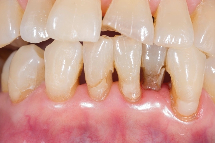
Marginal integrity is crucial for preventing microleakage, secondary caries, and structural weakness. Failures include:
Creep and corrosion (in amalgams)
Over time, amalgam can undergo slow deformation (creep) and corrosion, leading to:
- Ditching or gaps at the margin
- Accumulation of plaque in these areas
- Recurrent caries
Shrinkage in composites and glass ionomers
Composite resins shrink during polymerization, which can create tension at the interface and produce microscopic gaps. These gaps are entry points for bacteria and food debris, contributing to decay.
Marginal chipping
Chipping is especially common in:
- High-stress areas
- Patients with parafunctional habits
- Restorations with inadequate bulk
- Poorly bonded or poorly finished restorations
Clinical implications
Minor marginal defects are often harmless if the area remains plaque-free and there is no radiographic evidence of caries. Monitoring is often the best approach. However, intervention becomes necessary when:
- The lesion reaches dentine
- There is radiographic or clinical progression
- Cavitation is present
Early detection is key, and the dentist must counsel patients on preventive strategies.
3. Bulk Fracture
A bulk fracture refers to the breaking of a significant portion of the restoration. Common causes include:
- Heavy occlusal loading
- Poor cavity or crown design
- Insufficient material thickness
- Weak bonding between tooth and restoration
- Undetected cracks in the tooth
Posterior composite restorations, for example, may fracture if the occlusal surface is too thin or if the restoration extends across load-bearing cusps without adequate reinforcement.
Once bulk fracture occurs, repair may not be possible, and the restoration usually requires full replacement.
Strategies to Improve the Survival of Restorations
Prolonging the life of restorations requires a multifaceted approach involving both clinician and patient.
1. Improving Cavity Design and Preparation
- Preserve as much natural tooth as possible
- Ensure clean, well-defined margins
- Provide adequate resistance and retention form where needed
- Protect weakened cusps or walls through cusp coverage or onlays
2. Choosing the Correct Material
Material selection should consider:
- Location of the tooth
- Occlusal stresses
- Aesthetic needs
- Moisture control
- Patient habits
Examples:
- Amalgam or high-strength composite for load-bearing posterior teeth
- Glass ionomer for patients with high caries risk due to its fluoride release
- Ceramic inlays/onlays for aesthetics and wear resistance
3. Enhancing Clinical Technique
- Use rubber dam isolation
- Follow manufacturer instructions meticulously
- Ensure proper curing with a calibrated curing light
- Avoid over-contouring or leaving plaque-retentive areas
- Use incremental layering techniques to reduce polymerization shrinkage
4. Patient Education and Risk Management
- Emphasize the importance of oral hygiene
- Encourage fluoride use
- Advise dietary modifications
- Recommend night guards for bruxists
- Schedule regular maintenance visits
Patients who understand their role in maintaining restoration longevity experience fewer failures.
Conclusion
The survival and failure of dental restorations depend on a complex interplay of biological, mechanical, technical, and behavioral factors. A clinician’s ability to accurately diagnose the cause of failure, choose appropriate treatment strategies, and educate the patient plays a vital role in ensuring long-lasting restorations. Because each replacement tends to enlarge the cavity and further weaken the tooth, a conservative approach—repairing rather than replacing whenever possible—is fundamental in modern restorative dentistry.
A deep understanding of the causes and types of restoration failure allows clinicians to prevent common pitfalls, optimize outcomes, and promote long-term dental health.
References
- Opdam, N.J., van de Sande, F.H., Bronkhorst, E., Cenci, M.S., Bottenberg, P., Pallesen, U., et al. (2014). Longevity of posterior composite restorations: A systematic review and meta-analysis. Journal of Dental Research, 93(10), 943–949.
- Demarco, F.F., Corrêa, M.B., Cenci, M.S., Moraes, R.R., & Opdam, N.J. (2012). Longevity of posterior composite restorations: Not only a matter of materials. Dental Materials, 28(1), 87–101.
- Mjör, I.A., Moorhead, J.E., & Dahl, J.E. (2000). Reasons for replacement of restorations in permanent teeth in general dental practice. International Dental Journal, 50(6), 361–366.
- Burke, F.J.T., Lucarotti, P.S. (2009). Ten-year outcome of restorations placed in the general dental services in England and Wales. The Journal of Dentistry, 37(1), 12–24.
- Hickel, R., Brüshaver, K., & Ilie, N. (2013). Repair of restorations—Criteria for decision making and clinical recommendations. Dental Materials, 29(1), 28–50.
- Tyas, M.J., Anusavice, K.J., Frencken, J.E., & Mount, G.J. (2000). Minimal intervention dentistry—A review. International Dental Journal, 50(1), 1–12.
- Mondelli, R.F.L., Barbosa, W.F., Mondelli, J., Franco, E.B., Carvalho, R.M., & Ishikiriama, S.K. (2018). Clinical performance of composite restorations. In: Ferracane, J.L., Stansbury, J.W., & Ilie, N. (Eds.), Dental Composite Materials. Springer.
- Mount, G.J., & Hume, W.R. (1998). Preservation and Restoration of Tooth Structure. Mosby, St. Louis.
- Ritter, A.V., Swift Jr., E.J., Heymann, H.O., & Sturdevant, C.M. (2023). Sturdevant’s Art and Science of Operative Dentistry (7th ed.). Elsevier.
- Kidd, E.A.M., Fejerskov, O. (2016). Caries Diagnosis, Treatment, and Prevention. Wiley Blackwell.
- Heintze, S.D., Rousson, V. (2012). Clinical effectiveness of direct class II restorations—a meta-analysis. The Journal of Adhesive Dentistry, 14(5), 407–431.
- Bernardo, M., Luis, H., Martin, M.D., Leroux, B.G., Rue, T., Leitão, J., & DeRouen, T.A. (2007). Survival and reasons for failure of amalgam versus composite posterior restorations placed in a randomized clinical trial. Journal of the American Dental Association, 138(6), 775–783.
- van Dijken, J.W. (2010). Direct resin composite inlays/onlays: an 11-year follow-up. Journal of Dentistry, 38(7), 508–516.
- Davies, J.A. (1984). The longevity of dental restorations: A clinical study. British Dental Journal, 157, 322.
(This is the reference cited in the original textbook excerpt.)