
The management of dental caries and restoration of primary teeth in pediatric patients presents unique challenges. Unlike adult dentition, primary teeth have thinner enamel, larger pulp chambers, and less mineralized structures, making them more susceptible to rapid carious progression. Moreover, the behavioral management of children, especially those with dental anxiety or special health care needs, often limits the feasibility of complex restorative procedures.
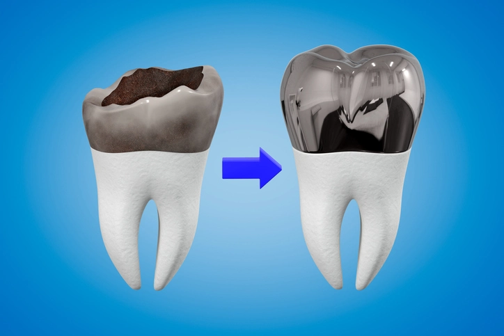
In this context, stainless steel crowns (SSCs) have emerged as one of the most durable, predictable, and long-lasting restorative options for primary molars that have undergone extensive carious destruction or pulpal therapy. Since their introduction by Humphrey in 1950, SSCs have remained a cornerstone in pediatric restorative dentistry.
Table of Contents
ToggleOverview and Composition of Stainless Steel Crowns
Stainless steel crowns are preformed metal restorations made from high-grade stainless steel, typically an iron-chromium-nickel alloy. These crowns are resistant to corrosion, provide superior durability under occlusal load, and maintain excellent marginal integrity over time.
However, due to their nickel content, SSCs are contraindicated in patients with known nickel allergies. Alternative esthetic options, such as pre-veneered or zirconia crowns, may be considered in such cases.
Unlike cast metal crowns, SSCs are preformed and require only minimal modification before cementation, making them highly efficient and economical in pediatric practice.
Indications for Stainless Steel Crowns
SSCs are indicated for a wide range of clinical scenarios in both primary and young permanent teeth. The most common indications include:
- Badly Broken-Down Primary Molars: Teeth that have extensive caries involving multiple surfaces, where conventional restorations (amalgam or composite) are likely to fail.
- Post-Pulp Therapy in Primary Molars: Following pulpotomy or pulpectomy, SSCs provide full coverage protection and prevent microleakage, thus maintaining the tooth until exfoliation.
- Interim Restoration for Permanent Molars: In young patients where the eruption of second permanent molars is incomplete, SSCs can serve as interim restorations to maintain space and function.
- Temporary Coverage During Orthodontic or Prosthodontic Procedures: They can protect prepared teeth while permanent restorations are fabricated.
- Developmental Anomalies: Conditions such as amelogenesis imperfecta, dentinogenesis imperfecta, or hypoplastic enamel often necessitate full coverage restorations.
- Severe Tooth Wear Due to Bruxism or Erosion: SSCs offer a durable solution that withstands parafunctional forces and prevents further tooth structure loss.
Instruments Required for SSC Placement
The clinical success of SSC placement depends heavily on proper armamentarium and technique. The following instruments are typically employed:
- High-speed tapered diamond bur and diamond occlusal wheel: for tooth preparation.
- Straight handpiece with a stone: for finishing or trimming.
- Slow-speed handpiece and various burs: for refining margins.
- Crown scissors: to adjust crown height.
- Dividers and calipers: to measure mesio-distal dimensions.
- Adam’s pliers and Johnson contouring pliers: to contour and crimp the crown margins for a snug fit.
- Band pliers (No. 112): for minor adjustments.
Techniques for Stainless Steel Crown Placement
There are two primary methods for SSC placement:
- Conventional Technique
- Hall Technique
Both aim to achieve a durable, retentive, and biologically acceptable full coverage restoration, yet they differ in their philosophy and clinical approach.
Conventional Stainless Steel Crown Technique
The conventional technique relies on mechanical retention achieved by close adaptation of the crown margins at the gingival level. This method requires minimal taper and precise tooth reduction to ensure crown fit and occlusal harmony.
Step-by-Step Procedure
1. Anesthesia and Isolation
- Local anesthesia (LA) is administered for patient comfort.
- Rubber dam placement is ideal but may be omitted if patient cooperation is limited.
2. Caries Removal and Tooth Preparation
- All carious tissue is removed before crown placement.
- The tooth is prepared to create adequate space for the crown material.
3. Occlusal Reduction
Approximately 1 mm of occlusal surface is reduced, following the natural contours of cuspal planes.
4. Proximal (Mesial and Distal) Reduction
Using a tapered diamond bur, about 20° from vertical, interproximal contacts are cleared to allow the crown to seat completely without producing ledges at the gingival margin.
5. Buccal and Lingual Reduction
Minimal reduction is required, limited to removing carious or unsupported enamel.
6. Crown Selection and Trial Fit
- Mesio-distal width is measured using dividers.
- The correct crown should “click” into place with light pressure.
- The margin should sit 1 mm subgingivally for optimal retention.
7. Adjustments and Crimping
- If the crown appears high or overextended, the gingival edge can be trimmed with scissors and re-contoured using pliers.
- Crimping the margin enhances adaptation to the tooth.
8. Cementation
- The crown is cemented using zinc polycarboxylate or glass ionomer cement (GIC).
- Excess cement is removed carefully after seating.
- The child is asked to bite gently on a cotton roll during cement setting.
Advantages of the Conventional Technique
- Excellent marginal fit and mechanical retention.
- Reliable long-term durability.
- Familiar technique for most clinicians.
Limitations
- Requires local anesthesia and tooth preparation.
- May be difficult for anxious or uncooperative children.
- Technique sensitive and time-consuming.
The Hall Technique
The Hall technique, introduced in Scotland by Dr. Norna Hall, represents a paradigm shift toward biological caries management. It is based on the principle of sealing caries beneath a stainless steel crown without removing carious tissue or performing any tooth preparation.
Rationale and Biological Basis
The Hall technique is predicated on the idea that caries is a bacterial infection that can be arrested by sealing the lesion from its nutrient source. By cutting off the bacterial supply of carbohydrates and oxygen, the carious process halts, allowing the tooth to remain asymptomatic until exfoliation.
This non-invasive approach aligns with minimal intervention dentistry (MID) principles and is particularly advantageous for children who are unable to tolerate traditional procedures.
Step-by-Step Hall Technique
1. Case Selection
- Indicated for asymptomatic primary molars with carious lesions confined to dentin.
- Should not be used where there is evidence of pulpal or periradicular pathology (e.g., abscess, fistula, or pain).
2. Pre-Separation
Orthodontic separators may be placed a few days before the appointment to ease crown placement by creating space between adjacent teeth.
3. Crown Selection
The correct crown size is chosen to fit snugly over the tooth with minimal resistance.
4. Cementation
The crown is filled with glass ionomer cement (GIC) or another suitable luting agent.
5. Crown Placement
- The child is instructed to bite firmly on the crown, seating it fully onto the tooth.
- The clinician and parent should be informed that a feeling of pressure or tightness is normal.
6. Cleanup
Excess cement is immediately cleared from the gingival margins.
7. Postoperative Occlusion
The crown may initially appear in premature occlusion, but this typically resolves within a few weeks through dentoalveolar compensation.
Advantages of the Hall Technique
- No local anesthesia or drilling required.
- Reduces anxiety and discomfort, making it ideal for young or behaviorally challenged children.
- Shorter chair time.
- Preserves sound tooth structure.
- Creates a tight coronal seal that cuts off carious lesion progression.
Limitations
- Not suitable for symptomatic teeth or those with pulpal involvement.
- Aesthetic limitations due to the metallic appearance.
- Potential for temporary occlusal discrepancies.
Biological and Clinical Rationale
The biological success of SSCs—especially the Hall technique—stems from the principle of sealing over carious lesions rather than removing them. The caries bacteria are deprived of fermentable substrates once the lesion is hermetically sealed beneath the crown.
In this oxygen-depleted, nutrient-poor environment, bacterial metabolism slows, and the lesion arrests. The tooth remains asymptomatic as long as there is no pre-existing pulpal pathology.
This concept has been validated by multiple microbiological studies showing a significant reduction in viable bacterial counts beneath sealed carious lesions over time.
Success Rates and Clinical Outcomes
Numerous clinical trials and systematic reviews have consistently demonstrated the superior longevity of stainless steel crowns compared to other restorative options in primary teeth.
- Conventional SSCs show survival rates of over 90–95% at five years, outperforming multi-surface amalgam and composite restorations, which often fail within 2–3 years due to marginal breakdown or secondary caries.
- Hall technique SSCs have shown comparable or even higher success rates in some studies, attributed to the creation of a robust pericoronal seal.
A landmark study by Innes et al. (2007, BMC Oral Health) found that Hall technique crowns performed equally well or better than conventionally placed SSCs, with fewer incidences of pain, infection, or restoration failure.
The UK FiCTION Trial
The FiCTION (Filling Children’s Teeth – Indicated or Not) trial, conducted in the UK, sought to evaluate three main treatment strategies for managing caries in primary teeth:
- Conventional caries removal and restoration.
- Biological management using the Hall technique.
- Non-restorative cavity control.
Preliminary findings suggest that biological approaches, such as the Hall technique, can achieve similar or better long-term outcomes compared to conventional methods, with significantly less discomfort and anxiety for pediatric patients.
Comparative Summary: Conventional vs. Hall Technique
| Feature | Conventional SSC | Hall Technique |
|---|---|---|
| Caries Removal | Complete removal required | None (sealed-in caries) |
| Tooth Preparation | Occlusal and proximal reduction | None |
| Local Anesthesia | Required | Not required |
| Rubber Dam Use | Recommended | Not required |
| Chair Time | Longer | Shorter |
| Child Comfort | Moderate | Excellent |
| Indications | Post-pulp therapy, severe breakdown | Asymptomatic carious teeth |
| Pulpal Health Requirement | Can restore after pulpotomy/pulpectomy | Must be vital and asymptomatic |
| Occlusal Adjustment | Immediate | Self-adjusts within weeks |
| Longevity | Very high | Equally high |
| Aesthetic Outcome | Metallic | Metallic |
Cementation Materials
Cement selection plays a crucial role in SSC retention and marginal seal. The commonly used cements include:
Zinc Polycarboxylate Cement
- Good adhesion to enamel and dentin.
- Mild pulpal irritation potential.
Glass Ionomer Cement (GIC)
- Chemically bonds to tooth structure.
- Fluoride release provides additional protection against secondary caries.
- Preferred for both conventional and Hall technique crowns.
Postoperative Considerations and Follow-Up
After crown placement, children and parents should be advised on the following:
- Mild discomfort or a feeling of tightness may occur initially.
- Occlusion typically normalizes within 2–3 weeks due to adaptive tooth movement.
- Maintain good oral hygiene, especially around the crown margins.
- Routine recall visits to monitor crown integrity and gingival health.
If the tooth becomes symptomatic (pain, swelling, or abscess), it should be re-evaluated for possible pulpal pathology.
Clinical Pearls and Tips
- Always ensure the selected crown covers all cusps and extends slightly subgingivally for optimal seal.
- Over-trimming the crown margin can compromise retention.
- In Hall technique, always confirm that the tooth is asymptomatic before cementation.
- Educate parents about the metallic appearance to prevent aesthetic concerns postoperatively.
- Use bitewing radiographs periodically to assess the integrity of crown margins and interproximal bone levels.
Conclusion
Stainless steel crowns remain the gold standard for the restoration of extensively carious or pulpotomized primary molars. Their proven durability, ease of placement, and biological effectiveness make them indispensable in pediatric dentistry.
While the conventional technique offers precision and control, the Hall technique revolutionizes pediatric restorative care by embracing a minimally invasive, biologically sound approach that is both child- and clinician-friendly.
With mounting evidence supporting their longevity and success, SSCs continue to represent one of the most effective and evidence-based solutions for managing primary molar caries—bridging the gap between restorative necessity and biological preservation.
References
- Innes NP, Evans DJ, Stirrups DR. The Hall technique: A randomized controlled clinical trial of a novel method of managing carious primary molars in general dental practice: Acceptability of the technique and outcomes at 23 months. BMC Oral Health. 2007;7:18.
- Randall RC. Preformed metal crowns for primary and permanent molar teeth: Review of the literature. Pediatr Dent. 2002;24(5):489–500.
- Chadwick BL, et al. The UK FiCTION Trial: Managing Carious Lesions in Primary Teeth. Br Dent J. 2019.