
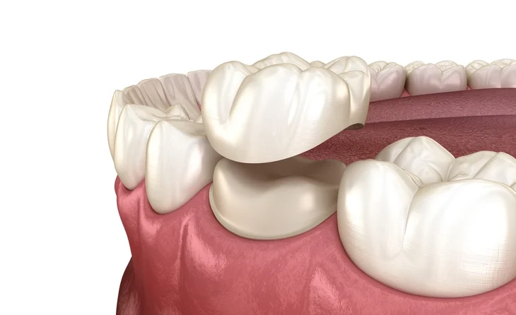
Restoration of posterior teeth presents a unique challenge to the clinician. Posterior teeth endure high occlusal loads, commonly suffer large structural defects from caries and fractures, and often require rehabilitation after endodontic treatment. As a result, indirect restorations—particularly crowns—play a critical role in restoring function, strength, and longevity.
Table of Contents
ToggleIndications for Posterior Crowns
Posterior crowns are typically indicated when insufficient tooth structure remains to support a durable direct restoration. Specifically, crowns may be required when:
1. Extensive Tooth Structure Loss
Posterior teeth with large carious lesions, fractured cusps, or those heavily restored with old amalgam/composite fillings may have compromised structural integrity. Large MOD cavities or teeth with three-surface involvement often need cuspal coverage to prevent future fracture.
2. Endodontically Treated Teeth
Endodontic treatment weakens tooth structure due to:
- Loss of internal dentine
- Removal of pulp chamber roof
- Reduced moisture content
Such teeth frequently require full-coverage restorations to prevent cusp deflection and vertical root fracture.
3. Tooth Wear
Attrition, abrasion, or erosion can reduce occlusal height and compromise tooth function. Crowns may help restore vertical dimension and protect remaining tooth tissue.
4. As Abutments for Fixed Prosthodontics
Crowns provide retention and resistance for bridge abutments. Strength and precision of margins are essential for long-term success.
5. When Aesthetic Enhancement Is Desired
Posterior aesthetics are increasingly important, especially for premolars visible during speech and smiling. Tooth-coloured crowns can improve appearance while maintaining function.
Before crown preparation begins, the clinician must:
- Assess vitality
- Replace defective restorations
- Ensure absence of active caries
- Obtain pre-operative radiographs
This ensures appropriate planning and minimizes the risk of pulpal complications.
Types of Posterior Crowns
Three primary categories of crowns are used in the posterior region:
- Full veneer gold (or non-precious metal) crowns
- Porcelain-fused-to-metal (PFM) crowns
- All-ceramic crowns, including zirconia and alumina-based systems
Selecting the appropriate crown involves balancing aesthetics, strength, tooth preparation considerations, and cost.
Full Veneer Gold or Non-Precious Metal Crowns
Overview
Full gold crowns (FGCs) are often considered the “gold standard” in restorative dentistry due to their:
- Exceptional longevity
- Minimal preparation requirements
- Superior marginal integrity
- High biocompatibility
- Excellent wear compatibility with opposing dentition
Despite their unmatched durability, FGCs are less aesthetic, making them more suitable for molars or for patients unconcerned with metal appearance.
Advantages
- Least destructive preparation: Gold requires minimal thickness (1–1.5 mm), preserving more natural tooth structure.
- Reduced pulpal risk: Less preparation → reduced heat and trauma.
- Excellent longevity: Studies show survival of 20–40 years.
- Easy to polish and repair.
- Minimal occlusal wear on opposing teeth.
Disadvantages
- Aesthetically unacceptable to some patients.
- Does not mask underlying discoloration.
- Metal allergies (rare).
Principles of Gold Crown Preparation
Achieving optimal retention, resistance form, and structural durability requires adherence to specific design principles.
Occlusal Reduction
- 1.5 mm on functional cusps (BUCCAL cusps of mandibular molars/premolars; PALATAL cusps of maxillary)
- 1 mm on non-functional cusps
The reduction should follow the natural cuspal anatomy to provide adequate space for gold thickness without compromising occlusal strength.
Bevel on Functional Cusps
A wide functional cusp bevel:
- Enhances structural durability
- Strengthens the margin
- Prevents thin unsupported areas of gold
Axial Wall Convergence (< 10°)
Slight taper ensures:
- Draw for the crown
- Retention and resistance form
Excessive taper drastically reduces retention.
Margin Design: Chamfer
A chamfer finish line:
- Provides enough space for metal
- Conserves tooth structure
- Minimizes unsupported enamel
Margins should ideally be supragingival for periodontal health unless aesthetics or caries require subgingival placement.
Steps in Gold Crown Preparation
Occlusal Preparation
Using a short diamond fissure bur:
- Reduce cusp height while maintaining anatomical form
- Maintain original ridge and fossa relationships
- Avoid flattening the occlusal surface excessively
Buccolingual Preparation
Using a torpedo-shaped bur:
- Remove undercuts
- Maintain 5° taper in the cervical two-thirds
- Preserve structural integrity
Approximal Preparation
Using a fine tapered diamond bur:
- Eliminate undercuts
- Create a slight convergence (≈5°)
- Maintain proximal contact position until it “breaks” for access
Finishing
Key aspects:
- Round axial line angles
- Ensure smooth surfaces for clean impressions
- Check for no undercuts
- Smooth margins thoroughly
Porcelain-Fused-to-Metal (PFM) Crowns
PFM crowns combine:
- Strength of metal substructure
- Aesthetics of porcelain veneering
They have long been the standard for posterior restoration where appearance is a concern.
Indications
- When a combination of strength + moderate aesthetics is required
- Premolars and visible molars
- Patients unable to afford full ceramic options
Advantages
- Better aesthetics than metal-only crowns
- Stronger than most all-ceramic options (except zirconia)
- Long track record and predictable outcomes
Disadvantages
- More tooth structure needs to be removed
- Porcelain chipping or fracture is possible
- Possible grey line at gingival margin
- Opposing tooth wear if porcelain is unglazed or rough
Preparation Guidelines for PFM Crowns
Proper design depends on whether the occlusal and buccal surfaces are to be metal, porcelain, or metal-ceramic combinations.
Occlusal Reduction
Two scenarios:
A. All-Metal Occlusal Surface
If acceptable to the patient:
- Remove LESS tooth structure
- 1–1.5 mm reduction
- Protects tooth vitality
B. All-Porcelain Occlusal Surface
Requires:
- 2 mm reduction over supporting cusps
- 1.5 mm over non-supporting cusps
This ensures sufficient porcelain thickness to resist fracture.
However, all-porcelain occlusal surfaces:
- Increase risk of pulp trauma
- May introduce occlusal interferences
- Have higher wear potential on opposing enamel
Buccal Reduction (1.2–1.5 mm)
Needed to accommodate:
- Metal coping
- Porcelain veneer
Unlike gold, porcelain requires more thickness to achieve strength and translucency.
Margin Design
Two options depending on aesthetic demand:
A. Metal Margin (preferred when aesthetics allow)
Advantages:
- Strong margin
- Conserves tooth structure
- Reduces risk of porcelain fracture
Finishing line:
Deep chamfer or bevelled shoulder
B. Porcelain Margin (for improved aesthetics)
Requires:
- 1.2–1.5 mm shoulder
- Natural transition to tooth
- Excellent impression technique
- More destructive preparation
Where no porcelain coverage is needed (lingual side, for example), a chamfer like that used for full gold crowns is ideal.
All-Ceramic Crowns
With improvements in ceramic technology, all-ceramic crowns have become viable options for posterior teeth. They offer superior aesthetics and sufficient strength when proper materials are selected.
Types of All-Ceramic Systems
Common systems include:
- Alumina-based: In-Ceram®, Procera®
- Leucite-reinforced ceramics: Empress® I
- Lithium disilicate ceramics: Empress® II, e.max®
- Zirconia: High-strength frameworks
Advantages
- Excellent aesthetics
- No metal → eliminates gingival greying
- High biocompatibility
- Zirconia offers extremely high strength
Disadvantages
- More tooth reduction required (typically 1.5–2 mm)
- Some ceramics are brittle when thin
- Zirconia is very strong but more difficult to bond
- Fracture risk if preparation guidelines aren’t followed
All-Ceramic Crown Preparation Principles
1. Occlusal Reduction
1.5–2 mm depending on ceramic type
Lithium disilicate needs less reduction than porcelain-fused-to-metal but more than gold.
2. Axial Reduction
1–1.5 mm
This ensures adequate ceramic thickness to avoid translucency issues or fracture.
3. Margin Design
A shoulder or deep chamfer is mandatory:
- Provides strong material support
- Prevents chipping of unsupported ceramic
- Necessary for optimal aesthetics
4. Avoid Sharp Internal Angles
Ceramics are brittle. Rounded internal line angles reduce stress concentrations and fracture risk.
Restoring Endodontically Treated Posterior Teeth
Single-Rooted Posterior Teeth
Can often be restored similarly to anterior teeth:
- Post-and-core systems may be used
- Ferrule effect is essential
- At least 2 mm of sound tooth structure above the margin improves resistance to fracture
Multirooted Teeth
Post space preparation differs:
Multiple canals diverge, making post placement impractical
Prefabricated posts may be placed only in straight canals
Coronal restoration typically involves:
Amalgam core buildup (Nayyar technique)
Resin composite buildup
RMGIC as a foundation material
Key Considerations
- Adhesive systems should be used with composite to maximize retention
- Core material must be compatible with final crown type
- Strong foundation is essential before crown fabrication
Choice of Crown Material for Endodontically Treated Teeth
Often:
- Full gold or PFM crowns are preferred for strength
- Zirconia for aesthetics combined with durability
Considerations for Margin Placement
Margin placement affects:
- Periodontal health
- Aesthetics
- Ease of impression taking
- Risk of caries
Supragingival Margins
Preferred whenever possible:
- Less irritation to gingiva
- Easier to clean
- Better impression accuracy
Subgingival Margins
Used when:
- Caries or existing restorations extend subgingivally
- Aesthetic demands require hiding junction
- Additional retention is needed
Clinical Workflow for Posterior Crowns
Diagnosis and Treatment Planning
Radiographs
Pulp vitality testing
Occlusal analysis
Pre-Crown Restoration Replacement
Remove all defective restorations
Build-up if needed
Tooth Preparation
Following principles for chosen material
Impression Taking
Digital or conventional
Ensure moisture control
Provisional Crown Placement
Protects pulp
Maintains position and occlusion
Try-In and Cementation
Verify margins, contacts, occlusion
Use appropriate luting cement
Conclusion
Posterior crowns remain a cornerstone in restorative dentistry, providing strength, function, and aesthetics when teeth have been significantly compromised. Understanding the principles of preparation, material requirements, and clinical indications is essential for achieving long-term success.
Whether opting for the durability of gold, the balanced aesthetics and strength of PFM, or the improved appearance and biocompatibility of modern ceramics, clinicians must carefully tailor treatment to the needs of each patient.
Mastery of these concepts ensures predictable outcomes and durable restorations that restore posterior teeth to optimal health and function.
References
- Shillingburg HT, Sather DA, Wilson EL, Cain JR, Mitchell DL, Blanco LJ, et al. Fundamentals of Fixed Prosthodontics. 4th ed. Quintessence Publishing; 2012.
– Gold standard reference for crown preparation principles, retention/resistance form, and margin design. - Rosenstiel SF, Land MF, Fujimoto J. Contemporary Fixed Prosthodontics. 5th ed. Mosby Elsevier; 2015.
– Comprehensive discussion on PFM, all-ceramic crowns, occlusal reduction guidelines, and preparation geometry. - Goodacre CJ, Campagni WV, Aquilino SA. Tooth preparations for complete crowns: an art form based on scientific principles. J Prosthet Dent. 2001;85(4):363-376.
– Evidence-based guidelines for occlusal and axial reduction, taper, and margin configurations. - Aquilino SA, Caplan DJ. Relationship between crown placement and the survival of endodontically treated teeth. J Prosthet Dent. 2002;87(3):256-263.
– Demonstrates the necessity of full-coverage restorations for endodontically treated posterior teeth. - Saunders WP, Saunders EM. Prevalence of coronal microleakage in endodontically treated teeth. Br Dent J. 1988;185:137–140.
– Addresses foundation restorations, coronal seal importance, and relevance to posterior crowns. - Patterson CJ, McLundie AC, Stirrups DR, Saunders WP. Comparison of marginal can adaptation of cast gold crowns prepared with three different finish lines. Br Dent J. 2000;188:148–152.
– Supports discussion on chamfer vs shoulder margins. - Kelly JR, Benetti P. Ceramic materials in dentistry: historical evolution and current practice. Aust Dent J. 2011;56:84–96.
– Overview of all-ceramic systems and material science. - Piddock V, Qualtrough AJE, Dummer PMH. Preformed metal posts and cores. Br Dent J. 1988;164:158–163.
– Background for post considerations in multirooted and single-rooted posterior teeth. - Dietschi D, Duc O, Krejci I, Sadan A. Biomechanical considerations for minimally invasive restorative procedures. Dent Clin North Am. 2011;55:489–519.
– Discusses biomechanics of cusp support, tooth reduction, and restorative strategies. - Magne P, Belser U. Bonded Porcelain Restorations in the Anterior Dentition: A Biomimetic Approach. Quintessence; 2002.
– Although anterior-based, includes fundamental ceramic bonding principles relevant to posterior ceramics. - Spear F, Kokich VG. Aesthetic considerations in restorative dentistry. J Am Dent Assoc. 2007;138(8):1139–1146.
– Supports aesthetic considerations for porcelain margins and ceramic selection. - Larson TD. The structural durability of full-coverage restorations. J Prosthet Dent. 2004;91(2):120–125.
– Summarizes evidence for long-term performance of gold, PFM, and ceramic crowns.