
The maxillary permanent canine plays a crucial role in the dental arch due to its function, aesthetics, and contribution to arch stability. It serves as a cornerstone of the dental arch, guiding occlusion during lateral excursions and providing essential lip support. Any disruption in its eruption path, especially palatal displacement, can lead to significant functional, aesthetic, and orthodontic challenges.
Palatally displaced maxillary canines (PDCs) represent one of the most commonly encountered anomalies in dental development. The early recognition and proper management of these teeth are critical for minimizing complications such as root resorption, prolonged orthodontic treatment, and potential tooth loss.
Table of Contents
TogglePrevalence
Palatal displacement of maxillary canines occurs in approximately 2% of the general population, with bilateral involvement reported in 17–25% of cases. The condition exhibits a higher prevalence in females than in males, typically in a ratio of about 2:1.
While buccal displacement of canines also occurs, palatal impaction is roughly twice as common. The difference in prevalence between the two directions of displacement suggests that different etiological factors may be at play. In orthodontic populations, the frequency of PDCs is higher due to the increased likelihood of crowding, malocclusion, and referral bias.
Aetiology
The aetiology of palatally displaced maxillary canines is multifactorial and has been debated for decades. Two main hypotheses are recognized: the guidance theory and the genetic theory.
1. Guidance Theory
This theory suggests that the eruption of the maxillary canine depends on proper guidance from the root of the adjacent lateral incisor. In normal development, the canine germ forms palatal to the deciduous canine and migrates labially, erupting distal to the lateral incisor.
If the lateral incisor is missing, peg-shaped, or short-rooted, the canine may lack an appropriate guide, causing it to deviate palatally. This theory is supported by the frequent association between palatally displaced canines and congenitally missing or malformed lateral incisors. Approximately 6% of palatally displaced canines occur in association with missing or short-rooted lateral incisors.
2. Genetic Theory
The genetic hypothesis proposes that palatal displacement is part of a broader genetically determined developmental anomaly. Studies have shown a higher frequency of PDCs among family members, indicating heritability. Moreover, PDCs often coexist with other dental anomalies such as tooth agenesis, microdontia, transposition, and enamel hypoplasia.
This theory aligns with the observation that palatally displaced canines may occur even when the lateral incisors are normal in form and position, suggesting a more systemic developmental influence.
3. Other Contributing Factors
Several additional local and environmental factors have been implicated:
- Space deficiency due to crowding or premature loss of deciduous teeth.
- Abnormal eruption sequence or delayed exfoliation of deciduous canines.
- Prolonged retention of the primary canine, preventing the normal eruption path.
- Trauma or infection affecting the developing canine follicle.
- Abnormal position of the tooth germ, either due to genetic predisposition or mechanical interference.
Ultimately, palatal displacement likely results from a complex interplay between local environmental influences and inherited developmental tendencies.
Clinical Assessment and Diagnosis
1. Clinical Examination
Early detection is fundamental. During the mixed dentition stage (ages 8–10 years), the clinician should palpate the buccal sulcus for the developing canine bulge. In normal cases, the canine can be palpated high in the buccal sulcus around age 9–10.
Absence of the bulge, asymmetry between the right and left sides, or the presence of a palatal swelling may indicate displacement.
Signs suggestive of PDC include:
- Delayed or asymmetrical eruption of the permanent canine.
- Prolonged retention of the primary canine.
- Absence of a palpable buccal canine bulge by age 10.
- Palatal swelling suggestive of an ectopic eruption path.
- Midline diastema or abnormal inclination of the lateral incisor.
2. Radiographic Assessment
Radiographic evaluation is essential to determine the position, orientation, and relationship of the displaced canine to adjacent teeth.
Common radiographic methods:
- Panoramic Radiograph (DPT/OPG): Provides an overall view of the maxillary arch and the position of both canines.
- Intraoral Periapical Views (IOPA): Taken with tube-shift or parallax technique to localize the canine in three dimensions.
- Parallax Technique: Utilizes two radiographs with altered horizontal or vertical angulation. The mnemonic “Same Lingual, Opposite Buccal (SLOB)” helps interpret the movement of the image relative to the X-ray tube shift.
- Cone Beam Computed Tomography (CBCT): Provides detailed 3D imaging and is particularly valuable in complex cases, especially when root resorption is suspected.
Radiographic findings that suggest palatal displacement include:
- Overlapping of the canine crown over the root of the lateral incisor.
- A more mesial angulation of the canine long axis.
- Higher vertical position of the canine crown.
- Delayed eruption compared to the contralateral side.
Management
Principles of Management
Early detection is key to successful management. The main objectives are to:
- Facilitate normal eruption of the canine whenever possible.
- Prevent damage to adjacent teeth.
- Maintain proper arch form, function, and aesthetics.
Management decisions depend on factors such as patient age, position of the displaced canine, amount of available space, and patient compliance.
Interceptive Management
Interceptive management is most effective between ages 9–13 years, before full root formation of the canine. The most common interceptive procedure is extraction of the primary canine (C) to encourage spontaneous eruption of the permanent canine.
Indications for interceptive extraction:
- The canine crown is positioned palatally or mesially to the root of the lateral incisor.
- There is sufficient space in the arch for the permanent canine to erupt.
- The patient is in early mixed dentition (root of canine less than two-thirds formed).
- The patient is cooperative and can attend regular follow-up appointments.
Outcomes and Success Rates:
Several studies (Ericson & Kurol, 1988; Power & Short, 1993) have demonstrated that extraction of the deciduous canine alone results in spontaneous correction in 60–80% of cases when performed early.
However, the success rate diminishes significantly in older patients or in cases where the canine is deeply impacted.
Adjunctive Measures:
In some cases, space creation using removable or fixed appliances (e.g., transpalatal arch with coil springs or active plates) can facilitate eruption. Regular monitoring every 6–9 months is crucial to assess progress radiographically.
Surgical and Orthodontic Management
If interceptive treatment fails or if the canine is too displaced, surgical exposure followed by orthodontic alignment becomes necessary.
1. Surgical Exposure
Two main techniques are employed depending on the position of the crown:
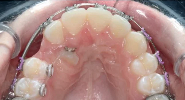
- Closed Eruption Technique: The canine crown is exposed surgically, and an orthodontic attachment (button or gold chain) is bonded. The flap is repositioned and sutured. Orthodontic traction then guides the tooth into the arch.
- Open Eruption Technique: The overlying tissue is removed to expose the crown, allowing the tooth to erupt naturally into the oral cavity before orthodontic alignment.
The closed eruption method is generally preferred for palatal impactions because it preserves attached gingiva and results in better periodontal outcomes.
2. Orthodontic Alignment
After exposure, light continuous forces (20–30 g) are applied to move the canine gradually into its proper position. The overall treatment may take 12–24 months, depending on the initial displacement and patient cooperation.
3. Considerations Before Surgery
- Adequate space must be available in the arch.
- Patient consent and motivation must be ensured.
- A realistic discussion regarding treatment duration and potential complications should be held.
Alternative Management Options
1. Autotransplantation
In cases where orthodontic alignment is not feasible, surgical transplantation of the canine into the correct position may be considered.
Success depends on:
- Open apex or incomplete root formation (ideal for revascularization).
- Minimal trauma to the periodontal ligament during extraction.
- Proper surgical technique and splinting.
Although promising, this method is technique-sensitive and carries a risk of ankylosis or root resorption.
2. Extraction of the Impacted Canine
If the impacted canine is severely displaced, or if orthodontic treatment is contraindicated (due to patient age, cost, or compliance issues), extraction may be the only option. In such cases, prosthetic replacement (fixed partial denture, implant, or orthodontic space closure) should be planned.
Resorption and Associated Complications
One of the most serious complications of impacted canines is root resorption of adjacent incisors. Studies indicate that up to 12% of lateral incisors and 4% of central incisors adjacent to impacted canines may exhibit resorption.
Mechanism
Resorption occurs due to a “head-on” collision between the canine crown and the incisor root during eruption. The resorptive process is often asymptomatic and can progress rapidly, necessitating radiographic vigilance.
Management of Resorption
If detected early, the clinician should consult a specialist orthodontist or oral surgeon to determine whether extraction of the impacted canine or the affected incisor is preferable.
In minor cases, the canine can be orthodontically redirected.
In severe cases, removal of the affected incisor may be required to allow eruption of the canine and to preserve arch integrity.
Follow-Up and Retention
Following exposure and alignment, long-term retention is essential to maintain results. The risk of relapse is minimal once the canine is properly positioned and functionally integrated, but retention may be required for:
- Maintenance of arch form.
- Prevention of spacing relapse.
- Stabilization of occlusion.
Retention may involve:
- Fixed bonded retainers.
- Removable retainers such as Hawley or Essix-type appliances.
Patients with unerupted canines that remain under observation should undergo periodic radiographic review every 12–18 months. If radiographs show cystic changes or progressive resorption, surgical intervention becomes necessary.
Prognosis
The prognosis of palatally displaced canines depends on several factors:
- Age of the patient: Earlier diagnosis yields better outcomes.
- Position and angulation: Canines positioned above the root apex of the lateral incisor have poorer prognosis.
- Root formation: Incomplete root formation is favourable for eruption.
- Available space and arch form.
- Patient cooperation and oral hygiene during treatment.
When managed appropriately, the majority of PDCs can be successfully aligned within the dental arch with good functional and aesthetic outcomes.
Prevention
Preventive orthodontic measures can reduce the risk of canine impaction:
- Routine palpation of the canine bulge during mixed dentition appointments (around 8–10 years).
- Timely extraction of retained deciduous canines when displacement is suspected.
- Space maintenance following premature tooth loss to prevent crowding.
- Regular radiographic screening when asymmetry or delayed eruption is observed.
Conclusion
Palatally displaced maxillary canines represent a common yet complex orthodontic problem. Early diagnosis, interceptive extraction of the primary canine, and appropriate surgical and orthodontic management are key to achieving optimal outcomes.
Failure to detect or manage these displacements in time can result in prolonged treatment, root resorption, and compromised aesthetics. Therefore, clinicians must remain vigilant during the mixed dentition phase and employ radiographic and clinical assessment judiciously.
References
- Becker, A., Smith, P. & Behar, R. (1981). The incidence of anomalous lateral incisors in relation to palatally displaced cuspids. Angle Orthodontist, 51(1), 24–29.
- Becker, A., Chaushu, S. (1996). Success rate and duration of orthodontic treatment for adult patients with palatally impacted maxillary canines. Angle Orthodontist, 66(4), 249–256.
- Ericson, S. & Kurol, J. (1988). Early treatment of palatally erupting maxillary canines by extraction of the primary canines. European Journal of Orthodontics, 10(4), 283–295.
- Ericson, S. & Kurol, J. (1987). Radiographic assessment of maxillary canine eruption in children with clinical signs of eruption disturbance. European Journal of Orthodontics, 9(3), 178–192.
- Power, S.M. & Short, M.B. (1993). An investigation into the response of palatally displaced canines to the removal of deciduous canines and an assessment of factors contributing to favourable eruption. British Journal of Orthodontics, 20(3), 215–223.
- Parkin, N., Benson, P.E., Thind, B., Shah, A. & Khalil, S. (2012). Extraction of primary (deciduous) teeth for unerupted palatally displaced permanent canines. Cochrane Database of Systematic Reviews, Issue 12, CD004621.
- Alqerban, A., Jacobs, R., Fieuws, S. & Willems, G. (2011). Comparison of two cone beam computed tomography systems versus panoramic imaging for localization of impacted maxillary canines and detection of root resorption. European Journal of Orthodontics, 33(1), 93–102.
- Baccetti, T., Leonardi, M. & Armi, P. (2008). A randomized clinical study of two interceptive approaches to palatally displaced canines. European Journal of Orthodontics, 30(4), 381–385.
- Lindauer, S.J., Rubenstein, L.K., Hang, W.M., Andersen, W.C. & Isaacson, R.J. (1992). Canine impaction identified early with panoramic radiographs. Journal of the American Dental Association, 123(3), 91–92.
- Dacre, J.T. (1983). The unerupted maxillary canine—A review of the literature and report of 28 cases. British Journal of Oral Surgery, 21(4), 212–218.