
Dental caries remains one of the most prevalent chronic diseases worldwide, affecting individuals across all age groups. The introduction of restorative dental materials has transformed the practice of operative dentistry by allowing clinicians to restore function, aesthetics, and structural integrity of carious or fractured teeth. Despite advancements in restorative techniques and materials, one persistent challenge in restorative dentistry is microleakage, the microscopic passage of bacteria, fluids, molecules, or ions between the cavity wall and the restorative material.
Microleakage is not just a laboratory curiosity; it has significant clinical implications, including postoperative sensitivity, marginal discoloration, recurrent caries, pulpal irritation, and ultimately, restoration failure. A restorative material that perfectly bonds to tooth structure and prevents microleakage has long been the “ideal,” but such perfection has not yet been achieved. Different restorative materials, including dental amalgam, resin composites, glass ionomer cements, resin-modified glass ionomers, and newer bulk-fill or bioactive materials, exhibit varying degrees of microleakage depending on their physical, chemical, and handling properties.
This article explores the concept of microleakage in detail, reviews the factors influencing it, and provides a comparative analysis of microleakage among commonly used restorative materials, with particular emphasis on composite resins and dental amalgam.
Table of Contents
ToggleDefinition of Microleakage
Microleakage is defined as the clinically undetectable passage of bacteria, fluids, molecules, or ions at the interface between the cavity wall and the restorative material. Unlike gross marginal gaps that can be seen visually or probed, microleakage is subtle and often detected only under laboratory conditions using dye penetration, radioactive isotopes, bacterial infiltration, or scanning electron microscopy.
Microleakage is distinct from nanoleakage, which refers to fluid and ion passage at the nanometer scale within the hybrid layer or resin tags, particularly in adhesive restorations. While microleakage relates more to marginal integrity, nanoleakage involves the microscopic porosities within the adhesive interface.
Clinical Significance of Microleakage
Microleakage poses several risks for the long-term success of dental restorations:
- Recurrent Caries – The seepage of oral fluids and bacteria beneath a restoration creates a niche for cariogenic bacteria to proliferate, leading to secondary caries, the most common cause of restoration failure.
- Postoperative Sensitivity – When bacterial toxins or fluids penetrate dentinal tubules, patients may experience transient or prolonged sensitivity to temperature changes, pressure, or sweet stimuli.
- Pulpal Inflammation – Long-standing microleakage may contribute to reversible or irreversible pulpitis due to continuous microbial irritation.
- Marginal Discoloration – Pigments from food or beverages can infiltrate marginal gaps, compromising aesthetics, especially in anterior teeth.
- Corrosion or Degradation of Materials – Some restorative materials undergo corrosion or breakdown when exposed to microleakage fluids, further aggravating marginal breakdown.
- Mechanical Failure – Weakening of the tooth-restoration bond can accelerate marginal chipping, fracture, or loss of retention.
Given these implications, reducing microleakage is a primary goal in restorative dentistry.
Factors Affecting Microleakage
Microleakage is influenced by numerous factors:
1. Material Properties
- Coefficient of thermal expansion (CTE) compared to tooth structure.
- Dimensional changes during setting (expansion or contraction).
- Adhesive ability and bonding mechanism.
- Solubility and water sorption.
2. Tooth Structure
- Enamel vs. dentin margins: enamel provides better bonding than dentin due to its high mineral content.
- Presence of sclerotic dentin or dentin permeability.
- Proximity to pulp, affecting dentinal tubule density.
3. Cavity Design and Preparation
- Beveling of enamel margins improves composite adaptation.
- Sharp internal line angles or unsupported enamel promote marginal breakdown.
- Depth and location (occlusal vs. cervical margins).
4. Restorative Technique
- Proper isolation and moisture control (saliva and blood contamination dramatically reduce adhesion).
- Incremental layering in composites to reduce polymerization shrinkage stress.
- Adequate condensation and carving in amalgam.
5. Oral Environment
- Thermal cycling from food and beverages causes expansion/contraction stresses.
- Mechanical stresses from mastication.
- pH fluctuations that may weaken adhesive bonds.
Methods of Assessing Microleakage
Laboratory studies employ various techniques to evaluate microleakage, as direct clinical observation is challenging:
- Dye Penetration – Immersing restorations in dye solutions (e.g., methylene blue, basic fuchsin) and sectioning teeth to evaluate dye penetration under a microscope.
- Radioisotope Penetration – Tracing radioactive isotopes (e.g., 45Ca, 32P) to detect microscopic penetration.
- Bacterial Infiltration – Using bacterial cultures to test whether microorganisms can penetrate marginal gaps.
- Scanning Electron Microscopy (SEM) – Provides high-resolution images of marginal adaptation.
- Fluid Filtration Technique – Measures the rate of fluid movement across the restoration-tooth interface.
Each method has limitations, but together they provide insights into the sealing ability of different restorative materials.
Microleakage in Amalgam Restorations
Properties of Dental Amalgam
Dental amalgam has been used for over 150 years and remains one of the most durable restorative materials. It is an alloy of mercury with silver, tin, copper, and sometimes zinc. Amalgam does not chemically bond to tooth structure; instead, it relies on mechanical retention from cavity design.
Microleakage Characteristics
- Initial Leakage: Freshly placed amalgam exhibits considerable microleakage because no chemical bond exists.
- Self-Sealing Effect: Over time, corrosion products (such as tin oxide and other metallic oxides) fill marginal gaps, reducing microleakage. This phenomenon explains the long-term clinical success of amalgam despite its initial poor sealing ability.
- Marginal Breakdown: With age, marginal fracture and ditching may occur, leading to recurrent leakage if corrosion cannot compensate.
Clinical Implications
Although amalgam is less aesthetic and non-adhesive, its unique ability to self-seal over time is an advantage. Microleakage in amalgam is usually highest immediately after placement and decreases within months. However, margins at cervical areas (cementum/dentin) are more prone to leakage compared to enamel margins.
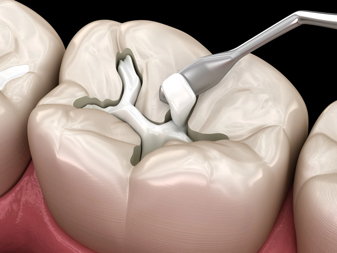
Microleakage in Composite Resin Restorations
Properties of Composite Resins
Composite resins are esthetic, tooth-colored materials composed of a resin matrix (commonly bis-GMA or UDMA), inorganic fillers (silica, glass), and a silane coupling agent. They are bonded to tooth structure using adhesive systems that create micromechanical retention through resin tags in etched enamel and a hybrid layer in dentin.
Microleakage Characteristics
- Polymerization Shrinkage: One of the main drawbacks of composites is volumetric shrinkage during polymerization (1.5–3%). This generates contraction stress, pulling the restoration away from cavity walls and creating marginal gaps.
- Adhesive Limitations: Despite advances in bonding agents (etch-and-rinse, self-etch, universal adhesives), the hybrid layer is susceptible to hydrolytic degradation and nanoleakage.
- Marginal Location: Restorations with margins on enamel show less leakage than those on dentin or cementum.
- C-factor (Configuration Factor): Cavities with high C-factor (ratio of bonded to unbonded surfaces, such as Class I cavities) exhibit more shrinkage stress and microleakage.
Techniques to Reduce Microleakage
- Incremental placement of composites to minimize polymerization stress.
- Use of flowable liners to absorb shrinkage stresses.
- Soft-start or pulse-delay curing methods.
- Development of bulk-fill composites with lower polymerization shrinkage.
- Use of bioactive composites with ion release that may promote remineralization.
Clinical Implications
Composites are more technique-sensitive than amalgam. Failure to achieve perfect bonding or control polymerization shrinkage often results in postoperative sensitivity and marginal staining. Despite continuous improvements, complete elimination of microleakage remains elusive.
Microleakage in Glass Ionomer and Resin-Modified Glass Ionomer Cements
Conventional Glass Ionomer Cement (GIC)
- Bonds chemically to tooth structure via ionic exchange between carboxyl groups of polyacrylic acid and calcium in hydroxyapatite.
- Releases fluoride, which may reduce secondary caries.
- Exhibits relatively high microleakage initially due to sensitivity to moisture and low mechanical properties.
Resin-Modified Glass Ionomer (RMGIC)
- Incorporates resin components to improve strength and reduce solubility.
- Shows better marginal sealing than conventional GIC but may still undergo polymerization shrinkage similar to composites.
Microleakage in Newer Restorative Materials
- Bulk-fill Composites: Designed to be placed in thicker increments with lower shrinkage stress. Studies show mixed results; some bulk-fills perform better, while others exhibit similar leakage to conventional composites.
- Bioactive Materials: Materials like giomers and “alkasite” composites (e.g., Cention N) claim to release ions (fluoride, calcium, phosphate) to strengthen tooth-restoration interfaces. Early studies suggest reduced secondary caries risk, though long-term microleakage outcomes are still under investigation.
- Adhesive Amalgam Restorations: Use of resin adhesives under amalgam may reduce initial microleakage, though clinical longevity varies.
Comparative Analysis
Amalgam
- High initial microleakage, but self-sealing over time due to corrosion products.
- Less sensitive to technique.
- Marginal ditching remains a weakness.
Composite Resin
- Strong aesthetic appeal but prone to polymerization shrinkage and nanoleakage.
- Performance highly technique-dependent.
- Better sealing on enamel margins than dentin/cementum.
Glass Ionomer Cements
- Chemical bonding and fluoride release reduce caries recurrence.
- Weaker mechanical properties, higher solubility, and more microleakage than composites.
Resin-Modified GICs
Better sealing than conventional GIC, but polymerization shrinkage remains an issue.
Strategies to Minimize Microleakage
- Proper case selection and cavity design.
- Use of rubber dam isolation to control moisture.
- Selection of adhesives with proven long-term stability.
- Incremental layering in composites.
- Light-curing protocols that minimize stress.
- Periodic clinical evaluation to monitor marginal integrity.
Conclusion
Microleakage remains one of the primary challenges in restorative dentistry, contributing to restoration failure, recurrent caries, and pulpal irritation. While dental amalgam demonstrates high initial microleakage but benefits from a self-sealing effect, composite resins—despite their aesthetic and adhesive advantages—are highly prone to microleakage due to polymerization shrinkage and adhesive degradation. Glass ionomer cements and resin-modified glass ionomers offer chemical bonding and fluoride release but remain less durable under mechanical stress.
Ongoing research into novel adhesives, bulk-fill composites, and bioactive restorative materials shows promise for reducing microleakage, but no material currently provides a completely hermetic seal. Clinicians must balance material properties, cavity location, patient needs, and restorative techniques to optimize outcomes. Ultimately, meticulous technique and an evidence-based approach to material selection are key to minimizing microleakage and maximizing the longevity of restorations.
References
- Aksornmuang, J., Foxton, R. M., Nakajima, M., & Tagami, J. (2004). Microtensile bond strength of a dual-cure resin core material to dentin with self-etching and total-etch adhesive systems. Journal of Dentistry, 32(2), 91–99.
- Al-Harbi, F., El Gedaily, M., & Sultan, A. (2020). Microleakage evaluation of bulk-fill resin composites in class II restorations: An in vitro study. Saudi Dental Journal, 32(3), 150–156.
- Borges, A. F. S., de Melo, M. A. S., Lima, J. P. M., & Rodrigues, L. K. A. (2018). Antimicrobial and remineralizing properties of bioactive glass and glass ionomer cement for dental applications. Dental Materials, 34(5), 710–718.
- Ferracane, J. L. (2011). Resin composite—State of the art. Dental Materials, 27(1), 29–38.
- Heintze, S. D., & Rousson, V. (2012). Clinical effectiveness of direct class II restorations—a meta-analysis. Journal of Adhesive Dentistry, 14(5), 407–431.
- Hegde, M. N., & Bhandary, S. (2008). An evaluation and comparison of microleakage in Class V cavities restored with different restorative materials: An in vitro study. Journal of Conservative Dentistry, 11(4), 127–131.
- Kumar, A., Kumar, V., & Kaushik, M. (2019). Comparative evaluation of microleakage of newer bulk-fill composites and conventional composites: An in vitro study. Journal of Conservative Dentistry, 22(1), 78–82.
- Mjör, I. A., & Gordan, V. V. (2002). Failure, repair, refurbishing and longevity of restorations. Operative Dentistry, 27(5), 528–534.
- Opdam, N. J., van de Sande, F. H., Bronkhorst, E., Cenci, M. S., Bottenberg, P., Pallesen, U., Gaengler, P., Lindberg, A., Huysmans, M. C. D. N. J. M., & van Dijken, J. W. V. (2014). Longevity of posterior composite restorations: A systematic review and meta-analysis. Journal of Dental Research, 93(10), 943–949.
- Reis, A. F., Giannini, M., & de Goes, M. F. (2004). Effect of water storage on microleakage of resin composite restorations. Operative Dentistry, 29(6), 667–672.
- Sidhu, S. K., & Nicholson, J. W. (2016). A review of glass-ionomer cements for clinical dentistry. Journal of Functional Biomaterials, 7(3), 16.
- Soares, C. J., Faria-E-Silva, A. L., Rodrigues, M. P., Fernandes-Neto, A. J., & Cavalcanti, A. N. (2019). Polymerization shrinkage stress of composite resins and resin cements—What do we need to know? Brazilian Oral Research, 33, e62.
- Tyas, M. J. (2005). Clinical evaluation of glass-ionomer cement restorations. Journal of Applied Oral Science, 13(4), 315–321.
- Van Meerbeek, B., Yoshihara, K., Van Landuyt, K., Yoshida, Y., & Peumans, M. (2020). From Buonocore’s pioneering acid-etch technique to self-adhering restoratives. Journal of Adhesive Dentistry, 22(1), 7–24.
- Yazici, A. R., Baseren, M., & Dayangaç, B. (2002). The effect of different types of flowable restorative resins on microleakage of Class V cavities. Operative Dentistry, 27(5), 500–505.
- Yilmaz, Y., & Bayrak, S. (2021). Evaluation of microleakage in different restorative materials: A systematic review. Clinical Oral Investigations, 25(10), 5985–6001.