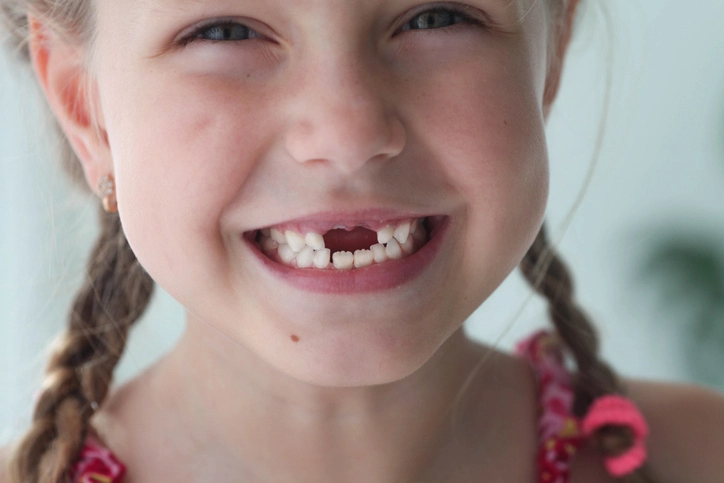
The management of missing incisors represents a significant clinical and psychological challenge in pediatric dentistry and orthodontics. Missing anterior teeth, particularly in the upper arch, can have a profound impact on a patient’s appearance, speech, and self-esteem. Because of the visibility of the incisors during speech and smiling, even a single missing incisor can dramatically affect facial aesthetics. In young patients, these concerns extend beyond cosmetics to issues of growth, occlusion, and long-term prosthetic planning.
Missing incisors may be congenitally absent—a developmental anomaly in which one or more teeth fail to form—or may be acquired, often following trauma, dilaceration, or extraction. Regardless of the cause, the clinician must carefully evaluate the individual’s occlusion, skeletal relationship, soft-tissue profile, and aesthetic expectations to determine the best management strategy. Options may include orthodontic space closure, space maintenance for future prosthetic replacement, or transplantation, depending on the patient’s age and developmental status.
Table of Contents
ToggleEtiology of Missing Incisors
Congenital Absence (Hypodontia)
Congenital absence of incisors is relatively uncommon, occurring in approximately 2% of the population. The upper lateral incisors are the most frequently missing anterior teeth. This developmental absence often occurs as part of a genetic pattern, sometimes associated with conditions such as ectodermal dysplasia, or it may occur sporadically. Congenitally missing teeth are more prevalent in females and may be unilateral or bilateral.
When an incisor fails to develop, compensatory dental and skeletal changes may occur, such as drifting of adjacent teeth, midline deviation, or altered crown morphology of the canines and central incisors. Early detection through radiographic evaluation allows for proactive planning of either orthodontic space closure or future prosthetic replacement.
Acquired Loss
Trauma is a major cause of missing incisors, particularly in children and adolescents. The upper central incisors are especially vulnerable due to their prominent position in the dental arch. Trauma can result in tooth avulsion, non-restorable fracture, or root resorption. In some cases, the developing tooth germ is damaged, leading to tooth dilaceration or non-eruption.
Following traumatic loss, the clinician must address both the aesthetic and functional concerns while considering the impact on alveolar bone development. Premature tooth loss can lead to alveolar bone resorption, complicating future implant placement and orthodontic mechanics.
Diagnostic and Planning Considerations
Successful management begins with a comprehensive evaluation that includes clinical examination, radiographic assessment, and aesthetic analysis. The following parameters are essential in the treatment planning process.
1. Skeletal Relationship
The underlying skeletal pattern significantly influences treatment choice.
- Class I relationship: Often allows flexibility between space closure or prosthetic replacement.
- Class II relationship: Space closure may accentuate the overjet, leading to an unbalanced facial profile.
- Class III relationship: Closing space in the upper arch may compromise the incisor relationship and negatively affect the overjet and overbite.
Patients with reduced lower facial height (LFH) may benefit from space closure to maintain facial balance, whereas those with increased LFH may require space maintenance and prosthetic replacement to preserve vertical proportions. Understanding the skeletal base and vertical dimensions ensures harmony between function and aesthetics.
2. Crowding and Spacing
Crowding or spacing within the arch dictates the feasibility of orthodontic space closure. In patients with adequate or excess spacing, space can be closed to achieve a symmetrical result. Conversely, in cases with crowding or minimal spacing, prolonged orthodontic retention may be necessary to prevent relapse.
Before initiating space closure, it is critical to ensure that adequate space remains for a prosthetic replacement if this is the chosen option. The minimum width required for an implant to replace an upper lateral incisor is approximately 6.5 mm, allowing for both the implant and appropriate bone support. Kesling set-ups or digital simulations are invaluable tools in visualizing final outcomes before treatment begins.
3. Colour and Form of Adjacent Teeth
The morphology and shade of the adjacent teeth—particularly the canines—play an essential role in achieving aesthetic balance. When lateral incisors are missing, orthodontists often consider moving the canine into the lateral incisor position. However, this can only be done successfully if the canine can be reshaped to mimic the lateral incisor form.
If the canine is significantly darker or larger than the natural lateral incisor, achieving aesthetic harmony may be difficult. Composite restorations, veneers, or enamel recontouring may be necessary to adjust shape and color. Similarly, when the central incisors are missing, the lateral incisors can sometimes be modified to simulate the central shape, provided their root length and gingival proportions are compatible.
4. Inclination of Adjacent Teeth
To achieve a natural appearance, the axial inclination of adjacent teeth must be carefully controlled during orthodontic treatment. The final inclination determines how the teeth reflect light, influencing both aesthetics and function. Misaligned axial positions can make replacement prostheses appear artificial or disrupt occlusal harmony.
5. Buccal Occlusion
The buccal occlusion must be preserved to maintain functional stability. In cases where a good Class I buccal interdigitation is present, orthodontic space closure should be approached cautiously to avoid compromising posterior occlusion. In contrast, if the buccal occlusion can accommodate mesial movement of the posterior teeth without detriment, space closure becomes a more viable option.
6. Unilateral vs. Bilateral Loss
When only one incisor is missing, achieving symmetry is often challenging. A symmetrical dental midline is aesthetically critical in anterior smile design. Therefore, in unilateral cases, it may sometimes be preferable to extract the contralateral tooth and treat both sides symmetrically. This approach provides better aesthetic balance and simplifies orthodontic alignment. If, however, the contralateral tooth is morphologically normal and the space is small, maintaining the natural tooth may be preferable with aesthetic restorative enhancement.
7. Smile Line and Gingival Contour
Patients with a high smile line expose the gingival margins when they smile, emphasizing discrepancies in gingival levels and crown length. In such cases, gingival recontouring or periodontal surgery may be required to achieve symmetry between the prosthetic or reshaped tooth and the natural dentition. Conversely, in patients with a low smile line, minor asymmetries are less noticeable and may not require surgical correction.
8. Patient’s Wishes and Expectations
The psychological impact of missing anterior teeth should not be underestimated. Adolescents, in particular, may experience embarrassment and self-consciousness. Therefore, patient preference and informed consent are vital in determining the treatment plan. The clinician must present all available options, including their benefits, limitations, costs, and long-term maintenance requirements, allowing the patient (and parents) to make an informed choice.
Treatment Modalities
1. Kesling Set-Up and Treatment Simulation
The Kesling set-up involves creating duplicate dental models and physically simulating tooth movement to visualize final outcomes. This process allows the clinician and patient to compare potential results for space closure, prosthetic replacement, or mixed approaches. Digital orthodontic planning tools now allow for virtual simulations, enabling precise prediction of both functional and aesthetic outcomes.
2. Orthodontic Space Closure
Space closure remains a preferred method in many cases due to its biological and aesthetic advantages. Studies show that patients are generally more satisfied with the results of space closure compared to prosthetic replacements, especially when tooth reshaping is performed skillfully.
Space closure can be achieved through controlled orthodontic movement of adjacent teeth. However, it is most feasible in cases with mild to moderate crowding or when there is a favorable occlusal and skeletal relationship. The clinician should pay careful attention to root angulation, crown morphology, and midline alignment.
When closing space to replace a lateral incisor, the canine is typically moved mesially and reshaped. The average difference in mesiodistal width between a canine and a lateral incisor is 1.2 mm, which can be adjusted by interproximal reduction. Composite additions to the mesial and distal aspects may also be used to refine the final tooth shape. Retention is crucial; a bonded retainer should be used to prevent reopening of space.
3. Space Maintenance and Opening
If space closure is not feasible or desirable, space maintenance becomes the priority. Following extraction or tooth loss, maintaining alveolar bone volume and interdental space is essential to allow for future prosthetic replacement or implant placement.
A palatal or acid-etch bridge can serve as an interim measure to maintain space while also restoring aesthetics. When a tooth is congenitally missing, orthodontic appliances may be used to open or preserve space. It is generally advisable to wait until skeletal growth is complete before placing a definitive prosthesis such as an implant.
The orthodontist must ensure sufficient occlusal clearance for any planned prosthesis. Following space creation, retention using a palatal retainer for 3–6 months helps stabilize the result before prosthetic work begins.
4. Resin-Bonded Bridges
Resin-bonded bridges (RBBs) are a conservative and aesthetic solution for replacing missing incisors, particularly in adolescents and young adults. They require minimal tooth preparation and rely on micromechanical retention through bonded metal or ceramic wings. Because they preserve enamel, RBBs are ideal for patients who are too young for implants.
Proper case selection is essential—adequate enamel surface, stable occlusion, and sufficient space are prerequisites. Advances in adhesive technology have improved the longevity of RBBs, making them a popular intermediate solution before definitive implant placement.
5. Autotransplantation
Tooth transplantation involves relocating a donor tooth—commonly a premolar—to the site of the missing incisor. This method is particularly beneficial in growing patients, as it allows preservation of alveolar bone and natural periodontal ligament function.
For example, a lower premolar can be transplanted to replace a missing upper incisor if the lower arch is crowded. The success of transplantation depends on careful case selection, atraumatic surgical technique, and rapid revascularization of the periodontal ligament. Long-term prognosis is generally favorable when root formation is incomplete at the time of transplantation.
6. Implants
Dental implants represent the gold standard for permanent tooth replacement once skeletal growth is complete. Implants provide superior aesthetics, stability, and longevity compared to removable or bonded prostheses. However, premature placement in growing individuals can lead to infraocclusion due to continued alveolar growth around the ankylosed implant.
Therefore, implants should only be considered after growth completion—usually in late adolescence. In the meantime, space must be preserved and bone volume maintained using temporary solutions such as RBBs or removable prostheses.
Retention and Long-Term Follow-Up
Retention is an indispensable part of post-treatment management. Whether space is closed or maintained, the risk of relapse remains high in anterior regions. Bonded retainers, fixed lingual wires, or removable retainers with aesthetic components help maintain stability.
Regular follow-up allows for assessment of gingival health, occlusal balance, and the integrity of prosthetic work. In implant cases, monitoring of peri-implant tissues and occlusal load distribution is critical for long-term success.
Aesthetic and Psychological Considerations
Beyond functional rehabilitation, restoring a patient’s confidence and self-image is a vital aspect of treatment. Missing incisors can have profound psychosocial effects, especially in children and adolescents who are highly sensitive to appearance-related concerns. Early intervention—through temporary prosthetics, cosmetic reshaping, or orthodontic adjustment—can help minimize these impacts during critical developmental years.
Furthermore, the clinician must consider factors such as lip support, facial symmetry, and gingival display to ensure a harmonious aesthetic outcome. Collaborative planning between the orthodontist, restorative dentist, and, where needed, a prosthodontist or oral surgeon is essential for achieving optimal results.
Conclusion
The management of missing incisors in pediatric dentistry is a multifaceted process requiring a delicate balance between aesthetics, function, and biological preservation. Each case demands individualized planning, taking into account the patient’s skeletal pattern, occlusal relationship, dental morphology, and personal expectations.
In growing patients, treatment decisions must be staged to accommodate ongoing development. Space closure offers a natural and stable solution in many cases, while prosthetic replacements—such as resin-bonded bridges, transplants, or implants—serve as excellent alternatives when orthodontic closure is not feasible.
Ultimately, the clinician’s goal is to restore not only the smile but also the confidence and oral health of the patient. By integrating orthodontic precision, restorative artistry, and patient-centered care, the management of missing incisors can achieve lasting aesthetic and functional harmony.
References
- Robertson, S., & Mohlin, B. (2000). Management of missing incisors. European Journal of Orthodontics, 22(6), 697–710.
- Polder, B. J., Van’t Hof, M. A., Van der Linden, F. P. G. M., & Kuijpers-Jagtman, A. M. (2004). A meta-analysis of the prevalence of dental agenesis of permanent teeth. Community Dentistry and Oral Epidemiology, 32(3), 217–226.
- Brook, A. H. (2009). Multilevel complex interactions between genetic, epigenetic and environmental factors in the aetiology of anomalies of dental development. Archives of Oral Biology, 54(Suppl 1), S3–S17.
- Björk, A., & Skieller, V. (1983). Normal and abnormal growth of the mandible: A synthesis of longitudinal cephalometric implant studies over a period of 25 years. European Journal of Orthodontics, 5(1), 1–46.
- Rosa, M., Zachrisson, B. U. (2001). Integrating esthetic dentistry and space closure in patients with missing maxillary lateral incisors. Journal of Clinical Orthodontics, 35(4), 221–234.
- Priest, G. (2011). Single-tooth implants and their role in preserving remaining teeth: A 10-year survival study. International Journal of Oral & Maxillofacial Implants, 26(4), 804–810.
- Kokich, V. O., Kinzer, G. A. (2005). Managing congenitally missing lateral incisors: Part I—Canine substitution. Journal of Esthetic and Restorative Dentistry, 17(1), 5–10.
- Kinzer, G. A., & Kokich, V. O. (2005). Managing congenitally missing lateral incisors: Part II—Tooth recontouring and implant considerations. Journal of Esthetic and Restorative Dentistry, 17(2), 76–84.
- Andersson, L., et al. (2012). International Association of Dental Traumatology guidelines for the management of traumatic dental injuries: 2. Avulsion of permanent teeth. Dental Traumatology, 28(2), 88–96.
- Czochrowska, E. M., Stenvik, A., Bjercke, B., & Zachrisson, B. U. (2002). Outcome of tooth transplantation: Long-term follow-up of 376 cases. American Journal of Orthodontics and Dentofacial Orthopedics, 121(2), 110–119.
- Garib, D. G., Alencar, B. M., Lauris, J. R. P., & Baccetti, T. (2010). Agenesis of maxillary lateral incisors and associated dental anomalies. American Journal of Orthodontics and Dentofacial Orthopedics, 137(6), 732.e1–732.e6.
- Malmgren, O., Czochrowska, E., & Zachrisson, B. U. (2012). Transplantation of teeth and implants in orthodontic treatment. Seminars in Orthodontics, 18(2), 110–120.
- Melsen, B., & Fiorelli, G. (2018). Orthodontic space closure and implant site development in patients with missing anterior teeth. Progress in Orthodontics, 19(1), 32.
- Papadopoulos, M. A. (Ed.). (2014). Skeletal Anchorage in Orthodontic Treatment of Class II Malocclusion: Contemporary Applications of Orthodontic Implants, Miniscrew Implants, and Mini Plates. Elsevier Health Sciences.
- Proffit, W. R., Fields, H. W., Larson, B., & Sarver, D. M. (2023). Contemporary Orthodontics (7th ed.). St. Louis: Elsevier.
- McDonald, R. E., Avery, D. R., & Dean, J. A. (2016). McDonald and Avery’s Dentistry for the Child and Adolescent (10th ed.). St. Louis: Elsevier.
- Zachrisson, B. U. (2007). Improving the esthetic outcome of orthodontic treatment by minimizing and camouflaging unavoidable soft tissue and tooth deficiencies. World Journal of Orthodontics, 8(3), 247–257.

