
Fixed Dental Bridges have long stood as one of the cornerstone treatments in restorative dentistry. They provide functional, aesthetic, and psychological benefits by replacing missing teeth and restoring a patient’s ability to chew, speak, and smile with confidence. Although implants have become increasingly popular in modern prosthodontics, bridges remain an essential component of dental treatment planning—particularly in cases where implant placement is contraindicated or undesirable.
Understanding the design, biomechanics, classification, and clinical requirements of dental bridges is vital for both dental students and practicing clinicians.
Table of Contents
ToggleFoundations of Bridgework
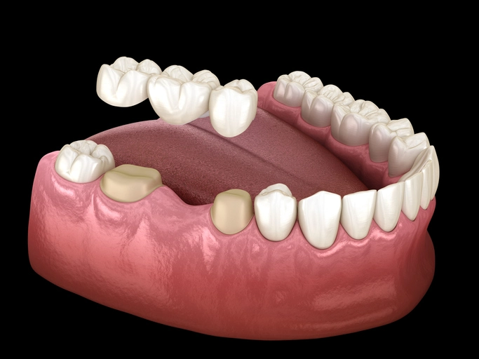
A bridge is a fixed partial denture designed to replace one or more missing teeth by joining artificial teeth to adjacent natural teeth or dental implants. Unlike removable dentures, bridges are cemented or bonded into place and cannot be removed by the patient.
To appreciate how bridges function, it is necessary to understand the primary components:
1. Abutment
An abutment is the supporting tooth or implant that bears the load of the bridge. The condition of the abutment, including its periodontal health, crown height, root morphology, and restorative status, determines whether it can successfully support prosthetic forces.
2. Retainer
The retainer is the restoration placed on the abutment tooth to provide retention for the bridge. Common retainers include full-coverage crowns, partial-coverage crowns, onlays, and inlays. The type chosen depends on the required retention, tooth structure, and design of the bridge.
3. Pontic
The pontic is the artificial tooth that replaces the missing natural tooth. It must restore aesthetics, function, and hygiene. Pontic design is a key factor in long-term periodontal health.
4. Connector
The connector is the element joining the pontic to the retainers. It may be rigid (commonly used in most bridges) or non-rigid (used in specific designs such as fixed–movable bridges to compensate for path-of-insertion mismatches).
5. Saddle (Ridge Area)
The saddle is the edentulous ridge area over which the pontic lies. Its anatomy influences pontic choice, emergence profile, and hygiene considerations.
6. Units
One unit corresponds to one tooth-sized component, whether abutment or pontic. A 3-unit bridge, for example, usually consists of 2 retainers and 1 pontic.
7. Retention, Support, and Resistance
- Retention prevents dislodgement in the occlusal/vertical direction.
- Resistance prevents movement in other directions (e.g., lateral or rotational).
- Support refers to the abutment tooth’s ability to sustain loading without biological or mechanical failure.
A successful bridge requires adequate retention and support, and its long-term stability depends on both the prosthesis and the oral environment.
Types of Bridges
Bridges can be categorized by their design, mode of attachment, and mechanical behaviour. Understanding each type helps clinicians choose an appropriate restoration for each clinical scenario.
1. Fixed–Fixed Bridge
A fixed–fixed design uses rigid connectors on both ends of the pontic. Both abutments contribute equally to retention and support.
Advantages
- Excellent stability
- Predictable load distribution
- Durable and widely applicable
Limitations
- Requires abutments with a common path of insertion
- Higher tooth reduction compared to adhesive bridges
Indications
- Robust abutments
- Long-term replacements
- Multi-unit restorations where rigidity is beneficial
2. Fixed–Movable Bridge
In this design, one end of the pontic attaches to a major retainer rigidly, while the other end uses a movable connector (e.g., a tenon–mortise design).
This is particularly useful when abutment teeth have non-parallel long axes.
Benefits
- Allows differential movement without stressing connectors
- Reduces need for aggressive tooth modification
- Provides good support in long-span bridges
Drawbacks
- More technically demanding
- Increased laboratory complexity
- Requires precise control of hinge mechanics
Clinically, fixed–movable bridges are often used when posterior abutments tilt mesially or when anatomical constraints prevent parallel preparations.
3. Cantilever Bridges
Direct Cantilever
Supported by only one abutment, the pontic extends from that single support tooth.
Pros:
- Minimal tooth preparation
- Useful in short-span, single-tooth replacements
Cons:
- Limited to areas with low occlusal forces (often anterior teeth)
- High risk of torque on abutment
Spring Cantilever
This design uses a long metal bar where the abutment and pontic are remote from each other—commonly used to replace an upper incisor when posterior teeth serve as abutments.
Rarely used today due to:
- Poor aesthetics
- Plaque retention
- Difficult cleaning
- Unfavourable leverage mechanics
4. Resin-Bonded Bridges (Minimal or No Preparation)
These bridges involve the bonding of metal or ceramic wings to enamel surfaces of abutment teeth.
Advantages
- Conservative
- Reversible in many cases
- High success rates when enamel bonding is optimal
Disadvantages
- Limited to specific occlusal situations
- Higher failure rates in posterior teeth
- Debonding is the most common complication
They are ideal for young patients, teeth with intact enamel, or temporary solutions.
5. Compound / Hybrid Bridges
A hybrid bridge combines multiple design philosophies—for example, combining a fixed–fixed and resin-bonded element within a single restoration. These are tailored to complex clinical scenarios.
Types of Retainers
1. Full-Coverage Crown
Provides the highest retention and resistance form.
Used when strong abutment support is needed or when existing restorations already require replacement.
2. Three-Quarter Crown
Covers most of the tooth while sparing the buccal enamel for aesthetics. Offers slightly less retention than full crowns.
3. Onlay
Covers one or more cusps. Retention is less than a full crown but conserves more tooth structure.
4. Inlay
Fits within the tooth; minimal coverage and least retentive.
Should only be used as a minor retainer in fixed–movable bridges.
Clinical Guideline:
Use the most retentive retainer compatible with the biological and structural condition of the abutment tooth.
Post crowns should be avoided unless absolutely necessary as they introduce a higher failure risk, especially in bridges.
Selecting Abutment Teeth
A meticulous assessment of the abutment teeth determines the success or failure of the bridge. Important considerations include:
1. Retention Factors
Retention depends largely on clinical crown height and surface area of prepared enamel.
Teeth with greater surface area provide better retention. Retention ranking (largest → smallest):
Maxillary Retention Ranking
6 > 7 > 4 > 5 > 3 > 1 > 2
Mandibular Retention Ranking
6 > 7 > 5 > 4 > 3 > 2 > 1
These rankings reflect differences in crown morphology, size, and accessibility for preparation.
2. Support Factors
Three key mechanical principles govern support:
A. Crown–Root Ratio
- Ideal: 2:3
- Acceptable: 1:1
Teeth with excessive bone loss (poor crown–root ratio) cannot withstand bridge loading.
B. Root Configuration
Wide, divergent roots resist forces better than fused or conical roots.
C. Periodontal Surface Area
Historically, Ante’s Law suggested that the support area of abutments must equal or exceed the missing teeth’s root surface area.
Modern dentistry rejects this rigid rule, recognizing that the periodontal ligament responds dynamically to load.
Support ranking:
Maxillary Support Ranking
6 > 7 > 3 > 4 > 5 > 1 > 2
Mandibular Support Ranking
6 > 7 > 5 > 4 > 2 > 3 > 1
3. Additional Factors
- Caries history and structural integrity
- Occlusal forces and parafunction
- Alignment and path of insertion
- Endodontic status
- Existing restorations
The best abutments are vital, healthy, structurally sound teeth with good alveolar support.
Taper and Parallelism in Abutment Preparation
Proper preparation geometry is crucial for optimal seating and retention of the bridge.
Ideal Taper
- 6° per wall
- 12° total convergence
Too much taper = insufficient retention
Too little taper = insertion impossible
Methods to Evaluate Parallelism
- Direct vision
- One-eye squint test (reduces stereoscopic depth perception)
- Surveyor use (most accurate)
When abutments are severely tilted, orthodontic correction, custom abutments, or a fixed–movable design may be required.
Pontic Design
Pontics must address aesthetics, function, phonetics, and hygiene. Poor pontic design can cause chronic inflammation, bad breath, and bridge failure.
Here are the main pontic designs:
1. Modified Ridge Lap Pontic
- Most popular design
- Minimal contact on buccal ridge surface
- Highly aesthetic
- Reasonably hygienic
Indication:
Anterior and premolar regions where aesthetics matter but hygiene must be maintained.
2. Bullet Pontic
- Point contact on ridge
- Excellent for hygiene due to minimal tissue contact
- Less aesthetic
Indication:
Posterior bridges where aesthetics are secondary.
3. Ovate Pontic
- Designed to sit into the soft tissue, creating a natural emergence profile
- Superior aesthetics
- Requires excellent oral hygiene
- Best used immediately after extraction or in implant-supported provisionalization to shape the gingival contour
Indication:
High-aesthetic anterior cases.
Modified ovate pontics are more labial and often used in cosmetic dentistry.
4. Hygienic (Sanitary) Pontic
- No tissue contact
- Easiest to clean
- Unesthetic
Indication:
Posterior molar areas.
5. Saddle (Ridge Lap) Pontic
- Covers ridge buccally and lingually
- Extremely difficult to clean
- Should be avoided due to high risk of inflammation and plaque accumulation
Biological and Mechanical Considerations
Bridges place unique demands on abutments. Even when periodontal tissues adapt via mechanoreception, excessive force can lead to mobility or failure.
Key principles include:
1. Distribution of Occlusal Forces
Avoid concentrating excessive forces on a single abutment, especially in cantilever designs.
2. Preservation of Tooth Structure
While full coverage provides superior retention, modern dentistry favours minimally invasive preparation wherever possible.
3. Soft Tissue Health
Pontic design, emergence profile, and finish line quality all influence gingival health.
4. Material Selection
Metal–ceramic, all-ceramic, and zirconia options each have unique advantages relating to strength, aesthetics, and bonding protocols.
Indications and Contraindications for Bridges
Indications
- Missing one or more teeth with suitable abutments
- Patient preference for a fixed option
- Insufficient bone for implants
- Medical contraindications to implant surgery
- Need for immediate aesthetic restoration
Contraindications
- Poor abutment prognosis
- Uncontrolled periodontal disease
- Extensive edentulous spans
- Severe parafunction (unless planned with metal occlusal surfaces)
- Poor oral hygiene compliance
Clinical Workflow for Bridge Fabrication
- Diagnosis and treatment planning
- Abutment evaluation and selection
- Tooth preparation
- Impression or digital scan
- Provisional restoration
- Laboratory fabrication
- Try-in (framework and full contour)
- Cementation or bonding
- Review and maintenance
Maintenance of Bridges
Patients should be instructed in:
- Interdental cleaning tools (superfloss, interdental brushes)
- Regular hygiene visits
- Awareness of signs of failure (looseness, sensitivity, odour)
Even the best-designed bridge can fail without meticulous home care.
Conclusion
Dental bridges remain a vital prosthodontic option and a cornerstone of restorative practice. Their success depends on careful case selection, thorough understanding of biomechanics, meticulous preparation, and patient compliance. By mastering the principles of bridge types, abutment selection, pontic design, and mechanical considerations, clinicians can deliver durable, functional, and aesthetic restorations tailored to each patient’s needs.
References
- Shillingburg HT, Hobo S, Whitsett LD, Jacobi R, Brackett SE. Fundamentals of Fixed Prosthodontics. 4th ed. Quintessence Publishing; 2012.
- Rosenstiel SF, Land MF, Fujimoto J. Contemporary Fixed Prosthodontics. 5th ed. Elsevier; 2015.
- Goodacre CJ, Bernal G, Rungcharassaeng K, Kan JYK. Clinical complications in fixed prosthodontics. J Prosthet Dent. 2003;90(1):31–41.
- Akkad A, Mendez R, Bomfim DI, Shakal KA. Fixed partial dentures: clinical considerations and factors influencing longevity. Journal of Dental Research and Review. 2018;5(3):87–94.
- Faucher RR, Nicholls JI. Distortion related to removable dies and impression materials in fixed partial denture fabrication. J Prosthet Dent. 1980;43(4):456–462.
- Jemt T. Fixed partial dentures supported by natural teeth and implants: a 10-year follow-up. Int J Prosthodont. 1991;4(4):347–354.
- Winkler S. Essentials of Complete Denture Prosthodontics. 3rd ed. Ishiyaku EuroAmerica; 2000.
(Pontic design principles relevant to fixed prosthodontics.) - Naylor WP. Introduction to metal-ceramic technology. In: Metal-Ceramic Technology for Dental Applications. Quintessence Publishing; 1992.
- Shillingburg HT, Kessler JC. Tooth Preparation. Quintessence Publishing; 2007.
(Detailed guidance on taper, convergence, and abutment design.) - Tylman SD, Malone WFP, Koth DL. Tylman’s Theory and Practice of Fixed Prosthodontics. 8th ed. Ishiyaku EuroAmerica; 1989.
- Young H, Kryshtalskyj B. Pontic design and hygiene considerations. Dent Clin North Am. 1989;33(4):815–839.
- Goodacre CJ, Naylor WP. Single-unit and fixed partial denture restorations: biomechanics and failure patterns. J Prosthodont. 1999;8(3):214–220.
- Meyenberg K, Lüthy H, Schärer P. Zirconia frameworks for fixed prosthodontics: performance and limitations. Int J Prosthodont. 1995;20(3):203–210.
(Material considerations for bridges.) - Pjetursson BE, Tan WC, Zwahlen M, Lang NP. A systematic review of the survival and complication rates of fixed partial dentures (FPDs). Clin Oral Implants Res. 2004;15(6):625–642.
- O’Brien WJ. Dental Materials and Their Selection. 4th ed. Quintessence Publishing; 2008.