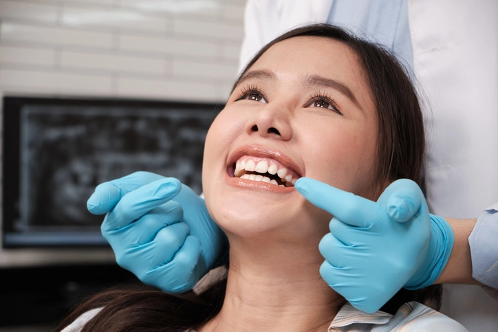
Orthodontic assessment is a vital part of dental examination and care. Its primary purpose is to ensure the early detection and management of any developmental abnormalities in the teeth and jaws. This assessment plays a pivotal role in guiding future orthodontic treatment, determining the prognosis of malocclusion, and preventing more complex problems later in life.
An effective orthodontic assessment combines systematic clinical examination, radiographic evaluation, and diagnostic record keeping. It also involves understanding the patient’s expectations, medical and dental history, and motivation for treatment. This article expands upon the standard procedures outlined in orthodontic assessment, delving deeper into each aspect—from screening to diagnosis and formulation of the problem list.
Table of Contents
TogglePurpose of Orthodontic Screening
The purpose of orthodontic screening is early detection and timely intervention. It helps the clinician identify deviations from normal growth and development, prevent the worsening of malocclusions, and guide the eruption of permanent teeth. Early screening allows clinicians to anticipate and manage issues such as delayed eruption, crowding, or skeletal discrepancies before they become more complex.
Key Objectives
- Early detection of abnormalities: Identifying potential orthodontic issues such as crowding, spacing, crossbites, or skeletal imbalances during mixed dentition.
- Timely referral and intervention: Recognizing when a condition requires specialist orthodontic management.
- Parental and patient education: Informing patients and guardians about the importance of maintaining oral hygiene and monitoring eruption sequences.
- Monitoring growth: Establishing a baseline record for evaluating changes in skeletal and dental development over time.
- Improving prognosis: Early recognition and correction of anomalies lead to more favorable long-term outcomes.
Timing and Frequency of Screening
Orthodontic screening should begin when the permanent incisors erupt and continue until the permanent dentition is fully established. At every dental visit, a basic orthodontic evaluation should be incorporated into the routine check-up.
Critical Observations
- Eruption sequence: The eruption sequence should follow normal developmental patterns. A delay of more than six months compared to the contralateral tooth or a deviation from the typical eruption order should prompt investigation.
- Eruption failure: Failure or delayed eruption might be due to crowding, impaction, or pathology.
- Overjet and overbite: Regularly observe for abnormal overjet or overbite, as they may indicate functional or skeletal imbalances.
- Quality of first permanent molars: These teeth are crucial anchors in orthodontic planning. Their condition must be monitored for caries, enamel defects, or premature loss.
- Asymmetry: Facial or dental asymmetry, especially a hollow or swelling in the buccal sulcus, warrants further investigation for pathology or displacement.
Gathering Information
An effective orthodontic assessment begins with thorough information gathering. This includes medical, dental, and social histories, as well as understanding the patient’s motivation and expectations.
Key Elements of Information Gathering
- Chief Complaint:
The clinician should understand what concerns brought the patient for evaluation. It may be esthetic concerns, difficulty chewing, speech problems, or parental observation of irregular teeth. - Patient and Parent Expectations:
Clarify what both the patient and parents hope to achieve through orthodontic treatment. This ensures realistic expectations and improves compliance. - Previous Dental or Orthodontic Treatment:
Knowledge of past extractions, restorations, or orthodontic procedures helps in treatment planning and understanding previous outcomes or complications. - Medical History:
Assess for systemic conditions that might affect treatment, such as growth disorders, cleft lip/palate, or medications that influence bone metabolism. - Oral Hygiene and Motivation:
Orthodontic success depends greatly on patient cooperation and oral hygiene. Poor motivation or oral hygiene can lead to complications such as decalcification or periodontal issues during treatment. - Lifestyle Factors:
Habits such as thumb sucking, mouth breathing, or tongue thrusting should be noted as they can influence malocclusion development.
Extraoral (EO) Examination
The extraoral examination focuses on assessing the patient’s facial symmetry, proportions, and soft tissue balance. It provides valuable insights into the skeletal and muscular framework that underlies dental occlusion.
Skeletal Pattern Assessment
Skeletal relationships are typically evaluated using the Frankfort plane, which runs horizontally from the lower border of the orbit to the upper margin of the external auditory meatus.
Anteroposterior (AP) Relationships
- Class I: Maxilla and mandible are harmoniously related.
- Class II: The maxilla is ahead of the mandible (retrusive mandible or protrusive maxilla).
- Class III: The mandible is ahead of the maxilla (protrusive mandible or retrusive maxilla).
Vertical Relationships
- The Frankfort–mandibular plane angle (FMPA) normally measures between 25–30°.
- A high FMPA indicates an increased lower facial height (long face), while a low FMPA suggests a short lower facial height.
- The lower face height is the distance from the base of the nose (subnasale) to the chin (menton) and should be proportional to the middle third of the face.
Transverse Relationships
Any facial asymmetry should be noted. Deviation of the chin point or midline discrepancies may indicate underlying skeletal or dental asymmetry.
Soft Tissue Evaluation
Soft tissue assessment includes observing lip competence, facial harmony, and profile balance.
- Lip competence: Evaluate whether lips can seal naturally without strain.
- Lip posture: Observe at rest—do lips meet or remain apart?
- Nasal and chin symmetry: Note any deviation or disproportion that affects facial balance.
- Smile analysis: Evaluate gingival display, symmetry, and incisal show.
- Muscular activity: Assess strain during lip closure or speech, as it may indicate an underlying discrepancy between skeletal and soft tissues.
Intraoral (IO) Examination
The intraoral examination focuses on the teeth, occlusion, oral hygiene, and supporting structures. It forms the foundation for diagnosing malocclusion and planning treatment.
Oral Hygiene and Gingival Health
Document oral hygiene status, gingival condition, and presence of any teeth with poor prognosis due to caries, mobility, or periodontal disease. Maintaining optimal oral health is a prerequisite for orthodontic treatment.
Dental and Occlusal Assessment
The occlusal examination involves analyzing the relationship of the upper and lower dental arches, alignment of individual teeth, and inter-arch relationships.
a. Lower Labial Segment (LLS)
- Assess inclination of incisors relative to the mandibular base.
- Evaluate crowding, spacing, and tooth rotations.
- Identify missing or displaced teeth.
b. Upper Labial Segment (ULS)
- Similar assessment for upper anterior teeth: inclination, alignment, spacing, rotations, or missing teeth.
- Examine the midline relationship between upper and lower central incisors.
c. Overjet and Overbite
Overjet (OJ): The horizontal distance between the labial surface of the lower incisors and the incisal edge of the upper incisors.
Normal OJ = 2–4 mm.
Increased OJ indicates proclined upper incisors or retroclined lower incisors (Class II tendency).
Negative OJ suggests anterior crossbite or Class III relationship.
Overbite (OB): The vertical overlap of upper incisors over lower incisors.
Normal OB ≈ one-third overlap.
Deep bite = excessive overlap; open bite = no vertical overlap.
d. Buccal Segments
Evaluate the relationship of molars and canines to determine the Angle classification:
- Class I, II (Division 1 or 2), or III.
Crowding should also be measured: - Mild (<4 mm), Moderate (4–8 mm), Severe (>8 mm).
e. Arch Symmetry and Alignment
Check whether the dental midlines coincide with facial midline. Any deviation could indicate skeletal asymmetry or localized dental shifts.
Diagnostic Records
Accurate diagnostic records are crucial for formulating treatment plans, monitoring progress, and evaluating outcomes. These records serve both clinical and medico-legal purposes.
Purpose of Diagnostic Records
- Aid in accurate diagnosis and case analysis.
- Facilitate planning and progress assessment.
- Serve as a medico-legal record and for audit or research.
Types of Diagnostic Records
a. Radiographs
Radiographs provide essential information on the teeth, jaws, and supporting structures.
1. DPT (Dental Panoramic Tomograph):
- Standard screening radiograph for most orthodontic cases.
- Reveals presence, position, and development of all teeth, including unerupted or impacted ones.
- Helps assess pathology, root morphology, and bone levels.
2. Cephalometric Radiographs:
- Used to evaluate skeletal and dental relationships in both the anteroposterior and vertical planes.
- Provides baseline measurements for treatment planning and growth analysis.
3. Periapical or Occlusal Views:
- Used when detailed information about specific teeth or regions is needed.
- Helpful for assessing impactions, root resorption, or anomalies.
b. Study Models
- Replicate the patient’s dentition and occlusion in three dimensions.
- Allow for accurate measurement of crowding, spacing, and arch symmetry.
- Digital 3D models are increasingly popular for their convenience and precision.
c. Clinical Photographs
- Extraoral photos: Frontal, profile, and smile views for facial analysis.
- Intraoral photos: Occlusal and buccal views to record dental alignment, arch form, and occlusion.
- High-quality color photographs are indispensable for diagnosis, monitoring, and documentation.
Diagnostic Analysis
Once clinical and radiographic data are collected, a thorough analysis is conducted to identify the nature and severity of the malocclusion. This includes:
- Skeletal analysis: Cephalometric measurements of maxilla and mandible.
- Dental analysis: Crowding, spacing, rotations, and inclination.
- Soft tissue analysis: Lip posture, profile harmony, and esthetics.
- Functional analysis: Examination of habits, mandibular movements, and occlusal interferences.
The integration of these findings allows for a comprehensive understanding of the patient’s orthodontic condition.
Problem List
After the diagnostic phase, information is systematically organized into a problem list, which serves as the foundation for treatment planning. The list is typically divided into pathological and developmental categories.
Pathological Problems
These are conditions requiring immediate or concurrent management before or during orthodontic treatment, such as:
- Caries or pulpal pathology.
- Periodontal disease.
- Missing or impacted teeth.
- Root resorption or trauma.
Developmental Problems
These relate to deviations in normal growth or alignment:
- Skeletal discrepancies (Class II or III relationships).
- Dental crowding or spacing.
- Malocclusion in AP, vertical, or transverse planes.
- Habits influencing development (e.g., thumb sucking, mouth breathing).
Formulation of the Treatment Plan
Once the problem list is complete, the clinician develops an individualized treatment plan. The plan should be realistic, evidence-based, and consider the patient’s motivation, growth potential, and cooperation.
Key Steps in Treatment Planning
- Define treatment objectives: Correct malocclusion, improve function, and enhance facial esthetics.
- Determine treatment timing: Early intervention (interceptive) vs. comprehensive orthodontic treatment.
- Select appliance type: Removable, fixed, or functional appliances based on diagnosis and age.
- Estimate duration and complexity: Provide a realistic treatment timeline.
- Evaluate retention phase: Plan retention strategy to maintain results.
Importance of Regular Monitoring
Orthodontic assessment is not a one-time process but a continuous one. Regular reviews are necessary to:
- Monitor eruption and occlusal development.
- Identify developing asymmetries or discrepancies early.
- Reassess treatment needs as the patient grows.
A systematic approach ensures that any deviation from normal development is addressed promptly, maintaining harmony between aesthetics, function, and health.
Conclusion
Orthodontic assessment forms the cornerstone of preventive and corrective orthodontics. It combines clinical observation, radiographic evaluation, and diagnostic analysis to provide a holistic view of the patient’s dental and facial development. Early and systematic assessment helps clinicians anticipate problems, formulate effective treatment plans, and achieve optimal functional and esthetic outcomes.
An ideal orthodontic assessment is comprehensive—it looks beyond teeth to consider the entire craniofacial complex, including soft tissues, skeletal bases, habits, and psychosocial factors. By maintaining a structured and patient-centered approach, orthodontists can deliver care that is both scientifically sound and personally satisfying for each individual.
References
- Proffit, W. R., Fields, H. W., Larson, B. E., & Sarver, D. M. (2018). Contemporary Orthodontics (6th ed.). St. Louis: Elsevier.
- Mitchell, L., Carter, N. E., Doubleday, B., O’Brien, K. D., & Sandler, P. J. (2013). An Introduction to Orthodontics (4th ed.). Oxford University Press.
- Graber, L. W., Vanarsdall, R. L., Vig, K. W. L., & Huang, G. J. (2017). Orthodontics: Current Principles and Techniques (6th ed.). Elsevier.
- Bishara, S. E. (2001). Textbook of Orthodontics. Philadelphia: W.B. Saunders Company.
- Moyers, R. E. (1998). Handbook of Orthodontics (4th ed.). Year Book Medical Publishers.
- British Orthodontic Society (BOS). (2022). Clinical Guidelines for Orthodontic Assessment and Treatment Planning. London: BOS Publications.
- Isaacson, R. J., & Murphy, T. D. (2015). Growth modification of the face: Current concepts and clinical applications. American Journal of Orthodontics and Dentofacial Orthopedics, 148(1), 1–16.
- Houston, W. J. B., Stephens, C. D., & Tulley, W. J. (2013). A Textbook of Orthodontics. Wiley-Blackwell.
- Kokich, V. G., & Spear, F. M. (2006). Guidelines for managing the orthodontic-restorative patient. American Journal of Orthodontics and Dentofacial Orthopedics, 130(1), 1–10.
- Littlewood, S. J., Mitchell, L., & Greenwood, D. C. (2019). Retention procedures in orthodontics: A contemporary review. British Dental Journal, 226(11), 835–843.
- McNamara, J. A. (2000). Components of Class II malocclusion in children 8–10 years of age. Angle Orthodontist, 70(3), 177–190.
- Riolo, M. L., Moyers, R. E., McNamara, J. A., & Hunter, W. S. (1974). An Atlas of Craniofacial Growth: Cephalometric Standards from the University School Growth Study. University of Michigan.
- Tulley, W. J. (1986). A Manual of Orthodontics (4th ed.). Wright PSG.
- Thilander, B., & Lennartsson, B. (2019). Clinical and diagnostic aspects of malocclusions. European Journal of Orthodontics, 41(1), 1–10.
- Ackerman, J. L., & Proffit, W. R. (1997). Soft tissue limitations in orthodontics: Treatment planning guidelines. Angle Orthodontist, 67(5), 327–336.

